This website uses cookies to ensure you get the best experience on our website.


- Table of Contents
RPL27A is a cytoplasmic component of the large ribosomal subunit (UniProt). This guide covers paraffin-section IHC planning and interpretation using the cytoplasmic staining profile reported across most tissues (HPA tissue IHC).

Expected localisation, validated protocols, controls and antibodies — the at-a-glance facts below, then the full design guide.
| Expected localisation | Cytoplasmic staining in most tissues (HPA tissue IHC) | |
| Staining pattern | Cytoplasmic signal in most tissues, including glandular cells (HPA tissue IHC) | |
| Antigen retrieval | Citrate pH 6.0 HIER, 95–98 °C, 20 min (rule: cytoplasmic / membrane antigen) | |
| Positive control | Cerebellum+4 more · see all | |
| Negative control | Skeletal muscle+1 more · see all |
| Fixation | Keep fixation consistent across paraffin sections (standard IHC practice; not target-specific); Matched tissue-IHC evidence does not establish the fixation claim. Validate the specimen-specific method before use. (selected-SKU IHC image A07864-1) | |
| Caveat | Medium staining–RNA concordance; verification pending (HPA tissue IHC) | |
| Regulation | Low tissue specificity (HPA tissue RNA) | |
| Isoform / epitope | No isoforms annotated; one chain spans residues 1–148 (UniProt) |
The catalog antibody's IHC-P protocol is paired with a published RPL27A tissue microarray protocol using HCC and peritumoral liver samples (PMC10360225).
| Sample | Paraffin-embedded human colorectal carcinoma tissue; fixative not specified (datasheet A07864-1) |
| Fixation | Image fixative and duration unreported (datasheet A07864-1); verify before use. |
| Sectioning | 4–5 µm sections on charged slides (standard) |
| Deparaffinisation | Xylene, graded ethanol series to water (standard) |
| Antigen retrieval | Heat-induced epitope retrieval in citrate buffer, pH 6.0, 20 min at 95–98 °C (standard rule: cytoplasmic / membrane antigen) |
| Peroxidase block | 3% H2O2, 10 min, room temperature (standard) |
| Blocking | 10% normal serum of the secondary host, 30 min, room temperature (standard) |
| Primary antibody | Rabbit anti-RPL27A, 1:50-1:200 (datasheet A07864-1) |
| Primary incubation | Overnight at 4 °C (standard) |
| Detection | HRP-polymer secondary, DAB chromogen 5–10 min (standard) |
| Counterstain | Hematoxylin, blue, dehydrate and mount (standard) |
| Expected result | RPL27A-positive staining in purkinje cells - cytoplasm/membrane of cerebellum (HPA tissue IHC: High). HPA tissue profile: Cytoplasmic expression in most tissues. No signal in the no-primary control. |
RPL27A is a cytoplasmic component of the large ribosomal subunit with no transmembrane segment (UniProt P46776). In paraffin tissue IHC, expect cytoplasmic staining across many tissues, especially the listed high-staining glandular cells and cerebellar Purkinje cells (HPA tissue IHC). HPA rates the tissue pattern Supported, with medium agreement between staining and RNA data and external verification pending (HPA tissue IHC).
| Glandular cytoplasm stains strongly in endometrium, pancreas, or salivary gland. | This matches HPA High staining in endometrial glandular cells, pancreatic exocrine glandular cells, and salivary glandular cells (HPA tissue IHC). Assess the named cells and their cytoplasm when judging whether a positive control has worked. |
| Purkinje cells show strong cytoplasmic staining, sometimes with a membrane pattern. | HPA records High staining in Purkinje cells as cytoplasm/membrane (HPA tissue IHC). A membrane component in these cells can fit that observation; interpret it alongside the expected cytoplasmic signal (UniProt P46776; HPA tissue IHC). |
| Signal is predominantly nuclear, or confined to cell borders outside the described Purkinje pattern. | That distribution does not match UniProt's cytoplasmic localisation or HPA's broad tissue profile (UniProt P46776; HPA tissue IHC). Treat it as a possible staining artefact and check the control slide before assigning RPL27A positivity. |
| Myocytes stain strongly while nearby expected positive cells do not. | HPA reports RPL27A as Not detected in skeletal myocytes and smooth muscle cells (HPA tissue IHC). Such a reversal raises concern for antibody cross-reactivity or endogenous chromogenic activity; the pattern alone cannot distinguish their causes. |
| Diffuse colour obscures cell boundaries, or a listed High tissue has no detectable signal. | Diffuse colour limits compartment scoring; absent signal in a listed positive tissue calls for a technical check (HPA tissue IHC). Because tissue IHC support has only medium staining/RNA consistency, one discordant section is not definitive evidence of target absence (HPA tissue IHC). |
| Cellular compartment | UniProt places RPL27A in the cytoplasm and identifies it as a large ribosomal subunit component (UniProt P46776). Score cell-associated cytoplasmic colour first; HPA also records cytoplasm/membrane staining specifically in Purkinje cells (HPA tissue IHC). |
| Reference tissue and cell type | HPA lists High staining in endometrial, pancreatic exocrine, and salivary glandular cells, but Not detected in skeletal and smooth muscle cells (HPA tissue IHC). Compare like cell types within each section; a whole-tissue average can hide the relevant pattern. |
| Strength of tissue evidence | Two listed rabbit polyclonal antibodies, HPA060776 and HPA063379, have Supported IHC status (HPA antibodies). The tissue profile itself has medium agreement with RNA data and awaits external verification (HPA tissue IHC), so use discordance as a reason to investigate. |
| IF/ICC: where should signal appear? | In the separate IF/ICC application, HPA reports enhanced localisation to endoplasmic reticulum and cytosol; both listed antibodies have Enhanced ICC status (HPA subcellular; HPA antibodies). This observation helps interpret fluorescence images without changing the tissue IHC scoring reference. |
| Chromogenic detection controls | Endogenous enzyme activity and nonspecific reagent binding can contribute colour in chromogenic IHC (standard IHC practice). Include an appropriate negative detection control and examine whether colour follows the expected cell and compartment pattern; these controls do not establish target specificity by themselves. |
| Situation | Likely cause | Next action |
|---|---|---|
| No cytoplasmic colour in a listed High control tissue. | The run may have failed, or the sampled section may differ from HPA's observed pattern (HPA tissue IHC). | Check section integrity, retrieval and detection steps, then repeat with an appropriate positive control (standard IHC practice). Interpret persistent absence cautiously because HPA tissue IHC has medium staining/RNA consistency. |
| Colour appears across the slide, including spaces without cells. | Diffuse reagent background or chromogen deposition can obscure the expected cytoplasmic pattern (standard IHC practice). | Review blocking, washes, detection reagents and a negative detection control; score RPL27A only where cell-associated colour can be assessed against HPA's cytoplasmic profile (HPA tissue IHC). |
| Skeletal or smooth muscle cells stain strongly. | HPA records these cells as Not detected; cross-reactivity or endogenous detection activity is possible (HPA tissue IHC; standard IHC practice). | Inspect the negative detection control and compare an expected positive cell population in the same run. Do not assign specificity from muscle staining alone. |
| Most colour is nuclear. | A dominant nuclear pattern conflicts with the reported cytoplasmic location (UniProt P46776; HPA tissue IHC). | Recheck counterstain and chromogen interpretation, inspect control slides, and reassess whether distinct cytoplasmic colour is present before calling the section positive. |
| Purkinje cell borders show colour alongside cytoplasm. | HPA describes High Purkinje staining as cytoplasm/membrane (HPA tissue IHC). | Check that cytoplasmic staining accompanies the border signal, then compare with other expected cells. Border colour alone cannot establish specificity. |
| IHC appears broadly cytoplasmic, while an IF/ICC image looks more structured. | HPA tissue IHC reports cytoplasmic expression in most tissues, whereas HPA ICC-IF places RPL27A in endoplasmic reticulum and cytosol (HPA tissue IHC; HPA subcellular). | Interpret each image using its application-specific HPA localisation. Use cytoplasmic cell staining for the paraffin IHC call; consult the separate IF/ICC guide for fluorescence experiments. |


Comprehensive Human Protein Atlas IHC scoring per tissue (reliability: Supported — Medium consistency between antibody staining and RNA expression data. Pending external verification.). Rows are taken directly from the HPA tissue chart — click any row's HPA link to view the source.
| Tissue | Cell type | Level | Evidence | Source |
|---|---|---|---|---|
| Cerebellum | Purkinje cells - cytoplasm/membrane | High | Protein (IHC) | HPA → |
| Endometrium | Glandular cells | High | Protein (IHC) | HPA → |
| Pancreas | Exocrine glandular cells | High | Protein (IHC) | HPA → |
| Salivary gland | Glandular cells | High | Protein (IHC) | HPA → |
| Stomach | Glandular cells | Medium | Protein (IHC) | HPA → |
Troubleshoot RPL27A staining in paraffin sections by checking retrieval, cytoplasmic distribution, tissue context and control staining before scoring.
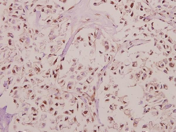
One anti-RPL27A antibody has real IHC data from paraffin-embedded human colorectal carcinoma; its listed reactivity includes human, mouse, and rat, with no IF data supplied (catalog: A07864-1).
A07864-1 has an IHC image of paraffin-embedded human colorectal carcinoma stained at 1:50 (image caption: A07864-1). It is listed for IHC and reacts with human, mouse, and rat; no IF image is supplied (catalog: A07864-1).
Which to pick: For paraffin-section tissue IHC, choose A07864-1: its own image shows human colorectal carcinoma at 1:50, within the listed 1:50–1:200 IHC range; the fixative is unreported (image caption: A07864-1; catalog: A07864-1). No SKU in this payload is listed for IF/ICC (catalog: A07864-1 applications). For mouse or rat tissue IHC, A07864-1 lists reactivity with both species, though its IHC image shows human tissue only; it is rabbit-hosted, and its caption identifies it as a pAb (catalog: A07864-1; image caption: A07864-1).