This website uses cookies to ensure you get the best experience on our website.


- Table of Contents
Plan RPN1 chromogenic IHC in paraffin sections using its cytoplasmic tissue staining profile (HPA tissue IHC) and ER membrane topology (UniProt). Compare strongly stained Paneth or pancreatic exocrine cells with weaker muscle cells (HPA tissue IHC), and start antibody A05063-2 at 2–5 μg/ml (datasheet).

Expected localisation, validated protocols, controls and antibodies — the at-a-glance facts below, then the full design guide.
| Expected localisation | Cytoplasmic staining (HPA tissue IHC); ER membrane (UniProt) | |
| Staining pattern | Cytoplasmic staining across tissues; intensity varies by cell type (HPA tissue IHC) | |
| Antigen retrieval | EDTA pH 8.0 HIER, heat-mediated (datasheet A05063-2) | |
| Positive control | Duodenum+4 more · see all | |
| Negative control | None in HPA (detected in all 45 tissues); use no-primary + isotype controls |
| Fixation | Keep fixation consistent across paraffin sections. (standard IHC practice; not target-specific) | |
| Caveat | Muscle cells may stain weakly despite broad tissue expression (HPA tissue IHC) | |
| Regulation | Broad tissue expression; no specific regulator reported (UniProt) | |
| Isoform / epitope | No isoforms annotated; lumenal 24–438, cytoplasmic 458–607 (UniProt) |
The catalog antibody protocol is paired with two published RPN1 IHC protocols using paraffin sections (PMC11693735; PMC11201601).
| Sample | Paraffin-embedded human liver cancer tissue; fixative not specified (datasheet A05063-2) |
| Fixation | Image fixative and duration unreported (datasheet A05063-2); verify before use. |
| Sectioning | 4–5 µm sections on charged slides (standard) |
| Deparaffinisation | Xylene, graded ethanol series to water (standard) |
| Antigen retrieval | Heat retrieval: EDTA pH 8.0 (datasheet A05063-2); 20 min, 95–100 °C (standard) |
| Peroxidase block | 3% H2O2, 10 min, room temperature (standard) |
| Blocking | 10% goat serum (datasheet A05063-2) |
| Primary antibody | Rabbit anti-RPN1, 2-5 μg/ml (datasheet A05063-2) |
| Primary incubation | Overnight at 4 °C (datasheet A05063-2) |
| Detection | HRP-conjugated secondary, DAB chromogen (datasheet A05063-2) |
| Counterstain | Hematoxylin, blue, dehydrate and mount (standard) |
| Expected result | RPN1-positive staining in paneth cells of duodenum (HPA tissue IHC: High). HPA tissue profile: Cytoplasmic expression in all tissues. No signal in the no-primary control. |
RPN1 is an endoplasmic reticulum membrane protein with a lumenal region and cytoplasmic tail (UniProt P04843 topology). In paraffin sections, expect cytoplasmic staining across tissues, with stronger staining in selected cell types including pancreatic exocrine glandular cells and intestinal Paneth cells (HPA tissue IHC). HPA rates the tissue pattern Enhanced, while reporting medium consistency between antibody staining and RNA expression (HPA tissue IHC).
| Cytoplasmic staining is strongest in pancreatic exocrine glandular cells or intestinal Paneth cells (HPA tissue IHC). | This matches HPA's High staining in those cells and its broader cytoplasmic tissue pattern (HPA tissue IHC). Judge the distribution across cells as well as intensity; a strong color reaction alone does not establish specificity (general IHC practice). |
| Staining is predominantly nuclear, with little cytoplasmic signal. | A nucleus-dominant result conflicts with the reported cytoplasmic IHC pattern (HPA tissue IHC) and ER membrane location (UniProt P04843). Treat it as suspect and compare with a known-positive section and a primary-antibody omission control (general IHC practice). |
| Strong staining appears only in a cell population expected to stain weakly. | For example, HPA reports Low staining in cardiomyocytes and myocytes (HPA tissue IHC). Check cell identity and controls before calling the signal RPN1; cross-reactivity or endogenous detection activity can mimic target staining (general IHC practice). |
| Color spreads across tissue and empty areas without a clear cellular pattern. | That distribution does not resemble HPA's cytoplasmic cellular staining (HPA tissue IHC). Background may arise from nonspecific antibody binding or the detection system; inspect a primary-antibody omission control and review blocking and washing (general IHC practice). |
| A known-positive section shows no discernible cytoplasmic staining. | Pancreatic exocrine glandular cells and intestinal Paneth cells are reported High (HPA tissue IHC). If both sample and positive control fail, review the staining workflow before interpreting the sample as RPN1-negative (general IHC practice). |
| Tissue and cell selection | RPN1 is expressed in all tissues tested (UniProt P04843), while HPA reports Low tissue RNA specificity and cytoplasmic IHC staining across tissues (HPA tissue IHC). Use a reported High cell population as a positive reference; weak staining elsewhere does not by itself establish absence. |
| ER membrane topology | RPN1 has a lumenal segment at residues 24–438, a transmembrane segment at 439–457, and a cytoplasmic region at 458–607 (UniProt P04843 topology). The payload does not identify the catalog antibody's epitope, so topology cannot predict its retrieval response. |
| Processing and modification | UniProt annotates a signal peptide at residues 1–23, a chain at 24–607, and one glycosylation site at 299 (UniProt P04843). These annotations do not establish how fixation or antigen retrieval affects staining. |
| Interpretation of melanosome evidence | UniProt also lists melanosomes based on mass spectrometry of stage I–IV fractions (UniProt P04843). That finding does not replace the mainly ER localization observed by ICC-IF or justify calling a distinct melanosomal IHC pattern here (HPA subcellular). |
| Strength of tissue evidence | HPA labels the tissue IHC pattern Enhanced but describes only medium consistency with RNA expression (HPA tissue IHC). Its High and Low calls are reference patterns for interpretation, not guaranteed intensities in every section or staining run. |
| IF/ICC Q: What pattern belongs on its separate guide page? | A: Mainly ER localization, with additional cytosolic signal, is reported by HPA ICC-IF (HPA subcellular). That cellular localization helps assess compartment agreement; the IF/ICC workflow and protocol choices belong on its own guide page. |
| Situation | Likely cause | Next action |
|---|---|---|
| Positive-control tissue is blank or much weaker than expected. | A failed staining run or insufficient detection can obscure a reported High population; HPA's High calls are reference observations, not run guarantees (HPA tissue IHC; general IHC practice). | Confirm the expected cell population is present, then review retrieval, antibody application, detection, and counterstain against the laboratory's validated IHC workflow (general IHC practice). Do not infer a target-specific fixation effect. |
| Only nuclei stain strongly. | The distribution disagrees with cytoplasmic tissue IHC and ER localization (HPA tissue IHC; UniProt P04843). Nonspecific staining or detection background is possible (general IHC practice). | Compare a reported High cell population with a primary-antibody omission control; review blocking and detection if nuclear color persists without primary antibody (HPA tissue IHC; general IHC practice). |
| Muscle cells appear stronger than the chosen High control. | HPA reports Low staining in cardiomyocytes and skeletal myocytes, versus High staining in several glandular or Paneth cell populations (HPA tissue IHC). Cell assignment or assay background may explain the reversal (general IHC practice). | Verify morphology and compare both populations in the same run; use a primary-antibody omission control to assess detection-derived color (general IHC practice). Avoid declaring the muscle signal specific from intensity alone. |
| Color is diffuse across tissue or appears outside cells. | Diffuse color lacks the reported cytoplasmic cellular pattern (HPA tissue IHC). Nonspecific binding, inadequate washing, or endogenous detection activity can contribute (general IHC practice). | Inspect the omission control, wash and block according to the validated workflow, and address endogenous activity when relevant to the chromogenic detection chemistry (general IHC practice). Reassess only cell-associated signal. |
| Staining is present, but the cell compartment is hard to judge. | Dense chromogen or counterstain can obscure cytoplasmic boundaries (general IHC practice); HPA describes the tissue signal as cytoplasmic (HPA tissue IHC). | Review a less saturated field and the counterstain, and compare with a reported High cell population (general IHC practice; HPA tissue IHC). Score localization only where cells remain distinguishable. |
| The slide suggests a distinct melanosomal pattern. | UniProt's melanosome annotation comes from fraction mass spectrometry, whereas HPA reports mainly ER localization in ICC-IF (UniProt P04843; HPA subcellular). | Prioritize the observed cytoplasmic IHC pattern and check controls before assigning a finer compartment (HPA tissue IHC; general IHC practice). Treat melanosomal identity as unresolved by this IHC evidence. |
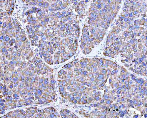
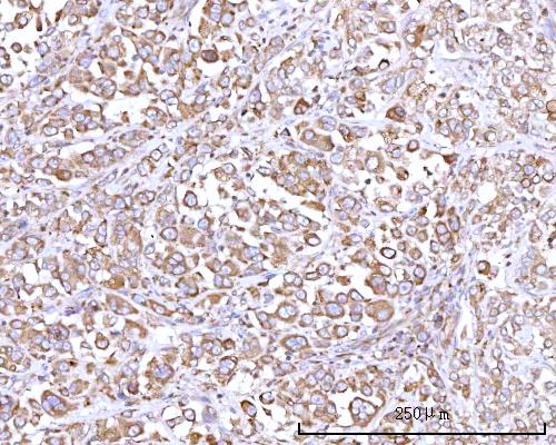


Comprehensive Human Protein Atlas IHC scoring per tissue (reliability: Enhanced — Medium consistency between antibody staining and RNA expression data.). Rows are taken directly from the HPA tissue chart — click any row's HPA link to view the source.
| Tissue | Cell type | Level | Evidence | Source |
|---|---|---|---|---|
| Duodenum | Paneth cells | High | Protein (IHC) | HPA → |
| Epididymis | Glandular cells | High | Protein (IHC) | HPA → |
| Pancreas | Exocrine glandular cells | High | Protein (IHC) | HPA → |
| Placenta | Trophoblastic cells | High | Protein (IHC) | HPA → |
| Skin | Fibrohistiocytic cells | High | Protein (IHC) | HPA → |
| Tissue | Cell type | Level | Evidence | Source |
|---|---|---|---|---|
| None in HPA: RPN1 is detected in all 45 scored tissues. Use a no-primary (secondary-only) and an isotype control instead. | ||||
Use the catalog antibody’s paraffin-section IHC conditions as the starting point, then judge staining against RPN1’s expected cellular distribution and tissue controls.
A05063-2 has human paraffin-section IHC images (A05063-2 IHC captions). M05063-1 lists human and rat reactivity and IF/ICC use, with no supplied image captions (M05063-1 catalog).
A05063-2 has IHC images from human liver cancer, lung adenocarcinoma, placenta and rectum adenocarcinoma paraffin sections (A05063-2 IHC captions). M05063-1 lists human and rat reactivity and IHC and IF/ICC applications; no IHC or IF image captions are supplied (M05063-1 catalog).
Which to pick: For human tissue IHC, choose A05063-2 for its paraffin-section images; its captions report EDTA retrieval at pH 8.0 and 2 μg/ml primary antibody, but do not report the fixative (A05063-2 IHC captions). For IF/ICC, choose M05063-1 because those applications are listed for the rabbit monoclonal clone 32R32, although no IF image is supplied (M05063-1 catalog). M05063-1 is also the listed option for rat samples; A05063-2 lists human reactivity only (M05063-1 catalog; A05063-2 catalog).