This website uses cookies to ensure you get the best experience on our website.


- Table of Contents
Plan chromogenic RPS28 IHC in paraffin sections using the cytoplasmic tissue pattern (HPA tissue IHC). Duodenal glandular cells show high staining, while adipocytes are not detected (HPA tissue IHC); the catalog antibody's IHC dilution is 2–5 μg/mL (datasheet A10672).

Expected localisation, validated protocols, controls and antibodies — the at-a-glance facts below, then the full design guide.
| Expected localisation | Cytoplasmic staining in several tissues (HPA tissue IHC) | |
| Staining pattern | Cytoplasmic staining in neuronal, glandular and squamous cells (HPA tissue IHC) | |
| Antigen retrieval | EDTA pH 8.0 HIER, heat-mediated (datasheet A10672) | |
| Positive control | Caudate+4 more · see all | |
| Negative control | Adipose tissue+4 more · see all |
| Fixation | Keep fixation conditions consistent across sections. (standard IHC practice; not target-specific) | |
| Caveat | Staining and RNA expression show medium consistency (HPA tissue IHC) | |
| Regulation | No specific expression regulator annotated (UniProt) | |
| Isoform / epitope | No annotated isoforms; one 1–69 chain (UniProt) |
The catalog antibody protocol uses EDTA pH 8.0 retrieval (datasheet A10672). Three published IHC protocols provide tissue-specific staining conditions (PMC9117278; PMC10776981; PMC13578613).
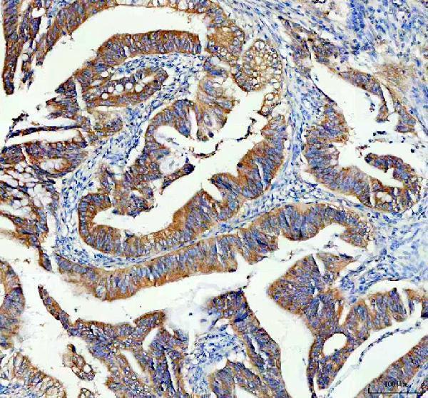
| Sample | Paraffin-embedded human colon cancer tissue; fixative not specified (datasheet A10672) |
| Fixation | Image fixative and duration unreported (datasheet A10672); verify before use. |
| Sectioning | 4–5 µm sections on charged slides (standard) |
| Deparaffinisation | Xylene, graded ethanol series to water (standard) |
| Antigen retrieval | Heat retrieval: EDTA pH 8.0 (datasheet A10672); 20 min, 95–100 °C (standard) |
| Peroxidase block | 3% H2O2, 10 min, room temperature (standard) |
| Blocking | 10% goat serum (datasheet A10672) |
| Primary antibody | Rabbit anti-RPS28, 2-5μg/ml (datasheet A10672) |
| Primary incubation | Overnight at 4 °C (datasheet A10672) |
| Detection | HRP-conjugated secondary, DAB chromogen (datasheet A10672) |
| Counterstain | Hematoxylin, blue, dehydrate and mount (standard) |
| Expected result | RPS28-positive staining in neuronal cells of caudate (HPA tissue IHC: High). HPA tissue profile: Cytoplasmic expression in several tissues. No signal in the no-primary control. |
RPS28 is a 40S ribosomal protein expected mainly in cytoplasm, including cytosol and rough endoplasmic reticulum; UniProt also lists the nucleolus (UniProt P62857). In tissue IHC, look for cytoplasmic staining in the cell types documented below (HPA tissue IHC). RPS28 has no transmembrane segment (UniProt P62857). HPA rates the tissue staining Approved, with medium consistency between antibody staining and RNA expression (HPA tissue IHC).
| Cytoplasmic staining in duodenal glandular cells or pancreatic exocrine glandular cells. | This matches HPA's High staining in those cell types and its overall cytoplasmic tissue profile (HPA tissue IHC). Score the specified cells, rather than treating the whole tissue as uniformly positive. |
| Predominantly nuclear or membrane-edge staining, with little cytoplasmic signal. | Recheck the result against HPA's cytoplasmic tissue pattern and UniProt's lack of a transmembrane segment (HPA tissue IHC; UniProt P62857). Nucleolar localization is listed by UniProt, so a nucleolar component alone does not establish an artefact (UniProt P62857). |
| Strong staining in adipocytes or cardiomyocytes. | HPA reports RPS28 as Not detected in those cells (HPA tissue IHC). Consider cross-reactivity or endogenous detection activity; compare a documented positive tissue and a control omitting the primary antibody before assigning the signal to RPS28. |
| Diffuse color across cells and surrounding tissue, without a clear cytoplasmic pattern. | The distribution is difficult to reconcile with HPA's cytoplasmic profile (HPA tissue IHC). Check a control omitting the primary antibody and inspect blocking, washes and detection conditions as general IHC troubleshooting steps. |
| No signal in duodenal glandular cells despite a visible counterstain. | HPA reports High staining in those cells (HPA tissue IHC). First confirm that the expected cells are present; then review the antibody's IHC validation and the assay's retrieval and detection steps. A failed positive control cannot establish true absence. |
| Cell type and tissue (HPA tissue IHC) | HPA reports High staining in several neuronal, glandular and squamous epithelial cell types, but Not detected in adipocytes, cardiomyocytes and others (HPA tissue IHC). Choose and score controls at the cell-type level; a tissue name alone is insufficient. |
| Subcellular distribution (UniProt P62857; HPA tissue IHC) | Cytosol and rough endoplasmic reticulum are listed by UniProt, while HPA summarizes tissue staining as cytoplasmic (UniProt P62857; HPA tissue IHC). UniProt also lists the nucleolus; interpret any nuclear component alongside the dominant tissue pattern. |
| Antibody evidence (HPA antibodies; HPA tissue IHC) | HPA047132 is Approved for IHC, and the tissue profile has medium consistency with RNA expression (HPA antibodies; HPA tissue IHC). These ratings support use of the reference pattern but do not make unexpected staining proof of RPS28. |
| IF/ICC Q: What localization should I expect? | A: HPA reports approved cytosolic localization and lists A-431 and U2OS images; HPA047132 is Approved for ICC (HPA subcellular; HPA antibodies). Use the separate IF/ICC guide for that application. |
| Antigen retrieval and fixation | Target-specific fixation sensitivity and retrieval effects are unreported in the supplied UniProt and HPA records. Follow the IHC-validated antibody's documented workflow, and interpret retrieval changes as assay checks rather than established RPS28 behavior. |
| Situation | Likely cause | Next action |
|---|---|---|
| Documented positive cells are blank. | The cells may be absent from the section, or the IHC assay may have failed; duodenal glandular cells are High in HPA (HPA tissue IHC). | Confirm cell identity and run a documented positive tissue alongside the sample. Check antibody, retrieval, detection and counterstain steps using the chosen IHC workflow. |
| Only nuclei stain strongly. | A nuclear-only result differs from HPA's cytoplasmic tissue profile, although UniProt lists nucleolar localization (HPA tissue IHC; UniProt P62857). | Inspect whether signal is nucleolar and whether cytoplasm also stains. Repeat with a documented positive tissue and controls before calling the nuclear signal specific. |
| Adipocytes or cardiomyocytes stain strongly. | Both are Not detected in HPA tissue IHC; nonspecific binding or endogenous detection activity is possible (HPA tissue IHC). | Compare the suspect cells with a positive control and a control omitting the primary antibody. Review blocking and detection conditions if color persists without primary antibody. |
| Color is diffuse across the section. | A uniform haze obscures the cytoplasmic pattern described by HPA (HPA tissue IHC); excess background can arise during general IHC detection. | Inspect the control omitting the primary antibody. Review washes, blocking and detection exposure, then reassess whether individual expected cells retain cytoplasmic staining. |
| A presumed negative tissue shows a few positive cells. | HPA levels apply to named cell types, and its tissue IHC profile has medium consistency with RNA expression (HPA tissue IHC). | Identify the stained cells before scoring. Compare their compartment and distribution with a documented positive; do not label an entire section negative from its tissue name. |
| IF/ICC localization differs from the IHC section. | HPA reports approved cytosolic ICC-IF localization and a cytoplasmic tissue IHC profile (HPA subcellular; HPA tissue IHC). | Score each application against its own HPA reference and controls. Use the separate IF/ICC guide for imaging and assay setup. |


Comprehensive Human Protein Atlas IHC scoring per tissue (reliability: Approved — Medium consistency between antibody staining and RNA expression data.). Rows are taken directly from the HPA tissue chart — click any row's HPA link to view the source.
| Tissue | Cell type | Level | Evidence | Source |
|---|---|---|---|---|
| Caudate | Neuronal cells | High | Protein (IHC) | HPA → |
| Cerebral cortex | Neuronal cells | High | Protein (IHC) | HPA → |
| Duodenum | Glandular cells | High | Protein (IHC) | HPA → |
| Esophagus | Squamous epithelial cells | High | Protein (IHC) | HPA → |
| Hippocampus | Neuronal cells | High | Protein (IHC) | HPA → |
| Tissue | Cell type | Level | Evidence | Source |
|---|---|---|---|---|
| Adipose tissue | Adipocytes | Not detected | Protein (IHC) | HPA → |
| Heart muscle | Cardiomyocytes | Not detected | Protein (IHC) | HPA → |
| Ovary | Follicle cells | Not detected | Protein (IHC) | HPA → |
| Smooth muscle | Smooth muscle cells | Not detected | Protein (IHC) | HPA → |
| Soft tissue | Chondrocytes | Not detected | Protein (IHC) | HPA → |
Troubleshoot RPS28 chromogenic IHC by checking retrieval, cellular location, controls, and scoring against the documented paraffin-section workflow.
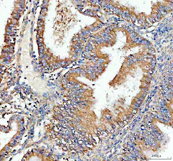
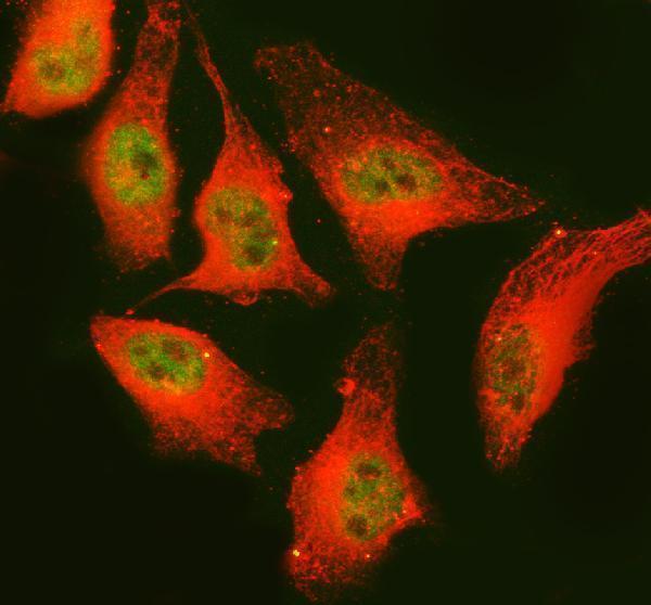
A10672 has real IHC data from human paraffin sections and IF/ICC data from A549 cells (catalog image captions); its listed reactivity includes human, mouse and rat (catalog reactivity).
A10672 has IHC images from human colon cancer and endometrial cancer paraffin sections (catalog IHC captions). Its IF/ICC image shows A549 cells, and its listed reactivity is human, mouse and rat (catalog IF caption; catalog reactivity).
Which to pick: Choose A10672 for paraffin-section chromogenic IHC: its human tissue captions report EDTA pH 8 retrieval, 2 μg/ml primary antibody and DAB detection; the fixative is unreported (catalog IHC captions). Choose A10672 for IF/ICC because its A549 image documents 5 μg/ml primary antibody and fluorescent detection (catalog IF caption). For mouse or rat samples, A10672 lists those species as reactive, but the supplied IHC and IF images document human samples only (catalog reactivity; catalog image captions).