This website uses cookies to ensure you get the best experience on our website.
- Table of Contents
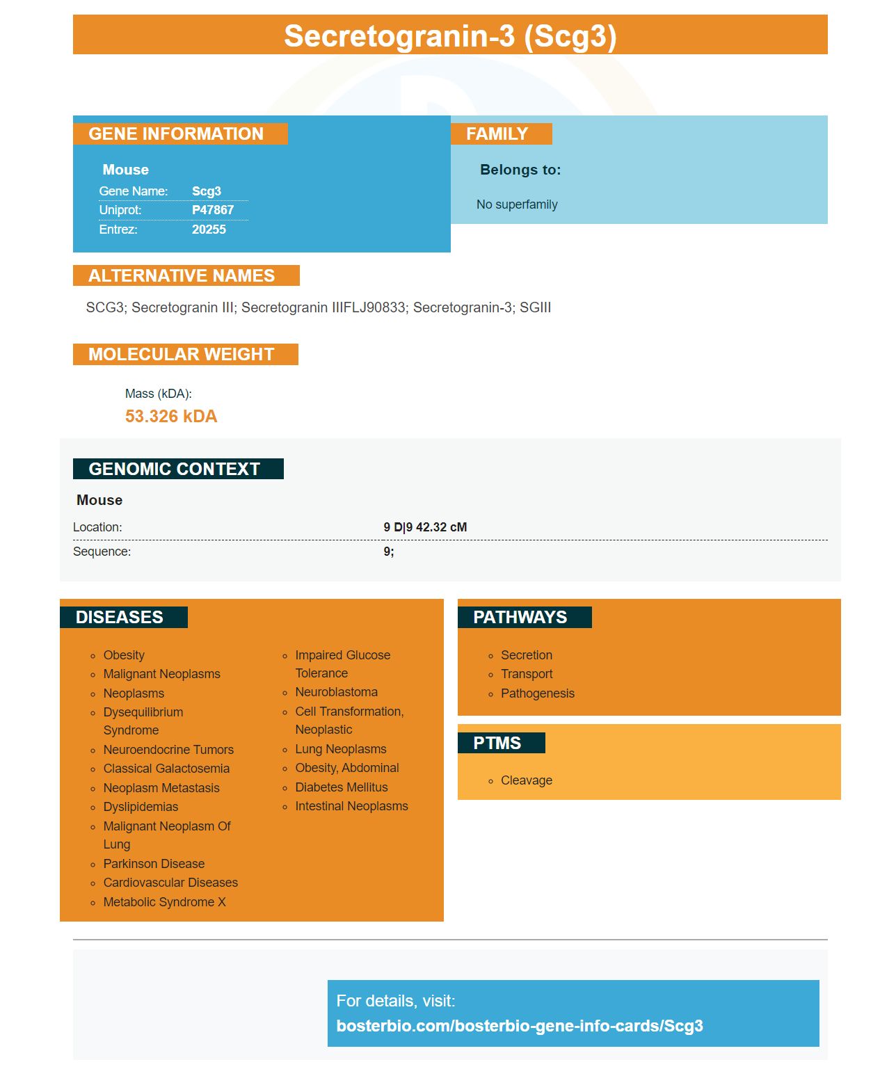
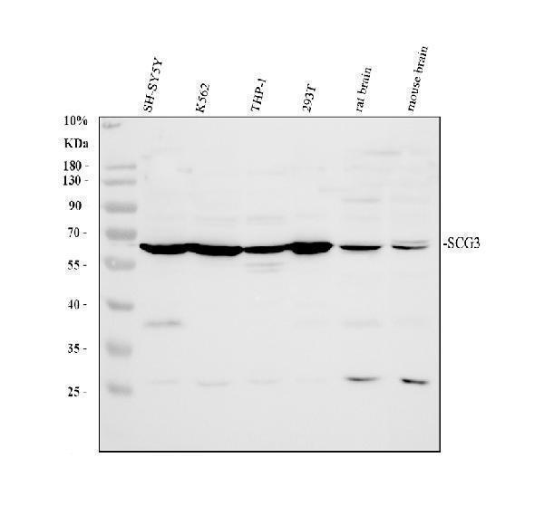
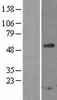
Facts about Secretogranin-3.
Mouse | |
---|---|
Gene Name: | Scg3 |
Uniprot: | P47867 |
Entrez: | 20255 |
Belongs to: |
---|
No superfamily |
SCG3; Secretogranin III; secretogranin IIIFLJ90833; Secretogranin-3; SGIII
Mass (kDA):
53.326 kDA
Mouse | |
---|---|
Location: | 9 D|9 42.32 cM |
Sequence: | 9; |
In this article we'll look at the Anti-Secretogranin 3 Marker Monoclonal Antibody and its applications. We'll examine the SCG3 marker from Boster Bio. These two monoclonal antibodies target the protein-secretion enzyme SCG3 that is found on erythrocytes.
Many suppliers offer Anti-Secretogranin/SCG3 Marker. This protein is encoded by the gene SCG3. Its predicted mass is 53 kDa . the two known isoforms are SCG3 and SGIII. It is found in humans and mice and canines or monkey, as well as rat.
Secretogranin III is a member of the granin protein family. It regulates the biogenesis of secretory granules. It has recently been discovered that it acts as an angiogenic factor. Studies have shown that Scg3 has the highest affinity for binding to diabetic eyes and the lowest background binding with the normal vasculature. Anti-Scg3 antibodies are also used to treat diabetic retinopathy in mice models of oxygen-induced retinal disease.
This protein is part of the secretory-granules family. It participates in many biological processes. SCG3 is responsible for releasing neurotransmitters. It also plays a part in the biogenesis secretory granules. Secretogranin III can be found in neuroendocrine tumors. It is associated with depression and anxiety, as well as atherosclerosis. It also encourages the proliferation of hepatocellular cancer cells.
Numerous studies have demonstrated that SCG3 is subcellularly localized within Astroglia that are activated with PQ. The subcellular distribution of SCG3 in U118MG astrocytes was assessed using immunoblotting. The levels of protein of SCG3 were then quantified by semi-quantitative densitometric analysis. In these studies, SCG3 secretion was compared with control cells.
The protein is commonly expressed in neuroendocrine tumors. While it isn't typically used to identify gliomas or other types, it is used in other methods of diagnosis. In a retrospective study of 223 patients, SCG3 protein expression was found. Its practical utility requires further research. The study also showed an association between SCG3 protein and the malignancy of glioma. It indicates a positive outcome for patients with GBM.
A preliminary proteomic study has identified the SCG3 protein, which has the 2.1-fold increase in expression in GBMs and the oligodendrogliomas. The SCG3 protein was identified with IHC in 267 primary glioma specimens. From 226 cases, 226 were quantified successfully. These cases represented 44 grade II, 38 grade III, and 144 grade IV gliomas.
A novel, humanized antigen called Anti-Glycophorin-A or CD235a was created to detect the expression of the erythrocyte marker B cells. The antibody is able to detect both endogenous and exogenous ZNF268 proteins. It is monoclonal and can detect specific types of tumors.
Boster Bio's monoclonal antibody against glycophorin-A/CD235 recognizes glycophorin-A/CD235a. It is a sialoglycoprotein in a single-pass membrane that is found on mature erythrocytes as in their precursor cells. This protein also carries blood group specificities, M, N S, U, and M. Recent studies have proven that CD235a is an adhesin receptor for Streptococcus, parvovirus and Sandei virus.
This monoclonal antibody recognizes CD235a as a type I sialoglycoprotein (10 kDa) which is located on the cell membrane of erythroid precursors. It is known to contain antigenic determinants of the MN and Ss blood groups and has been implicated in leukemia as well as hemopoiesis.
The cytotoxic activity in the cells that are the effectors is essential to ADCC treatment's efficacy. Although the (51)Cr Release Assay is the most widely used method to evaluate ADCC activity however, it isn't sensitive enough for distinguishing cell types within the target population. Therefore, flowcytometry is suggested to conduct this study.
Boster Bio's Antiglucophorin A and CD235a antibodies have been tested for use in various tests, including PCR and ELISA. In this assay, the antibodies were used in K562 cells. Negative control was created using IgG antibody.
The anti-Glycophorin antibody /CD235a (Erythrocyte Markers), is compatible with a wide variety of experimental models and exhibits excellent reproducibility. The stability of the antibody was evaluated in murine experiments as well as in tests using cord blood from humans. Boster scientists are able to submit their results by submitting their findings for specific species and applications.
This antibody is able to recognize blood type variants, including LU4 and LU8. It also blocks cell adhesion I+-5. It is also used for research to study the effects of sickle cell disease on vaso-occlusion. This antibody can be used in the treatment and detection of sickle cells.
We are currently studying the clinical effects of various antibodies against RBC. The most frequently reported adverse effects were in women who had antibodies to Db and anti-K. The combination of antibodies was more dangerous than the individual specificities. The results revealed that antibodies against the same antigens showed synergistic effects.
The immunoprecipitated fragments of immunoprecipitated Chin from K562 cells were immunoprecipitated with monoclonal antibodies against GATA-1. The GATA-1 complex was biotin-labeled and bound to the G1 site of competitive EMSAs. FOG-labeled competition could not bind to the G1 sequence. The DNA was amplified with specific primers for the promoter region, which contains the G1 site.
The SCG3 marker has many applications that include the detection of neuroblastoma in children as well as the distinction between NE and PGC in prostate cancer. It also plays a crucial function in the metabolism of catecholamines and its expression has been confirmed through IHC analysis. The marker has also been found to be highly expressed in gastroenteropancreatic tumors. Its use isn't restricted to a specific area of study. However it is intended for research purposes.
Although SCG3 is present in mature neurons, it is downregulated in glioma cells without a differentiation process. It is therefore crucial to investigate the role of SCG3 and its effect on the pathogenesis and diagnosis of gliomas. The SCG3 expression of the protein is a reliable biomarker for detecting gliomas and may be used as an adjunctive diagnostic tool. Further research is needed to determine whether SCG3 is effective in detecting gliomas.
SCG3 belongs to the secretory granule protein family. It is found in many tissues including the brain. It is responsible for controlling secretory granule biogenesis and transporting neurotransmitters. It could also play an important role in neuroendocrine functions, and is associated with a variety of diseases like obesity, atherosclerosis and depression. The SCG3 marker was recently used to detect tumors that involve the secretory system (e.g. liverocellular carcinoma).
A preliminary proteomic study identified the SCG3 marker, with an inverse correlation with the presence of glioma. The study employed the immunohistochemical staining method to identify SCG3 protein in glioma tissue. 267 primary glioma specimens were used for the study. The SCG3 protein was identified in 226 cases, including 44 grade II 38 grade III and 144 grade IV tumors.
A full marker application is due by July 1 of each year. A committee is able to review these applications during their annual meeting in late summer. An applicant can submit a second application in the event that they are not selected for this particular cycle. However it is not a guarantee. Every application will be scrutinized by the IHB to determine eligibility. Only the most reliable and top-quality applications will be accepted. It is essential that applicants provide reliable primary source materials.
The application form is the most straightforward part. It is all you need to do is provide your contact details, as well as the text. It will take about a year to complete the process. It will take approximately a year to complete the process. If you move and need to update the information, you'll need to contact the THC again. Once the application process is completed, the applicant will be issued an SCG3 marker. The SCG3 marker is a well-known method of recognizing an individual's achievements.
PMID: 7917832 by Dopazo A., et al. Primary structure of mouse secretogranin III and its absence from deficient mice.
PMID: 12388744 by Hosaka M., et al. Identification of a chromogranin A domain that mediates binding to secretogranin III and targeting to secretory granules in pituitary cells and pancreatic beta-cells.