This website uses cookies to ensure you get the best experience on our website.


- Table of Contents
Plan SEC63 paraffin IHC using the high staining in pancreatic exocrine glandular cells as a positive reference (HPA tissue IHC). This guide covers fixation consistency, chromogenic detection and scoring of the observed cytoplasmic pattern (HPA tissue IHC).

Expected localisation, validated protocols, controls and antibodies — the at-a-glance facts below, then the full design guide.
| Expected localisation | General cytoplasmic staining (HPA tissue IHC) | |
| Staining pattern | Exocrine glandular cells show cytoplasmic staining (HPA tissue IHC) | |
| Antigen retrieval | EDTA pH 8.0 HIER, heat-mediated (datasheet A04852-1) | |
| Positive control | Pancreas+4 more · see all | |
| Negative control | Oral mucosa+2 more · see all |
| Fixation | Keep fixation consistent across sections (standard IHC practice; not target-specific). Selected-image fixative and duration unreported (datasheet A04852-1); verify before use. | |
| Caveat | Hepatocytes stain low despite reported high liver expression (HPA tissue IHC; UniProt) | |
| Regulation | No specific expression regulator reported (UniProt) | |
| Isoform / epitope | 0 isoforms; epitopes may face the ER lumen or cytoplasm (UniProt) |
The catalog antibody protocol uses EDTA pH 8.0 retrieval (datasheet A04852-1). Two published SEC63 IHC protocols provide additional tissue-specific examples (PMC3508994; PMC2655295).
| Sample | Paraffin-embedded human thyroid cancer tissue; fixative not specified (datasheet A04852-1) |
| Fixation | Image fixative and duration unreported (datasheet A04852-1); verify before use. |
| Sectioning | 4–5 µm sections on charged slides (standard) |
| Deparaffinisation | Xylene, graded ethanol series to water (standard) |
| Antigen retrieval | Heat retrieval: EDTA pH 8.0 (datasheet A04852-1); 20 min, 95–100 °C (standard) |
| Peroxidase block | 3% H2O2, 10 min, room temperature (standard) |
| Blocking | 10% goat serum (datasheet A04852-1) |
| Primary antibody | Rabbit anti-SEC63, 2-5 μg/ml (datasheet A04852-1) |
| Primary incubation | Overnight at 4 °C (datasheet A04852-1) |
| Detection | HRP-conjugated secondary, DAB chromogen (datasheet A04852-1) |
| Counterstain | Hematoxylin, blue, dehydrate and mount (standard) |
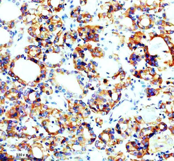
| Expected result | SEC63-positive staining in exocrine glandular cells of pancreas (HPA tissue IHC: High). HPA tissue profile: General cytoplasmic expression. No signal in the no-primary control. |
SEC63 is an endoplasmic reticulum membrane protein with three transmembrane segments (UniProt Q9UGP8 topology). In paraffin sections, expect cytoplasmic staining in pancreatic exocrine glandular cells, salivary glandular cells and testicular Leydig cells, each scored High by HPA (HPA tissue IHC). HPA describes the overall tissue profile as general cytoplasmic expression and rates its agreement with RNA expression as Supported (HPA tissue IHC).
| Cytoplasmic staining in pancreatic exocrine or salivary glandular cells. | This fits the reported High staining in those cells (HPA tissue IHC) and the expected ER location (UniProt Q9UGP8; HPA subcellular). Assess intensity in the named cells rather than treating every cell in the section as an equivalent positive control (HPA tissue IHC). |
| Predominantly nuclear staining, with little cytoplasmic signal. | A nuclear dominant pattern conflicts with the reported ER location and general cytoplasmic IHC profile (UniProt Q9UGP8; HPA tissue IHC). Treat it as suspect; inspect controls and detection background before assigning it to SEC63 (general IHC practice). |
| Strong staining in oral mucosal squamous cells or parathyroid glandular cells. | HPA reports SEC63 as Not detected in these cell types (HPA tissue IHC). Check cell identity and controls; an unexpected signal may reflect cross-reactivity or endogenous detection activity (general IHC practice). HPA's result does not establish absolute absence in every specimen. |
| Diffuse color across cells, stroma and empty areas. | Color without a cell associated cytoplasmic pattern is poor evidence of SEC63 localization (UniProt Q9UGP8; HPA tissue IHC). Review blocking, washes, chromogen exposure and a control omitting primary antibody (general IHC practice). |
| No staining in pancreatic exocrine glandular cells. | That conflicts with HPA's High tissue IHC result for those cells (HPA tissue IHC). First check that the cells are present and the run's positive control worked; a negative slide alone cannot establish SEC63 absence (general IHC practice). |
| Choice of positive and comparison tissue | Pancreatic exocrine glandular cells and salivary glandular cells are reported High; oral mucosal squamous cells are Not detected (HPA tissue IHC). Compare the specified cell populations, because a tissue level label does not score every constituent cell (HPA tissue IHC). |
| Antibody evidence | HPA053295 and HPA060535 each have Supported IHC status (HPA antibodies). This supports using their reported patterns as references, but it does not supply a dilution or show that a different catalog antibody has the same specificity (HPA antibodies). |
| Epitope location and accessibility | SEC63 has three membrane spans, a lumenal segment at residues 91–188 and a cytoplasmic region at 210–760 (UniProt Q9UGP8 topology). Interpret a known antibody epitope against that map; the supplied evidence gives no epitope for the catalog antibody or SEC63-specific retrieval requirement. |
| Processing and variants | UniProt lists one chain spanning residues 1–760, no signal peptide, no propeptide and no isoforms in this record (UniProt Q9UGP8). These entries offer no basis to predict a shed SEC63 signal or an isoform-specific tissue pattern. |
| Retrieval and chromogenic detection | For paraffin IHC, use the IHC-validated antibody's stated retrieval and dilution conditions if available, then assess controls alongside the section (general IHC practice). No SEC63-specific retrieval, fixation sensitivity or dilution is established by the supplied UniProt and HPA records. |
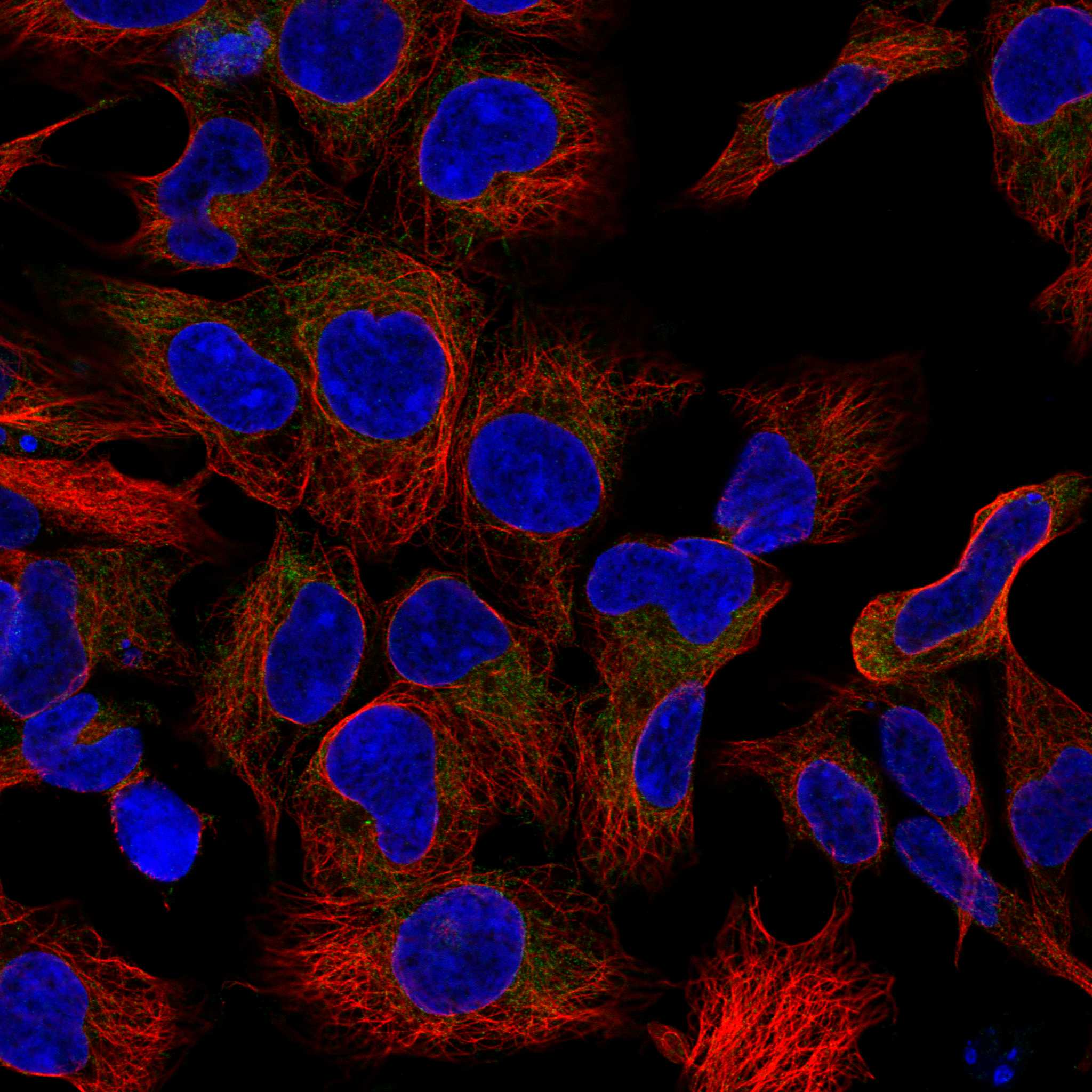
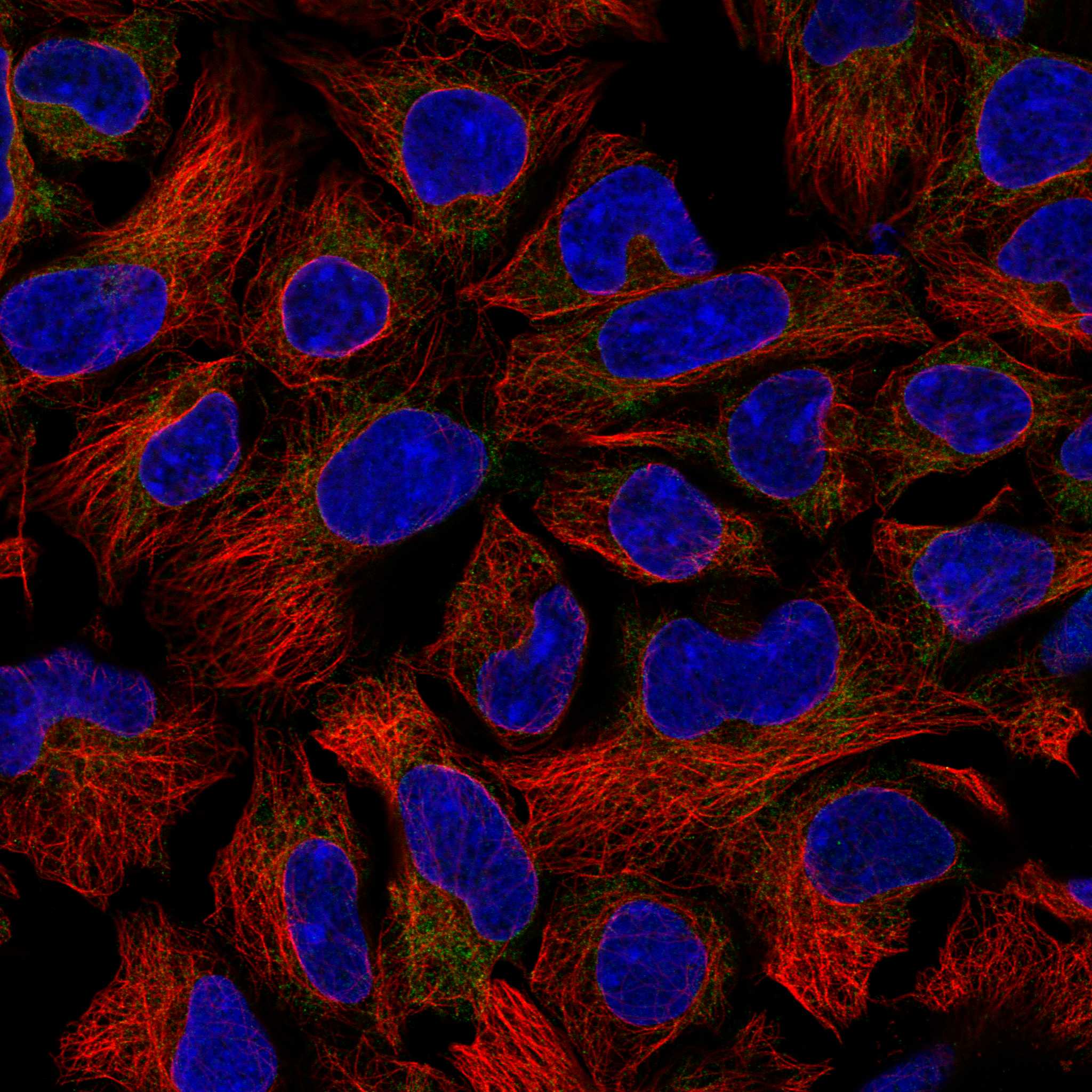
| IF/ICC Q: What localization should an IF experiment check? | A: Check for ER localization; HPA reports a supported ER main location in ICC-IF, consistent with UniProt's ER membrane annotation (HPA subcellular; UniProt Q9UGP8). IF/ICC has its own guide; this IHC section supplies no IF protocol option. |
| Situation | Likely cause | Next action |
|---|---|---|
| A known positive area is blank. | Pancreatic exocrine glandular cells are reported High, so a blank result needs investigation (HPA tissue IHC). | Confirm those cells are present; review the run's positive control, antibody preparation and detection steps before interpreting the section as negative (general IHC practice). |
| Signal is mostly nuclear. | That compartment conflicts with SEC63's ER membrane annotation and HPA's cytoplasmic IHC profile (UniProt Q9UGP8; HPA tissue IHC). | Compare with the expected cytoplasmic control and a no-primary control; review chromogen and counterstain before scoring (general IHC practice). |
| Oral mucosal squamous cells stain strongly. | HPA reports SEC63 as Not detected in those cells; cross-reactivity or endogenous detection activity is possible (HPA tissue IHC; general IHC practice). | Verify the cell type and inspect no-primary and detection controls. Do not infer specificity from intensity alone (general IHC practice). |
| Color covers stroma and empty areas. | This lacks the cellular cytoplasmic distribution expected from the HPA profile (HPA tissue IHC). Excess background is a possible technical cause (general IHC practice). | Inspect the no-primary control and review blocking, washing and chromogen exposure using the validated IHC workflow (general IHC practice). |
| Liver hepatocytes stain weakly despite UniProt's high liver expression statement. | The sources describe different measures: UniProt reports high levels in liver, while HPA scores hepatocyte IHC staining Low (UniProt Q9UGP8 tissue specificity; HPA tissue IHC). | Use the named pancreatic or salivary glandular cells as the stronger IHC reference; record hepatocyte staining against HPA's cell-specific Low result (HPA tissue IHC). |
| Two antibodies give different cell patterns. | HPA lists Supported IHC status for HPA053295 and HPA060535, but that status does not establish agreement for every specimen or a separate catalog antibody (HPA antibodies). | Compare matched sections and controls, and prioritize staining that tracks the reported cytoplasmic pattern in the named positive cells (HPA tissue IHC; general IHC practice). |


Comprehensive Human Protein Atlas IHC scoring per tissue (reliability: Supported — High consistency between antibody staining and RNA expression data.). Rows are taken directly from the HPA tissue chart — click any row's HPA link to view the source.
| Tissue | Cell type | Level | Evidence | Source |
|---|---|---|---|---|
| Pancreas | Exocrine glandular cells | High | Protein (IHC) | HPA → |
| Salivary gland | Glandular cells | High | Protein (IHC) | HPA → |
| Testis | Leydig cells | High | Protein (IHC) | HPA → |
| Adrenal gland | Glandular cells | Medium | Protein (IHC) | HPA → |
| Appendix | Glandular cells | Medium | Protein (IHC) | HPA → |
Troubleshoot SEC63 staining in paraffin sections by checking retrieval, cellular pattern, controls and scoring against its expected ER localisation (UniProt Q9UGP8; HPA subcellular).
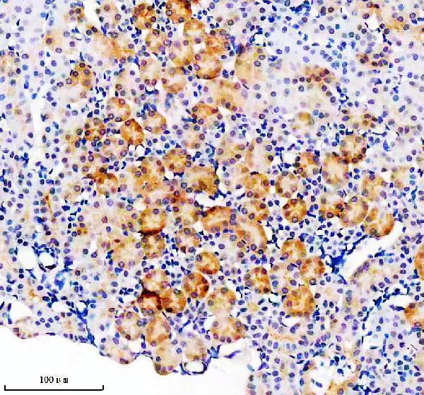
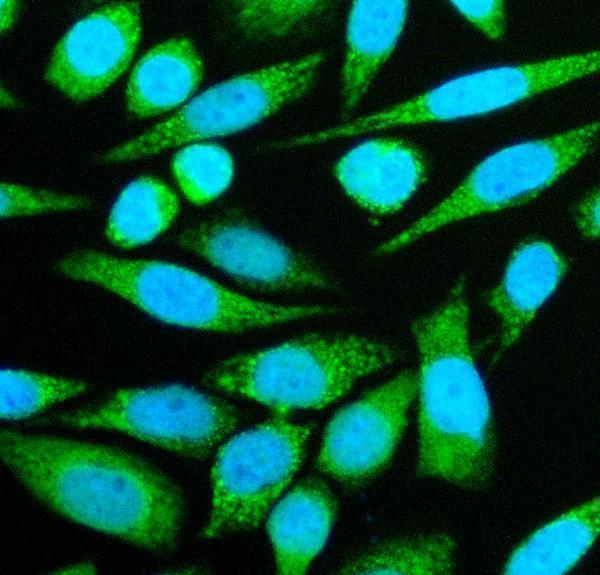
The catalog antibody has IHC images from paraffin sections of human thyroid cancer and mouse kidney, plus IF images from SIHA cells and human thyroid cancer sections (catalog image captions).
A04852-1 has IHC images from paraffin sections of human thyroid cancer and mouse kidney (catalog IHC captions). A04852-1 also has IF images from SIHA cells and paraffin sections of human thyroid cancer (catalog IF captions).
Which to pick: Choose A04852-1 for paraffin section IHC: its own captions document both tissue examples, although the fixative is unreported (catalog IHC captions). Choose the same SKU for IF/ICC because those applications are listed and its IF captions document SIHA cells and human thyroid cancer sections (catalog applications; catalog IF captions). For work across species, the catalog lists human, mouse and rat reactivity, but its IHC dilution listing covers human and mouse only; rat IHC is unverified here (catalog reactivity; catalog dilution listing).