SEL1L / Protein sel-1 homolog 1 · IHC design guide

Design Immunohistochemistry for SEL1L

Plan chromogenic SEL1L IHC using pancreatic exocrine glandular cells, which show high staining (HPA tissue IHC). Start the catalog antibody at 2–5 μg/mL (datasheet A03343-2) and assess cytoplasmic staining with appropriate controls (HPA tissue IHC; standard IHC practice).

Evidence assembled Oct 2026 · For research use; verify linked source records and product datasheet before use
Immunohistochemistry protocol sheet for SEL1L (IHC for SEL1L): expected localisation Cytoplasmic tissue staining (HPA tissue IHC); ER membrane localisation (UniProt), antibody A03343-2, validated IHC image, and IHC protocol steps
Printable SEL1L IHC protocol sheet — expected localisation Cytoplasmic tissue staining (HPA tissue IHC); ER membrane localisation (UniProt), antibody A03343-2, controls and protocol steps. Open the full SEL1L IHC guide →

SEL1L Immunohistochemistry Experimental Design Guide

Expected localisation, validated protocols, controls and antibodies — the at-a-glance facts below, then the full design guide.

Must know before staining
Expected localisation Cytoplasmic tissue staining (HPA tissue IHC); ER membrane localisation (UniProt)
Staining pattern Ubiquitous cytoplasmic staining (HPA tissue IHC)
Antigen retrieval EDTA pH 8.0 HIER, heat-mediated (datasheet A03343-2)
Positive control ⓘ Pancreas+4 more · see all
Negative control ⓘ None in HPA (detected in all 43 tissues); use no-primary + isotype controls
Important caveats
Reasons your staining may differ from the expected pattern.
Fixation Keep fixation consistent across sections (standard IHC practice; not target-specific)
Caveat Staining and RNA show medium consistency (HPA tissue IHC)
Regulation Pancreas enriched (UniProt)
Isoform / epitope 2 isoforms; map epitopes to lumenal or cytoplasmic regions (UniProt)
Section 1

Recommended SEL1L IHC & IF Protocols

The catalog antibody protocol uses EDTA retrieval (datasheet: A03343-2). The published IHC protocols below cover colorectal, pancreatic, and cartilage samples (PMC3345950; PMC8155860; PMC8782459).

Recommended immunohistochemistry (IHC-P) protocol parameters
SampleParaffin-embedded mouse pancreas tissue; fixative not specified (datasheet A03343-2)
FixationImage fixative and duration unreported (datasheet A03343-2); verify before use.
Sectioning4–5 µm sections on charged slides (standard)
DeparaffinisationXylene, graded ethanol series to water (standard)
Antigen retrievalHeat retrieval: EDTA pH 8.0 (datasheet A03343-2); 20 min, 95–100 °C (standard)
Peroxidase block3% H2O2, 10 min, room temperature (standard)
Blocking10% goat serum (datasheet A03343-2)
Primary antibodyRabbit anti-SEL1L, 2-5 μg/ml (datasheet A03343-2)
Primary incubationOvernight at 4 °C (datasheet A03343-2)
DetectionHRP-conjugated secondary, DAB chromogen (datasheet A03343-2)
CounterstainHematoxylin, blue, dehydrate and mount (standard)
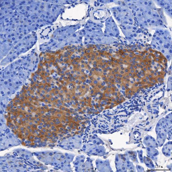
Expected resultSEL1L-positive staining in exocrine glandular cells of pancreas (HPA tissue IHC: High). HPA tissue profile: Ubiquitous cytoplasmic expression. No signal in the no-primary control.
💡Decision noteStart with heat-mediated EDTA, pH 8.0 (datasheet: A03343-2). Citrate retrieval is a published alternative for colorectal and pancreatic samples (PMC3345950; PMC8155860).
Section 2

What Is the Expected SEL1L Staining Pattern?

SEL1L is an endoplasmic reticulum membrane protein with a lumenal region spanning residues 22–738 and a transmembrane segment at 739–759 (UniProt Q9UBV2 topology). In paraffin section IHC, expect cytoplasmic staining, strongest in pancreatic exocrine glandular cells (HPA tissue IHC: High). HPA describes the tissue pattern as ubiquitous cytoplasmic expression and rates its IHC evidence Enhanced, with medium consistency between staining and RNA expression (HPA tissue IHC).

What am I looking at on my slide?
Strong cytoplasmic staining in pancreatic exocrine glandular cells (HPA tissue IHC: High).This matches the clearest supplied tissue positive and the expected compartment (HPA tissue IHC; UniProt Q9UBV2: endoplasmic reticulum membrane). Chromogenic IHC shows a cellular pattern; cytoplasmic staining alone cannot resolve the endoplasmic reticulum membrane.
Moderate cytoplasmic staining in adipocytes or the listed glandular, ciliated, or neuronal cells (HPA tissue IHC: Medium).Expression outside the pancreas can be expected: HPA reports a ubiquitous cytoplasmic profile and Medium staining in these listed cell populations (HPA tissue IHC). Compare intensity within each tissue; pancreatic intensity is not a universal cutoff for calling another tissue positive.
Predominantly nuclear staining, with little corresponding cytoplasmic signal.Treat this as a compartment mismatch, since the supplied localisation is an endoplasmic reticulum membrane protein and the observed tissue profile is cytoplasmic (UniProt Q9UBV2 localisation; HPA tissue IHC). Check staining controls and morphology before assigning the signal to SEL1L (general IHC practice).
Prominent signal in an unexpected cell population, especially if the expected pancreatic exocrine pattern is absent.Consider nonspecific antibody binding or endogenous detection activity (general IHC practice). HPA reports Low staining in several named cell populations, but supplies no negative tissue; staining in those cells alone cannot establish cross-reactivity (HPA tissue IHC).
Diffuse stain across cells and surrounding section, or no signal in pancreatic exocrine cells.Diffuse staining limits interpretation and can reflect background from detection or blocking (general IHC practice). Absent pancreatic exocrine signal conflicts with the reported High staining; check assay controls and the tested section before concluding the tissue lacks SEL1L (HPA tissue IHC; general IHC practice).
💡Expected SEL1L appearanceCall a convincing positive when pancreatic exocrine glandular cells show High cytoplasmic staining (HPA tissue IHC); prominent nuclear signal or indiscriminate section-wide colour is suspect against that pattern (UniProt Q9UBV2 localisation; general IHC practice).
How each factor affects the staining
Cell population and comparison tissue (HPA tissue IHC).Pancreatic exocrine glandular cells are reported High; adipocytes and several listed glandular, ciliated, and neuronal populations are Medium, while listed Low populations can retain faint staining (HPA tissue IHC). Choose the pancreas as a clear positive comparator without treating other tissues as universal negatives.
Compartment and topology (UniProt Q9UBV2 topology).SEL1L has a lumenal segment at residues 22–738, one membrane-spanning segment at 739–759, and a cytoplasmic tail at 760–794 (UniProt Q9UBV2 topology). These features support a cytoplasmic cellular readout in IHC; they do not identify the catalog antibody’s epitope or prove that any particular retrieval condition will work.
Strength of tissue evidence (HPA tissue IHC; HPA antibody record).The HPA IHC profile is rated Enhanced, and antibody HPA024267 is listed as IHC Enhanced (HPA tissue IHC; HPA antibody record). HPA also reports only medium consistency between antibody staining and RNA expression, so a divergent result deserves review rather than automatic rejection.
Isoforms and processing (UniProt Q9UBV2).UniProt lists two isoforms and a signal peptide at residues 1–21, with the annotated protein chain beginning at residue 22 (UniProt Q9UBV2). Without a supplied antibody epitope, neither isoform coverage nor any effect of processing on IHC detection can be assigned.
Antigen retrieval and chromogenic detection (general IHC practice).Retrieval conditions and detection controls can affect paraffin section IHC interpretation (general IHC practice). The supplied HPA tissue profile and UniProt topology provide no SEL1L-specific retrieval recipe or fixation sensitivity; assess candidate conditions against a positive tissue and background controls.
IF/ICC Q&A: Is an organelle-resolved IF pattern established here?No. HPA gives a Membrane summary but no main subcellular location or ICC-IF images, and its listed antibody has no ICC status (HPA subcellular; HPA antibody record). The endoplasmic reticulum membrane assignment comes from UniProt; this evidence does not establish an IF image pattern (UniProt Q9UBV2 localisation).
Why is my staining missing, weak or wrong?
SituationLikely causeNext action
Pancreatic exocrine cells show no chromogenic signal (HPA tissue IHC: High expected).A technical failure in the staining or detection run is possible; the result alone does not establish loss of SEL1L (general IHC practice; HPA tissue IHC).Review a run control, section integrity, antibody and detection steps, and retrieval conditions before interpreting the negative result (general IHC practice).
Pancreatic signal is weak while section-wide background is low (HPA tissue IHC: High expected).The assay may be underdeveloped or its working conditions may be unsuitable; no SEL1L-specific dilution or retrieval setting is supplied here (general IHC practice).Compare a controlled adjustment of the IHC-validated antibody’s working conditions with the pancreatic positive tissue, recording both signal and background (general IHC practice; HPA tissue IHC).
Brown colour appears broadly in cells and tissue spaces.Nonspecific staining or endogenous detection activity can obscure cellular localisation in chromogenic IHC (general IHC practice).Inspect an appropriate negative detection control, then review blocking, washing, and detection conditions; score SEL1L only where cellular staining remains interpretable (general IHC practice).
Nuclear staining dominates the section.The dominant compartment conflicts with SEL1L’s endoplasmic reticulum membrane assignment and HPA’s cytoplasmic IHC profile (UniProt Q9UBV2 localisation; HPA tissue IHC).Check morphology and control sections, and reassess the antibody and detection conditions before calling the nuclear colour specific (general IHC practice).
A listed Low population stains strongly while pancreatic exocrine cells do not.The relative pattern conflicts with the supplied HPA tissue observations, although those observations do not establish a true negative tissue (HPA tissue IHC).Verify tissue and cell identification, compare sections stained in the same run, and investigate nonspecific or endogenous signal with controls (general IHC practice).
An IF image seems to show a location different from the IHC cytoplasmic profile.The supplied HPA subcellular entry has a Membrane summary but no ICC-IF images or main location; its antibody entry has no ICC status (HPA subcellular; HPA antibody record).Keep the paraffin section IHC call tied to its tissue pattern, and evaluate IF localisation separately with appropriate imaging controls (HPA tissue IHC; general IF practice).

Sample controls for SEL1L IHC & IF

🧪Run pancreas first and assess staining in exocrine glandular cells, the highest positive group in the supplied tissue rows (HPA: High in pancreatic exocrine glandular cells). HPA detects SEL1L in all 43 scored tissues, so there is no supported negative tissue; use no-primary and isotype controls, and treat any less-stained cells within the pancreas section as relative comparators rather than confirmed internal negatives (HPA: no negative tissue rows).
Positive control tissue: Pancreas (Exocrine glandular cells, HPA High)
Negative control tissue: None in HPA: SEL1L is detected in all 43 scored tissues. Use a no-primary (secondary-only) and an isotype control instead.
ICC-IF cell lines (HPA subcellular resource): HPA carries no ICC-IF cell line for SEL1L; derive a cell-line control from the positive tissue's cell type (Exocrine glandular cells) and confirm it by RNA or western blot first.
Technical controls: Include a no-primary, secondary-only section and a concentration-matched rabbit isotype control for the rabbit catalog antibody (selected IHC caption: rabbit anti-SEL1L). A confirmed SEL1L knockout specimen can provide a biological negative; block endogenous peroxidase before HRP/DAB detection in pancreas (selected IHC caption: mouse pancreas, HRP/DAB).
⚠️Feasibility: The selected-SKU paraffin-section caption reports heat retrieval in EDTA at pH 8.0, but does not establish whether retrieval is required (A03343-2 tissue-IHC caption). Its fixative is unreported, and the supplied evidence reports no target-specific fixation window or fixation effect (A03343-2 tissue-IHC caption: fixative not stated). Comparative ease of frozen-section IHC or IF is unreported; assess pancreatic tissue preservation carefully because autolysis can obscure staining (standard histology practice).

HPA tissue IHC evidence for SEL1L

Comprehensive Human Protein Atlas IHC scoring per tissue (reliability: Enhanced — Medium consistency between antibody staining and RNA expression data.). Rows are taken directly from the HPA tissue chart — click any row's HPA link to view the source.

Positive expression · recommended positive controls

TissueCell typeLevelEvidenceSource
Pancreas Exocrine glandular cells High Protein (IHC) HPA →
Adipose tissue Adipocytes Medium Protein (IHC) HPA →
Adrenal gland Glandular cells Medium Protein (IHC) HPA →
Appendix Glandular cells Medium Protein (IHC) HPA →
Breast Glandular cells Medium Protein (IHC) HPA →

Undetected expression · recommended negative controls

TissueCell typeLevelEvidenceSource
None in HPA: SEL1L is detected in all 43 scored tissues. Use a no-primary (secondary-only) and an isotype control instead.
Section 3

Advanced SEL1L IHC Tips

Troubleshoot SEL1L staining in paraffin sections by checking retrieval, controls, cellular distribution, and scoring against its expected endoplasmic reticulum localisation (UniProt Q9UBV2 location).

Which retrieval condition should I try first for SEL1L in paraffin sections?
Start with heat-mediated retrieval in EDTA at pH 8.0 (datasheet A03343-2). This condition produced staining in a paraffin-embedded mouse pancreas section using 2 μg/ml primary antibody overnight at 4°C (caption A03343-2). On matched sections, vary heating and cooling while holding antibody concentration and DAB development constant, and inspect tissue morphology alongside signal (standard IHC practice). If staining remains weak, test another buffer only as a documented fallback and compare background with the EDTA condition (standard IHC practice). Prioritise cytoplasmic staining in exocrine glandular cells when assessing pancreas sections (HPA tissue IHC; UniProt Q9UBV2 location).
How should I assess whether fixation is affecting SEL1L staining?
SEL1L-specific fixation sensitivity is unknown because the selected pancreas caption does not report a fixative or fixation interval (caption A03343-2). It documents a paraffin-embedded section, heat retrieval in EDTA at pH 8.0, and overnight primary incubation at 4°C (caption A03343-2). For a new series, record fixative, time to fixation, fixation duration, and section thickness; compare matched sections under the same retrieval and detection conditions (standard IHC practice). Inspect both tissue preservation and the distribution of cytoplasmic signal, using a no-primary control to identify detection background (HPA tissue IHC; standard IHC practice). UniProt topology and glycosylation annotations do not establish an optimal fixation condition (UniProt Q9UBV2 topology and glycosylation).
Where should convincing SEL1L staining appear in pancreas IHC?
Expect predominantly cytoplasmic staining consistent with an endoplasmic reticulum membrane protein, rather than isolated nuclear staining (HPA tissue IHC; UniProt Q9UBV2 location). SEL1L has a lumenal region at residues 22–738, a membrane span at 739–759, and a cytoplasmic tail at 760–794 (UniProt Q9UBV2 topology). Human pancreatic exocrine glandular cells show high tissue staining, making their pattern useful for reviewing a stained pancreas section (HPA tissue IHC). Compare intact cells across several fields with a no-primary control before calling faint diffuse colour positive (standard IHC practice). Record nuclear, extracellular, or sharply peripheral deposits separately for investigation rather than adding them to the cytoplasmic score (UniProt Q9UBV2 location; standard IHC practice).
Could isoforms or epitope placement explain a discordant SEL1L IHC result?
UniProt lists 2 SEL1L isoforms, but the supplied caption does not map the catalog antibody’s epitope or establish isoform coverage (UniProt Q9UBV2 isoforms; caption A03343-2). The protein has a lumenal segment at 22–738, a transmembrane segment at 739–759, and a cytoplasmic tail at 760–794 (UniProt Q9UBV2 topology). It also has annotated glycosylation sites at 195, 217, 272, 431, and 608; these positions alone do not demonstrate masking in fixed tissue (UniProt Q9UBV2 glycosylation). Check available epitope documentation before assigning an isoform-specific interpretation, then compare matched retrieval conditions and morphology (standard IHC practice). Keep any unresolved epitope coverage explicit in the scoring notes (standard IHC practice).
How can IF help check the SEL1L pattern seen by chromogenic IHC?
Use IF as a separate validation experiment and pair SEL1L with a marker identifying the expected exocrine glandular cell population in pancreas (HPA tissue IHC; standard IF practice). Choose spectrally separated fluorophores, favouring a longer wavelength SEL1L channel if tissue autofluorescence obscures shorter wavelength signal, and include single-channel controls (standard IF practice). Permeabilisation should be assessed against the antibody’s epitope: access to the lumenal 22–738 region and cytoplasmic 760–794 tail can differ, while this antibody’s epitope is unspecified (UniProt Q9UBV2 topology; caption A03343-2). Compare intracellular staining with the ER-associated distribution expected for SEL1L, and avoid treating colour overlap alone as proof of molecular interaction (UniProt Q9UBV2 location; standard IF practice).
What should I adjust when SEL1L DAB staining is widespread or patchy?
First compare the stained section with a no-primary control and inspect whether DAB follows intact cell cytoplasm or tissue folds, edges, and damaged areas (standard IHC practice). The selected mouse pancreas example used 10% goat serum, a peroxidase-conjugated secondary antibody, and DAB development (caption A03343-2). For a new run, check blocking, washing, primary concentration, and DAB development on matched sections while changing one variable at a time (standard IHC practice). Include a peroxidase block as a general chromogenic IHC control step, particularly when the no-primary section develops colour (standard IHC practice). Judge improvement against the expected cytoplasmic distribution and preserved tissue detail, rather than overall darkness alone (HPA tissue IHC; UniProt Q9UBV2 location; standard IHC practice).
How should I score SEL1L staining across pancreas sections? ⚠ ANSWER MARKED FOR VERIFICATION
Define the compartment and cell population before scoring: use cytoplasmic staining in exocrine glandular cells for pancreatic tissue (HPA tissue IHC; UniProt Q9UBV2 location). Report the percentage of positive eligible cells and an intensity-based H-score from 0–300, with thresholds fixed before comparing groups (standard IHC practice). Normalise each result to the number of evaluable exocrine cells or the evaluable exocrine tissue area, and exclude folds and damaged regions by a consistent rule (standard IHC practice). Keep retrieval, illumination, DAB development, and scoring criteria consistent across slides (standard IHC practice). Record no-primary background separately so it cannot inflate the positive-cell fraction or H-score (standard IHC practice).
How do I distinguish true SEL1L signal from artefact in pancreas IHC?
Give greatest weight to reproducible cytoplasmic staining in intact pancreatic exocrine glandular cells: that cell population stains highly in human tissue, and SEL1L is an ER membrane protein (HPA tissue IHC; UniProt Q9UBV2 location). Treat isolated nuclear colour, section-edge accentuation, and staining confined to necrotic or damaged areas as patterns requiring control review (UniProt Q9UBV2 location; standard IHC practice). Check a no-primary section for background and endogenous enzyme contribution before interpreting weak DAB deposits (standard IHC practice). Compare staining after the documented EDTA retrieval at pH 8.0 with tissue morphology and replicate sections (datasheet A03343-2; standard IHC practice). HPA reports only medium consistency between antibody staining and RNA expression, so assess discordance with controls and cell-level morphology (HPA tissue IHC).
Boster reagents

Best SEL1L / Protein sel-1 homolog 1 IHC Antibodies

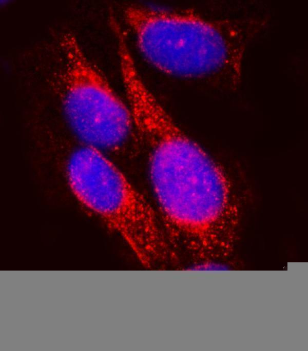
A03343-2 has paraffin-section IHC images from mouse and rat pancreas and IF images from U20S cells and mouse and rat pancreas (catalog image captions). Human reactivity is listed (catalog reactivity).

Real IHC data IHC analysis of SEL1L using anti-SEL1L antibody (A03343-2). SEL1L was detected in a paraffin-embedded section of mouse pancreas tissue. Heat mediated antigen retrieval was performed in EDTA buffer (pH 8.0, epitope retrieval solution). The tissue section was blocked with 10% goat serum. The tissue section was then incubated with 2 μg/ml rabbit anti-SEL1L Antibody (A03343-2) overnight at 4°C. Peroxidase Conjugated Goat Anti-rabbit IgG was used as secondary antibody and incubated for 30 minutes at 37°C. The tissue section was developed using HRP Conjugated Rabbit IgG Super Vision Assay Kit (Catalog # SV0002) with DAB as the chromogen.
Anti-SEL1L Antibody ®
Cat # A03343-2

A03343-2 is listed for IHC, IF and ICC, with IHC images from paraffin-embedded mouse and rat pancreas (catalog applications and IHC captions). Its IF images show U20S cells and paraffin-embedded mouse and rat pancreas (catalog IF captions).

Which to pick: Choose A03343-2 for tissue IHC starting with paraffin sections: its mouse and rat pancreas captions document EDTA retrieval at pH 8.0 and 2 μg/ml primary antibody (catalog IHC captions). For IF/ICC, the same SKU has cell and tissue IF images and a listed 5 μg/ml dilution (catalog IF captions and dilution guidance). It lists human, mouse and rat reactivity, while the tissue IHC images demonstrate mouse and rat; the clone and tissue fixative are unreported (catalog reactivity, product record and IHC captions).

Each figure is that product's own IHC / IF validation image from its datasheet.

References

  1. UniProt Consortium. UniProt entry Q9UBV2 (SE1L1_HUMAN, Protein sel-1 homolog 1).
  2. Human Protein Atlas. SEL1L tissue IHC expression (reliability: Enhanced).
  3. Human Protein Atlas. SEL1L subcellular location (ICC-IF): Membrane.
  4. Human Protein Atlas. SEL1L antibody validation summary (1 antibodies).
  5. SEL1L, an UPR response protein, a potential marker of colonic cell transformation. Digestive diseases and sciences 2012 — PMC3345950.
  6. MicroRNAs Deregulated in Intraductal Papillary Mucinous Neoplasm Converge on Actin Cytoskeleton-Related Pathways That Are Maintained in Pancreatic Ductal Adenocarcinoma. Cancers 2021 — PMC8155860.
  7. Biallelic Cys141Tyr variant of SEL1L is associated with neurodevelopmental disorders, agammaglobulinemia, and premature death. The Journal of clinical investigation 2024 — PMC10786703.
  8. Augmented ERAD (ER-associated degradation) activity in chondrocytes is necessary for cartilage development and maintenance. Science advances 2022 — PMC8782459.
  9. PubMed PMID:10496078 — UniProt-cited evidence.
  10. PubMed PMID:10746565 — UniProt-cited evidence.
  11. PubMed PMID:12975309 — UniProt-cited evidence.