This website uses cookies to ensure you get the best experience on our website.


- Table of Contents
Plan SH3GL2 chromogenic IHC in paraffin sections using CNS neuropil as a positive reference (HPA tissue IHC). This guide covers the catalog antibody’s 2–5 μg/ml IHC range (datasheet A05430-2), fixation consistency, and cell-aware scoring.

Expected localisation, validated protocols, controls and antibodies — the at-a-glance facts below, then the full design guide.
| Expected localisation | Cytoplasmic staining in CNS tissue (HPA tissue IHC) | |
| Staining pattern | Strong cytoplasmic staining in CNS neuropil (HPA tissue IHC) | |
| Antigen retrieval | EDTA pH 8.0 HIER, heat-mediated (datasheet A05430-2) | |
| Positive control | Cerebellum+4 more · see all | |
| Negative control | Adipose tissue+4 more · see all |
| Fixation | Matched tissue-IHC evidence does not establish the fixation claim. Validate the specimen-specific method before use. (selected-SKU IHC image A05430-2) | |
| Caveat | Placental Hofbauer cells also stain strongly (HPA tissue IHC) | |
| Regulation | High expression in fetal cerebellum (UniProt) | |
| Isoform / epitope | No isoforms reported; chain spans aa 1–352 (UniProt) |
The catalog antibody’s IHC-P protocol is paired with three published SH3GL2 IHC protocols covering gerbil hippocampus and human tumor tissue (PMC7916150; PMC3651136; PMC5661104).
| Sample | Paraffin-embedded human laryngeal squamous cell carcinoma tissue; fixative not specified (datasheet A05430-2) |
| Fixation | Image fixative and duration unreported (datasheet A05430-2); verify before use. |
| Sectioning | 4–5 µm sections on charged slides (standard) |
| Deparaffinisation | Xylene, graded ethanol series to water (standard) |
| Antigen retrieval | Heat retrieval: EDTA pH 8.0 (datasheet A05430-2); 20 min, 95–100 °C (standard) |
| Peroxidase block | 3% H2O2, 10 min, room temperature (standard) |
| Blocking | 10% goat serum (datasheet A05430-2) |
| Primary antibody | Rabbit anti-SH3GL2, 2-5 μg/ml (datasheet A05430-2) |
| Primary incubation | Overnight at 4 °C (datasheet A05430-2) |
| Detection | Streptavidin-biotin complex (SABC), DAB chromogen (datasheet A05430-2) |
| Counterstain | Hematoxylin, blue, dehydrate and mount (standard) |
| Expected result | SH3GL2-positive staining in molecular layer - neuropil of cerebellum (HPA tissue IHC: High). HPA tissue profile: Selective cytoplasmic expression in CNS. No signal in the no-primary control. |
SH3GL2 should give predominantly cytoplasmic staining in selected CNS structures, strongest in cerebellar molecular layer neuropil and cerebral cortex neuropil; placenta Hofbauer cells are another documented positive population (HPA: Enhanced tissue IHC; High in these populations). UniProt places SH3GL2 in cytoplasm, at membranes, early endosomes and presynapses, with no transmembrane segment (UniProt Q99962: subcellular location and topology). Read the slide by compartment and cell type, not overall tissue darkness.
| Strong staining in cerebellar molecular layer neuropil or cerebral cortex neuropil, with little nuclear signal. | This matches the documented High neuropil pattern (HPA: cerebellum and cerebral cortex tissue IHC). Cytoplasmic and presynaptic localization makes a neuropil centered result plausible (UniProt Q99962: subcellular location). Judge intensity against a matched control section, since this record does not set an absolute chromogen threshold. |
| Moderate cytoplasmic staining in glial cells of caudate or hippocampus, or strong staining in placental Hofbauer cells. | These are documented cell specific results, even though the tissue profile is described as selective cytoplasmic expression in CNS (HPA: Medium in caudate and hippocampus glial cells; High in placenta Hofbauer cells). Do not reject the placental population solely because it is outside the CNS. |
| Dominant nuclear staining, or a pattern confined to a compartment unrelated to the expected cytoplasmic signal. | Treat this as a localization mismatch requiring control review: nucleus is absent from the listed SH3GL2 locations (UniProt Q99962: cytoplasm, membrane, early endosome, presynapse). Check counterstain separation and antibody dependent signal before calling the compartment positive (standard IHC practice). |
| Strong staining in a cell population documented as Not detected, such as adipocytes in adipose tissue. | This conflicts with the supplied tissue observation (HPA: adipose adipocytes Not detected). Cross reactivity or endogenous detection activity are possibilities, not proven diagnoses; compare an antibody omission control and, if available, an independently validated SH3GL2 antibody (standard IHC practice; HPA: multiple IHC Enhanced antibodies). |
| Uniform haze across cells and extracellular space, or no signal in cerebellar molecular layer neuropil. | Haze obscures the selective cell and compartment pattern (HPA: selective cytoplasmic tissue IHC). A blank known positive section conflicts with the documented High neuropil signal (HPA: cerebellum). Inspect control sections and the IHC detection workflow before interpreting either result as SH3GL2 expression (standard IHC practice). |
| Which tissue and cell population is being scored? | The reference level depends on the precise population: High in cerebellar and cortical neuropil and placental Hofbauer cells, Medium in caudate and hippocampal glial cells, and Not detected in listed populations including adipose adipocytes (HPA: tissue IHC). Score those populations separately rather than assigning one expected intensity to every cell in an organ. |
| Which compartment should carry the chromogen? | Expect a cytoplasmic pattern compatible with membrane associated, early endosomal and presynaptic SH3GL2; no transmembrane segment is annotated (UniProt Q99962: subcellular location and topology). These annotations guide interpretation but do not establish that individual vesicles can be resolved by chromogenic IHC. |
| How strong is the tissue pattern evidence? | HPA rates the tissue profile Enhanced because antibody staining is highly consistent with RNA expression data (HPA: tissue IHC reliability). HPA also lists 3 IHC Enhanced antibodies: HPA026685, HPA063573 and CAB010056 (HPA: antibody validation). This supports the reference pattern; it does not validate a new specimen's staining without its own controls. |
| Q: Does an IF/ICC microtubule pattern change the IHC call? | A: HPA reports approved microtubule localization, plus cytokinetic bridge and mitotic spindle localization, in ICC-IF (HPA: subcellular ICC-IF). That is a distinct assay observation; use the HPA tissue IHC cell and compartment pattern to judge a paraffin section (HPA: tissue IHC). |
| Situation | Likely cause | Next action |
|---|---|---|
| Known positive cerebellar molecular layer neuropil is blank or much weaker than the matched control (HPA: High). | The run may have lost signal during retrieval, primary antibody incubation or chromogenic detection (standard IHC practice); no SH3GL2 specific fixation sensitivity is supplied. | Review the positive control, retrieval record, antibody dilution and detection reagents; repeat with the catalog antibody's validated IHC-P conditions where available (standard IHC practice). Do not infer absence of SH3GL2 from a failed control. |
| Chromogen appears mainly in nuclei rather than neuropil or cytoplasm (HPA: tissue IHC; UniProt Q99962: location). | A nuclear dominant result conflicts with the supplied localization; counterstain confusion or nonspecific staining may account for the appearance (standard IHC practice). | Compare the stained slide with an antibody omission control, confirm which color is chromogen, and reassess the same cell population on a known positive section (standard IHC practice). |
| Adipose adipocytes or another HPA Not detected population stains strongly (HPA: tissue IHC). | Cross reactivity or endogenous detection activity is possible; the supplied record cannot identify which explains a particular slide. | Check an antibody omission control for detection derived signal, inspect the cell identity, and compare an independent IHC Enhanced antibody if available (standard IHC practice; HPA: antibody validation). |
| Diffuse brown haze makes neuropil and cell boundaries hard to distinguish (HPA: selective cytoplasmic tissue IHC). | Background from nonspecific binding, inadequate washing or detection chemistry can reduce contrast (standard IHC practice). | Inspect omission and positive controls; review blocking, wash steps and development time, then repeat under validated IHC-P conditions (standard IHC practice). |
| Placental Hofbauer cells stain despite an expectation of CNS restricted staining (HPA: High in Hofbauer cells). | The shorthand CNS profile overlooks a documented placental positive cell population (HPA: selective cytoplasmic CNS profile; placenta Hofbauer cells High). | Score Hofbauer cells separately from surrounding placental cells and retain them as a documented positive population when morphology and controls agree (HPA: placenta tissue IHC; standard IHC practice). |
| IF/ICC shows microtubules while the paraffin IHC section highlights neuropil (HPA: subcellular ICC-IF; tissue IHC). | HPA reports microtubules in ICC-IF and neuropil staining in tissue IHC; the observations come from different preparations (HPA: subcellular ICC-IF; tissue IHC). | Interpret each result against its own HPA reference and controls; do not require visible microtubules to accept the documented chromogenic neuropil pattern (HPA: tissue IHC; standard IHC practice). |
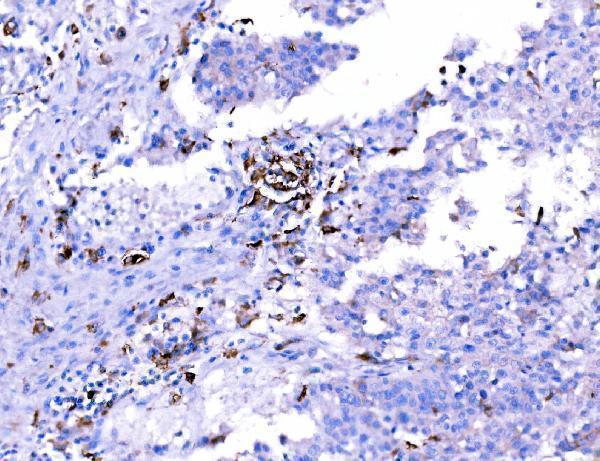
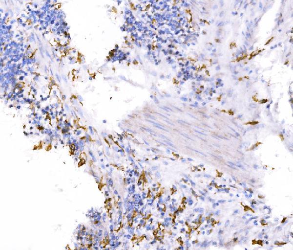
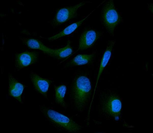


Comprehensive Human Protein Atlas IHC scoring per tissue (reliability: Enhanced — High consistency between antibody staining and RNA expression data.). Rows are taken directly from the HPA tissue chart — click any row's HPA link to view the source.
| Tissue | Cell type | Level | Evidence | Source |
|---|---|---|---|---|
| Cerebellum | Molecular layer - neuropil | High | Protein (IHC) | HPA → |
| Cerebral cortex | Neuropil | High | Protein (IHC) | HPA → |
| Placenta | Hofbauer cells | High | Protein (IHC) | HPA → |
| Caudate | Glial cells | Medium | Protein (IHC) | HPA → |
| Hippocampus | Glial cells | Medium | Protein (IHC) | HPA → |
| Tissue | Cell type | Level | Evidence | Source |
|---|---|---|---|---|
| Adipose tissue | Adipocytes | Not detected | Protein (IHC) | HPA → |
| Adrenal gland | Glandular cells | Not detected | Protein (IHC) | HPA → |
| Appendix | Glandular cells | Not detected | Protein (IHC) | HPA → |
| Bone marrow | Hematopoietic cells | Not detected | Protein (IHC) | HPA → |
| Breast | Adipocytes | Not detected | Protein (IHC) | HPA → |
Troubleshoot SH3GL2 chromogenic IHC in paraffin sections using the catalog antibody’s tissue example and the reported tissue and subcellular patterns.
A05430-2 has IHC images from human tissue and mouse brain paraffin sections, plus IF/ICC data from HeLa cells (catalog image captions).
A05430-2 is listed for IHC in human, mouse and rat; its IHC captions show human laryngeal squamous cell carcinoma, gastric cancer, Hashimoto thyroiditis and mouse brain paraffin sections (catalog applications and IHC captions). A05430-2 is also listed for IF/ICC, with an IF image from HeLa cells (catalog applications and IF caption).
Which to pick: Choose A05430-2 for paraffin-section IHC: its image captions document EDTA retrieval at pH 8.0 and primary antibody at 2 μg/ml (A05430-2 IHC captions); the fixative is unreported (A05430-2 IHC captions). For IF/ICC, A05430-2 has a HeLa-cell image using 5 μg/ml primary antibody (A05430-2 IF caption). For cross-species work, its catalog lists human, mouse and rat reactivity, while the supplied IHC images show human and mouse samples; clonality is unreported (A05430-2 catalog and IHC captions).