This website uses cookies to ensure you get the best experience on our website.


- Table of Contents
Plan chromogenic SIAH2 IHC in paraffin sections with the catalog antibody at 1:50–1:200 (datasheet: IHC dilution). Use testis spermatogonia as a positive reference and adipocytes as an unstained comparison, while accounting for low staining and RNA concordance (HPA tissue IHC).

Expected localisation, validated protocols, controls and antibodies — the at-a-glance facts below, then the full design guide.
| Expected localisation | Nuclear tissue staining observed; cytoplasm expected (HPA tissue IHC; UniProt) | |
| Staining pattern | Nuclear staining in several tissue types (HPA tissue IHC) | |
| Antigen retrieval | Tris-EDTA pH 9.0 HIER, 95–98 °C, 20 min (rule: nuclear antigen) | |
| Positive control | Testis+4 more · see all | |
| Negative control | Adipose tissue+4 more · see all |
| Fixation | Keep fixation consistent across sections. (standard IHC practice; not target-specific) | |
| Caveat | Staining and RNA expression show low consistency (HPA tissue IHC) | |
| Regulation | Widely expressed at low level (UniProt) | |
| Isoform / epitope | No annotated isoforms; single 1–324 chain (UniProt) |
Compare the catalog antibody’s IHC-P protocol with published SIAH2 IHC protocols from prostate and lung cancer specimens (PMC8085430; PMC4651316; PMC4102802).
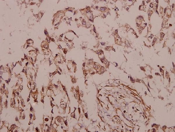
| Sample | Paraffin-embedded human colorectal carcinoma tissue; fixative not specified (datasheet A03379) |
| Fixation | Image fixative and duration unreported (datasheet A03379); verify before use. |
| Sectioning | 4–5 µm sections on charged slides (standard) |
| Deparaffinisation | Xylene, graded ethanol series to water (standard) |
| Antigen retrieval | Heat-induced epitope retrieval in Tris-EDTA buffer, pH 9.0, 20 min at 95–98 °C (standard rule: nuclear antigen) |
| Peroxidase block | 3% H2O2, 10 min, room temperature (standard) |
| Blocking | 10% normal serum of the secondary host, 30 min, room temperature (standard) |
| Primary antibody | Rabbit anti-SIAH2, 1:50-1:200 (datasheet A03379) |
| Primary incubation | Overnight at 4 °C (standard) |
| Detection | HRP-polymer secondary, DAB chromogen 5–10 min (standard) |
| Counterstain | Hematoxylin, blue, dehydrate and mount (standard) |
| Expected result | SIAH2-positive staining in spermatogonia cells of testis (HPA tissue IHC: High). HPA tissue profile: Nuclear expression in several different tissue types. No signal in the no-primary control. |
In paraffin-section IHC, expect chiefly nuclear staining in selected cells, including high staining in testicular spermatogonia and medium staining in glandular cells of the appendix and colon (HPA tissue IHC). SIAH2 is predominantly cytoplasmic and partly nuclear, with no transmembrane segment (UniProt O43255). Treat the tissue pattern cautiously: HPA rates its IHC reliability Approved but reports low consistency with RNA expression (HPA tissue IHC).
| Distinct nuclear staining in spermatogonia, with weaker or absent staining in neighboring cells. | This fits the high spermatogonial signal reported in testis (HPA tissue IHC). Score the identified cell population and nuclear compartment, rather than treating the whole section as uniformly positive (general IHC practice). The match supports interpretation but does not independently prove antibody specificity (general IHC practice). |
| Nuclear signal in appendix or colon glandular cells; some cytoplasmic signal may also be visible. | HPA reports medium staining in these glandular cells and describes nuclear expression across several tissues (HPA tissue IHC). Cytoplasmic signal is biologically plausible because UniProt describes SIAH2 as predominantly cytoplasmic and partly nuclear (UniProt O43255). Assess whether any cytoplasmic signal is cell-restricted and above background (general IHC practice). |
| Staining confined to cell borders, extracellular material, or an otherwise unexpected compartment. | This is discordant with the reported nuclear IHC pattern and cytoplasmic or nuclear localisation (HPA tissue IHC; UniProt O43255). SIAH2 has no transmembrane segment (UniProt O43255). Consider artefact or off-target staining, then check morphology and staining controls before assigning the signal to SIAH2 (general IHC practice). |
| Strong staining in adipocytes or cardiomyocytes, where HPA reports no detected signal. | The cell-type mismatch warrants scrutiny; HPA reports these cells as not detected, while also noting limited agreement between antibody staining and RNA data (HPA tissue IHC). Cross-reactivity or endogenous detection activity are possible explanations, not established diagnoses; compare no-primary and detection controls (general IHC practice). |
| Haze across the section obscures cell boundaries and nuclei. | Diffuse background cannot establish the cell-restricted pattern reported by HPA (HPA tissue IHC). It may arise from nonspecific antibody binding, detection activity, or incomplete washing (general IHC practice). Interpret staining only after background is low enough to distinguish positive cells from nearby tissue (general IHC practice). |
| Tissue and cell selection | Testicular spermatogonia provide a reported high signal; appendix, colon and other listed positive cell populations show medium signal (HPA tissue IHC). Use the named cells when judging a section, since HPA levels do not apply uniformly to every cell in a tissue (HPA tissue IHC). |
| Compartment evidence | HPA describes nuclear tissue IHC, while UniProt describes predominantly cytoplasmic, partly nuclear localisation (HPA tissue IHC; UniProt O43255). Record nuclear and cytoplasmic scores separately when both appear (general IHC practice); do not force agreement between the two sources. |
| Antibody evidence | The HPA tissue antibody CAB069930 has an Approved IHC label, alongside a low-consistency warning for staining versus RNA (HPA tissue IHC; HPA antibodies). This supports cautious use of the reported pattern, not an Enhanced or independently reproduced IHC claim (HPA antibodies; general IHC practice). |
| Antigen retrieval and fixation | Target-specific fixation effects are not established by the supplied assay evidence. Verify with a matched IHC source before attributing a result to fixation. |
| IF/ICC Q&A: where should signal appear? | HPA reports mainly nucleoplasmic signal, with additional vesicles, in ICC-IF; HPA061293 is Supported for ICC (HPA subcellular ICC-IF; HPA antibodies). That observation informs an IF/ICC interpretation, but it does not establish a paraffin-section IHC pattern or supply an IF/ICC protocol (HPA tissue IHC; HPA subcellular ICC-IF). |
| Situation | Likely cause | Next action |
|---|---|---|
| No staining in testicular spermatogonia. | The expected high cell population may be absent from the field, or the IHC workflow may have failed (HPA tissue IHC; general IHC practice). | First locate spermatogonia on the counterstained section; then check the positive-control section, antibody application, retrieval, and detection steps against the IHC-P instructions (general IHC practice). |
| Nuclei and cytoplasm both stain. | HPA's tissue IHC description favors nuclei, whereas UniProt allows cytoplasmic and nuclear SIAH2 (HPA tissue IHC; UniProt O43255). Background can also blur compartment boundaries (general IHC practice). | Score each compartment separately and compare signal with adjacent cells and the no-primary control (general IHC practice). Report the source discrepancy rather than calling all cytoplasmic staining artefactual. |
| Adipocytes or cardiomyocytes stain strongly. | These cells are listed as not detected by HPA; off-target binding or endogenous detection activity may explain unexpected color (HPA tissue IHC; general IHC practice). | Verify cell identity and compare no-primary and detection controls (general IHC practice). If those controls are clean, treat the result as an unresolved disagreement with HPA and seek independent specificity evidence. |
| Diffuse chromogen obscures the expected cells. | Nonspecific binding, incomplete washing, or endogenous detection activity may raise section-wide background (general IHC practice). | Inspect no-primary and detection controls, review blocking and wash steps, and optimize the IHC-validated antibody under its IHC-P instructions (general IHC practice). Re-score only when nuclei and cell boundaries are discernible. |
| Signal appears only on cell borders or outside cells. | This distribution conflicts with HPA's nuclear tissue pattern and UniProt's cytoplasmic or nuclear localisation; no transmembrane segment is annotated (HPA tissue IHC; UniProt O43255). | Check section morphology, counterstain, no-primary control, and detection background (general IHC practice). Do not count border-only or extracellular color as a SIAH2-positive cell without independent support. |
| An HPA negative cell population stains faintly. | HPA's not detected calls describe its observations, and its tissue IHC record warns of low consistency with RNA data (HPA tissue IHC). Faint color may also be background (general IHC practice). | Compare intensity and compartment with a reported positive population and a no-primary control (HPA tissue IHC; general IHC practice). Record the discrepancy without converting a faint signal into a confirmed positive result. |


Comprehensive Human Protein Atlas IHC scoring per tissue (reliability: Approved — Low consistency between antibody staining and RNA expression data.). Rows are taken directly from the HPA tissue chart — click any row's HPA link to view the source.
| Tissue | Cell type | Level | Evidence | Source |
|---|---|---|---|---|
| Testis | Spermatogonia cells | High | Protein (IHC) | HPA → |
| Appendix | Glandular cells | Medium | Protein (IHC) | HPA → |
| Bone marrow | Hematopoietic cells | Medium | Protein (IHC) | HPA → |
| Bronchus | Basal cells | Medium | Protein (IHC) | HPA → |
| Cervix | Squamous epithelial cells | Medium | Protein (IHC) | HPA → |
| Tissue | Cell type | Level | Evidence | Source |
|---|---|---|---|---|
| Adipose tissue | Adipocytes | Not detected | Protein (IHC) | HPA → |
| Caudate | Glial cells | Not detected | Protein (IHC) | HPA → |
| Cerebellum | Cells in granular layer | Not detected | Protein (IHC) | HPA → |
| Cerebral cortex | Endothelial cells | Not detected | Protein (IHC) | HPA → |
| Heart muscle | Cardiomyocytes | Not detected | Protein (IHC) | HPA → |
Troubleshoot SIAH2 staining in paraffin sections by checking retrieval, compartment, cell type, and controls before comparing chromogenic signal across samples.
The IHC-validated SIAH2 antibody has real staining data from human paraffin-embedded colorectal carcinoma tissue (IHC image caption) and listed Human, Mouse, and Rat reactivity (catalog: A03379).
A03379 is listed for IHC with Human, Mouse, and Rat reactivity (catalog: A03379). Its IHC figure shows human paraffin-embedded colorectal carcinoma tissue at 1:50 (IHC image caption: A03379).
Which to pick: Choose A03379 for paraffin-section tissue IHC: its figure shows human colorectal carcinoma tissue, and the catalog lists an IHC dilution of 1:50–1:200 (IHC image caption and catalog: A03379). The caption does not report a fixative (IHC image caption: A03379). For cross-species work, A03379 lists Mouse and Rat reactivity alongside Human, with a Rabbit host and no clone specified (catalog: A03379); no IF/ICC option is listed (catalog: A03379 applications).