This website uses cookies to ensure you get the best experience on our website.


- Table of Contents
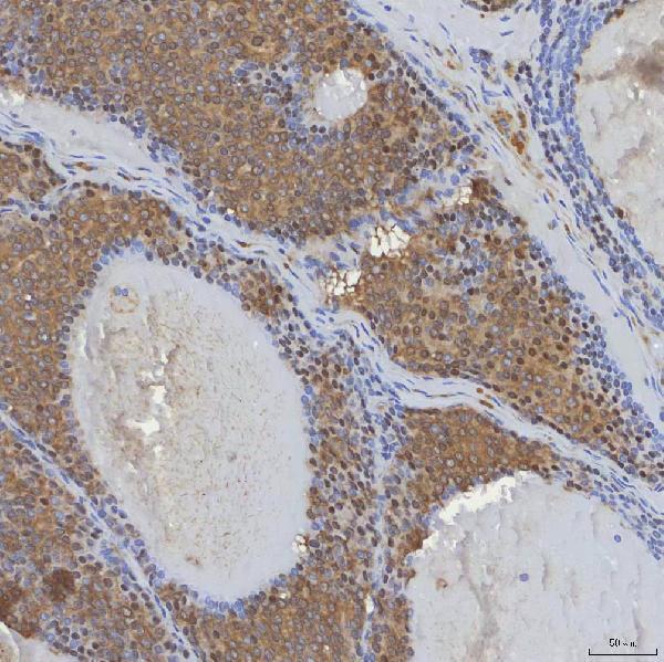
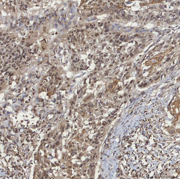
Plan chromogenic SIN3A IHC around its widespread nuclear staining, strongest in testis (HPA tissue IHC). The catalog antibody was used at 2 μg/ml overnight at 4°C on paraffin sections, with DAB detection (datasheet A01203-2).

Expected localisation, validated protocols, controls and antibodies — the at-a-glance facts below, then the full design guide.
| Expected localisation | Nuclear staining in tissue (HPA tissue IHC) | |
| Staining pattern | Widespread nuclear staining, strongest in testis (HPA tissue IHC) | |
| Antigen retrieval | EDTA pH 8.0 HIER, heat-mediated (datasheet A01203-2) | |
| Positive control | Nasopharynx+4 more · see all | |
| Negative control | Adipose tissue+2 more · see all |
| Fixation | Keep fixation consistent across sections. (standard IHC practice; not target-specific) | |
| Caveat | Staining varies by cell type; some cells are undetected (HPA tissue IHC) | |
| Regulation | Developing brain: highest in ventricular zone (UniProt) | |
| Isoform / epitope | No annotated isoforms; one 1–1273 chain (UniProt) |
The catalog antibody’s IHC protocol is paired with four published SIN3A staining protocols for liver, breast and testis sections (PMC11077365; PMC9177717; PMC6207735; PMC3423586).
| Sample | Paraffin-embedded human breast cancer tissue; fixative not specified (datasheet A01203-2) |
| Fixation | Image fixative and duration unreported (datasheet A01203-2); verify before use. |
| Sectioning | 4–5 µm sections on charged slides (standard) |
| Deparaffinisation | Xylene, graded ethanol series to water (standard) |
| Antigen retrieval | Heat retrieval: EDTA pH 8.0 (datasheet A01203-2); 20 min, 95–100 °C (standard) |
| Peroxidase block | 3% H2O2, 10 min, room temperature (standard) |
| Blocking | 10% goat serum (datasheet A01203-2) |
| Primary antibody | Rabbit anti-SIN3A, 2-5 μg/ml (datasheet A01203-2) |
| Primary incubation | Overnight at 4 °C (datasheet A01203-2) |
| Detection | HRP-conjugated secondary, DAB chromogen (datasheet A01203-2) |
| Counterstain | Hematoxylin, blue, dehydrate and mount (standard) |
| Expected result | SIN3A-positive staining in respiratory epithelial cells of nasopharynx (HPA tissue IHC: High). HPA tissue profile: Ubiquitous nuclear expression with highest expression in testis. No signal in the no-primary control. |
SIN3A should appear predominantly in nuclei across many cell types (HPA: ubiquitous nuclear tissue expression; UniProt Q96ST3: nucleus). Strong examples are testis preleptotene spermatocytes, placental cytotrophoblasts and nasopharyngeal respiratory epithelial cells (HPA: High in each). The HPA tissue pattern has Enhanced reliability, meaning high consistency between antibody staining and RNA expression (HPA: tissue reliability). SIN3A has no transmembrane segment (UniProt Q96ST3 topology).
| Distinct nuclear staining in preleptotene spermatocytes, cytotrophoblasts or nasopharyngeal respiratory epithelium. | This matches three reported High cell populations (HPA: testis, placenta and nasopharynx). Judge intensity within the named cells, alongside slide controls; a positive field need not have identical staining in every cell (general IHC practice). |
| Strong staining sits chiefly in cytoplasm or along cell membranes, while nuclei remain faint. | That compartment conflicts with the reported nuclear tissue pattern (HPA: ubiquitous nuclear expression) and nuclear localisation (UniProt Q96ST3). Review staining specificity and tissue morphology before calling it SIN3A; a membrane pattern has no support from its topology (UniProt Q96ST3: no transmembrane segment). |
| The dominant signal is in adipocytes, cholangiocytes or parathyroid glandular cells. | Those named populations are reported Not detected (HPA: adipose tissue, liver and parathyroid gland). Consider cross-reactivity or endogenous detection activity, particularly when the signal lacks nuclear definition (general IHC practice). These are cell-level comparisons, not claims that the entire organs lack SIN3A. |
| A broad, hazy deposit covers nuclei and surrounding tissue without clear cell boundaries. | Diffuse background cannot establish the expected nuclear pattern (HPA: ubiquitous nuclear expression). Compare a no-primary control, examine blocking and washing, and assess whether detection reagent or chromogen contributes background (general IHC practice). |
| No nuclear signal appears in a testis section containing identifiable preleptotene spermatocytes. | Those cells are a reported High population (HPA: testis). First confirm their presence and section quality; then check the IHC-validated antibody, retrieval and detection workflow against its documented IHC-P procedure (general IHC practice). A blank slide alone does not show biological absence. |
| Cell-specific reference populations (HPA: tissue IHC). | High staining is reported in three named populations, while bronchial respiratory epithelium and several glandular populations are Medium (HPA: tissue IHC). Choose comparisons by cell type; Low or Not detected observations should not be promoted to whole-tissue negative controls. |
| Subcellular assignment (UniProt Q96ST3; HPA: tissue IHC). | Both sources support a nuclear result. UniProt also lists the nucleolus, with recruitment by SAP30L (UniProt Q96ST3: subcellular location). The HPA tissue profile establishes ubiquitous nuclear staining, so a distinct nucleolar IHC signal is not required for a positive call. |
| Antibody-level support (HPA: antibody validation). | HPA047213 and HPA062123 have Enhanced IHC validation; CAB004506 has Supported IHC validation (HPA: antibody validation). These statuses support their reported applications but do not establish that an unrelated catalog antibody gives the same intensity in every tissue. |
| Target structure and modifications (UniProt Q96ST3). | SIN3A has no signal peptide, propeptide or transmembrane segment; its listed chain spans residues 1–1273 (UniProt Q96ST3). Reported phosphorylation and acetylation sites do not, by themselves, establish an IHC epitope effect or a different expected compartment. |
| Situation | Likely cause | Next action |
|---|---|---|
| Known-positive testis cells show no nuclear staining (HPA: High in preleptotene spermatocytes). | The relevant cells may be absent from the examined field, or an IHC workflow step may have failed (general IHC practice). | Confirm cell identity and section quality, then check the catalog antibody's IHC-P instructions for retrieval, dilution, incubation and detection; compare a same-run positive control (general IHC practice). |
| Signal is widespread and hazy rather than nuclear (HPA: expected nuclear pattern). | Background from blocking, washing, detection reagents or chromogen can obscure localisation (general IHC practice). | Inspect a no-primary control and adjust the documented blocking, washing or detection workflow as indicated by that control (general IHC practice). |
| Cytoplasmic or membranous staining dominates (HPA: nuclear tissue pattern). | Nonspecific binding or background is possible; SIN3A is nuclear and lacks a transmembrane segment (UniProt Q96ST3). | Compare nuclear detail with a known-positive cell population and check a no-primary control before scoring the extranuclear signal (general IHC practice; HPA: High reference populations). |
| Adipocytes or cholangiocytes stain strongly (HPA: Not detected in those cells). | Cell-level discordance can indicate cross-reactivity or endogenous detection activity (general IHC practice). | Check whether staining is nuclear, compare the no-primary control and confirm the same-run positive population before assigning specificity (general IHC practice; HPA: nuclear tissue pattern). |
| A Low reference population appears faint or negative, such as cortical neurons (HPA: Low). | Weak staining is consistent with that reported level; it cannot by itself distinguish low expression from a weak run (HPA: cerebral cortex; general IHC practice). | Interpret it beside a High reference population processed in the same run, such as preleptotene spermatocytes (HPA: testis; general IHC practice). |
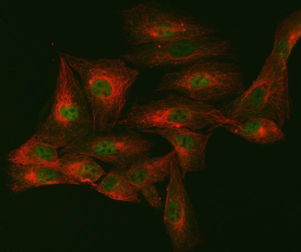
| IF/ICC question: where should the signal appear in imaged cells? | HPA reports enhanced nucleoplasmic localisation in ICC-IF (HPA: subcellular localisation). | Expect nucleoplasmic signal when interpreting IF/ICC images (HPA: subcellular localisation). Use the separate IF/ICC guide for its workflow; the tissue IHC pattern does not specify an IF/ICC protocol. |


Comprehensive Human Protein Atlas IHC scoring per tissue (reliability: Enhanced — High consistency between antibody staining and RNA expression data.). Rows are taken directly from the HPA tissue chart — click any row's HPA link to view the source.
| Tissue | Cell type | Level | Evidence | Source |
|---|---|---|---|---|
| Nasopharynx | Respiratory epithelial cells | High | Protein (IHC) | HPA → |
| Placenta | Cytotrophoblasts | High | Protein (IHC) | HPA → |
| Testis | Preleptotene spermatocytes | High | Protein (IHC) | HPA → |
| Bronchus | Respiratory epithelial cells | Medium | Protein (IHC) | HPA → |
| Cerebellum | Cells in granular layer | Medium | Protein (IHC) | HPA → |
Troubleshoot SIN3A staining in paraffin section IHC by checking nuclear localisation, the catalog antibody’s documented retrieval conditions, and matched controls.
A01203-2 has pictured IHC staining in human paraffin sections and IF staining in human ovarian cancer sections and U20S cells (catalog image captions); M01203 lists IHC and IF/ICC for human, mouse, and rat (catalog applications and reactivity).
A01203-2 has pictured IHC results in human breast, larynx, lung, and ovarian cancer paraffin sections, plus IF results in human ovarian cancer sections and U20S cells (catalog image captions). M01203 lists IHC and IF/ICC applications and human, mouse, and rat reactivity, but has no IHC or IF image caption in the payload (catalog applications, reactivity, and image captions).
Which to pick: Choose A01203-2 for human tissue IHC: its pictured paraffin sections used EDTA retrieval at pH 8.0 and 2 μg/ml primary antibody; the fixative is unreported (A01203-2 IHC image captions). A01203-2 also has pictured IF in an ovarian cancer paraffin section and U20S cells, with 5 μg/ml primary antibody (A01203-2 IF image captions). For mouse samples, consider rabbit monoclonal M01203, which lists mouse reactivity and IHC and IF/ICC applications, while A01203-2 lists human and rat reactivity (catalog title, applications, and reactivity).