This website uses cookies to ensure you get the best experience on our website.


- Table of Contents
Plan SLAIN2 IHC in paraffin sections using cytoplasmic staining as the expected tissue pattern (HPA tissue IHC). Compare high-staining myocytes and smooth muscle cells with an unstained cell population, and optimize the catalog antibody from its stated 5 μg/mL starting concentration (HPA tissue IHC; datasheet: 5 μg/mL).

Expected localisation, validated protocols, controls and antibodies — the at-a-glance facts below, then the full design guide.
| Expected localisation | Cytoplasmic staining in most tissues (HPA tissue IHC) | |
| Staining pattern | Cytoplasmic staining, high in myocytes and smooth muscle cells (HPA tissue IHC) | |
| Antigen retrieval | Citrate pH 6.0 HIER, 95–98 °C, 20 min (rule: cytoplasmic / membrane antigen) | |
| Positive control | Skeletal muscle+4 more · see all | |
| Negative control | Adipose tissue+4 more · see all |
| Fixation | Keep fixation consistent across sections. (standard IHC practice; not target-specific) | |
| Caveat | Staining varies by cell type; adipocytes were unstained (HPA tissue IHC) | |
| Regulation | Stimulus-linked regulation is unreported (UniProt) | |
| Isoform / epitope | No annotated isoforms; one 1–581 chain, with no transmembrane segment (UniProt) |
The catalog antibody’s IHC-P protocol (datasheet) is accompanied by a published SLAIN2 IHC workflow in colorectal cancer tissue (PMC6444028: IHC methods).
| Sample | Tissue sections; selected-image fixative not specified (standard IHC workflow) |
| Fixation | Image fixative and duration unreported (datasheet A11030); verify before use. |
| Sectioning | 4–5 µm sections on charged slides (standard) |
| Deparaffinisation | Xylene, graded ethanol series to water (standard) |
| Antigen retrieval | Heat-induced epitope retrieval in citrate buffer, pH 6.0, 20 min at 95–98 °C (standard rule: cytoplasmic / membrane antigen) |
| Peroxidase block | 3% H2O2, 10 min, room temperature (standard) |
| Blocking | 10% normal serum of the secondary host, 30 min, room temperature (standard) |
| Primary antibody | Rabbit anti-SLAIN2, 5 μg/mL (datasheet A11030) |
| Primary incubation | Overnight at 4 °C (standard) |
| Detection | HRP-polymer secondary, DAB chromogen 5–10 min (standard) |
| Counterstain | Hematoxylin, blue, dehydrate and mount (standard) |
| Expected result | SLAIN2-positive staining in myocytes of skeletal muscle (HPA tissue IHC: High). HPA tissue profile: Cytoplasmic expression in most tissues, high expression in testis and smooth muscle. No signal in the no-primary control. |
SLAIN2 localizes to the cytoplasm and microtubule cytoskeleton and has no transmembrane segment (UniProt Q9P270: localization/topology). In tissue IHC, expect cytoplasmic staining in many tissues, with High staining in skeletal myocytes and smooth muscle cells among the listed examples (HPA tissue IHC). HPA rates the tissue staining Approved, with external verification pending (HPA tissue IHC: reliability).
| Strong cytoplasmic staining in skeletal myocytes or smooth muscle cells, with recognizable cell boundaries (HPA tissue IHC: High). | This matches the reported cell types and compartment (HPA tissue IHC: High; UniProt Q9P270: cytoplasm). Judge intensity against nearby tissue structures and the staining control; a brown deposit alone does not establish cell identity (standard IHC practice). |
| Predominantly nuclear staining with little cytoplasmic signal in otherwise intact cells. | A nuclear-only pattern conflicts with the reported cytoplasmic localization (UniProt Q9P270; HPA tissue IHC). Check morphology, counterstain and detection controls before assigning it to SLAIN2; the supplied sources do not establish a nuclear tissue-IHC pattern. |
| Strong staining in adipocytes from adipose tissue or breast, or in another cell type listed as not detected (HPA tissue IHC). | Treat this as unexpected for that specified cell type, with cross-reactivity or endogenous chromogen activity among the possibilities (HPA tissue IHC; standard IHC practice). HPA's not-detected entries describe named cells, not every cell in the tissue. |
| Diffuse brown haze across cells and extracellular spaces, without a discernible cytoplasmic pattern. | This is difficult to score as specific SLAIN2 staining because the expected tissue pattern is cellular and cytoplasmic (HPA tissue IHC). Background from detection reagents or insufficient blocking is possible; compare a no-primary control (standard IHC practice). |
| No staining in skeletal myocytes or smooth muscle cells that are present and well preserved on the section (HPA tissue IHC: High). | This fails the supplied positive-tissue expectation, but one blank section cannot identify the cause (HPA tissue IHC). Check tissue identity, antibody and detection controls, and the documented IHC-P conditions before concluding that the sample lacks SLAIN2 (standard IHC practice). |
| Compartment and topology | UniProt places SLAIN2 in the cytoplasm and microtubule cytoskeleton, including growing microtubule plus ends, and reports no transmembrane segment (UniProt Q9P270). Interpret tissue IHC primarily at the cytoplasmic level; the record does not require visible plus-end puncta in chromogenic sections. |
| Choice of tissue and cell type | HPA reports High staining in skeletal myocytes and smooth muscle cells, Medium in several other listed cell types, and not-detected staining in specified cells such as adipocytes (HPA tissue IHC). Select and score controls by cell type; whole-tissue labels can conceal mixed cell populations. |
| Strength of antibody evidence | The tissue-IHC profile is Approved but pending external verification (HPA tissue IHC: reliability). HPA063599 is IHC Approved; HPA035824 has no IHC status listed (HPA antibodies). A missing status is not evidence of failed staining, and the supplied record does not establish two-antibody IHC confirmation. |
| IF/ICC Q: What localization may appear? | A: HPA reports supported cytosol and centrosome localization, plus primary cilium tip and basal body localization, in ICC-IF (HPA subcellular). Those finer structures are IF observations; their visibility is not a requirement for a positive chromogenic tissue-IHC section. |
| Chromogenic background | Endogenous detection activity and nonspecific reagent binding can create signal unrelated to the primary antibody (standard IHC practice). Use an appropriate no-primary or detection control to help distinguish such signal from the HPA-reported cytoplasmic cell pattern (HPA tissue IHC; standard IHC practice). |
| Situation | Likely cause | Next action |
|---|---|---|
| Known positive muscle cells are blank (HPA tissue IHC: High). | Tissue identification, staining execution, or antibody and detection performance may be responsible; the supplied sources provide no SLAIN2-specific fixation effect (standard IHC practice). | Confirm the muscle cell type and section quality, then check controls and the catalog antibody's documented IHC-P retrieval and dilution conditions before changing one workflow variable at a time (standard IHC practice). |
| Nuclei dominate while cytoplasm is weak. | This conflicts with cytoplasmic localization and may reflect background, counterstain interpretation, or nonspecific binding (UniProt Q9P270; standard IHC practice). | Inspect the no-primary control and cell morphology, then reassess blocking and detection conditions; withhold a positive SLAIN2 call until cytoplasmic staining is distinguishable (standard IHC practice). |
| Adipocytes stain strongly in an HPA not-detected context (HPA tissue IHC). | Unexpected cell staining may arise from cross-reactivity or endogenous detection activity; the HPA result applies to the named cell population (HPA tissue IHC; standard IHC practice). | Verify cell identity on the counterstained section, compare an appropriate detection control, and assess staining in listed positive muscle cells on the same run (HPA tissue IHC; standard IHC practice). |
| Brown haze obscures cell boundaries. | Nonspecific binding or background from the chromogenic detection system can prevent reliable compartment scoring (standard IHC practice). | Compare a no-primary control, check blocking and washes, and adjust the antibody or detection conditions within the documented IHC-P workflow; rescore only when cytoplasm can be distinguished (standard IHC practice). |
| The IHC section lacks centrosomal or ciliary puncta seen in IF/ICC. | HPA's supported centrosome, cilium-tip and basal-body localizations come from ICC-IF; the tissue-IHC profile describes cytoplasmic expression (HPA subcellular; HPA tissue IHC). | Score tissue IHC using cell identity and cytoplasmic signal. Do not require IF-resolved puncta as an IHC acceptance criterion (HPA tissue IHC; HPA subcellular). |


Comprehensive Human Protein Atlas IHC scoring per tissue (reliability: Approved — Antibody staining in cells/structures not annotated, view images. Pending external verification.). Rows are taken directly from the HPA tissue chart — click any row's HPA link to view the source.
| Tissue | Cell type | Level | Evidence | Source |
|---|---|---|---|---|
| Skeletal muscle | Myocytes | High | Protein (IHC) | HPA → |
| Smooth muscle | Smooth muscle cells | High | Protein (IHC) | HPA → |
| Bone marrow | Hematopoietic cells | Medium | Protein (IHC) | HPA → |
| Cervix | Squamous epithelial cells | Medium | Protein (IHC) | HPA → |
| Esophagus | Squamous epithelial cells | Medium | Protein (IHC) | HPA → |
| Tissue | Cell type | Level | Evidence | Source |
|---|---|---|---|---|
| Adipose tissue | Adipocytes | Not detected | Protein (IHC) | HPA → |
| Adrenal gland | Glandular cells | Not detected | Protein (IHC) | HPA → |
| Breast | Adipocytes | Not detected | Protein (IHC) | HPA → |
| Caudate | Glial cells | Not detected | Protein (IHC) | HPA → |
| Cerebellum | Cells in granular layer | Not detected | Protein (IHC) | HPA → |
Troubleshoot SLAIN2 staining in paraffin section IHC by checking retrieval, controls and the expected cytoplasmic pattern before scoring (UniProt Q9P270; HPA tissue IHC).
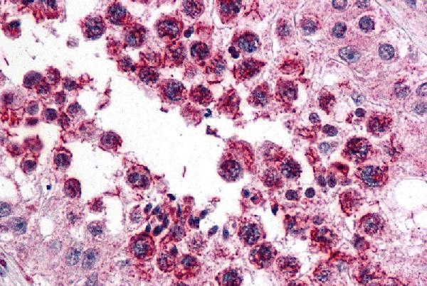
The IHC-validated anti-SLAIN2 antibody A11030 has real IHC data from human testis (catalog image caption); its listed application is IHC-P (catalog applications).
A11030 is listed for human IHC-P (catalog applications; catalog reactivity). Its IHC image shows human testis stained at 5 μg/mL (catalog image caption).
Which to pick: Choose A11030 for paraffin-section tissue IHC: it is a rabbit polyclonal antibody listed for human IHC-P, with an image from human testis (catalog host; catalog dilution description; catalog applications; catalog image caption). The fixative used for that image is unreported (catalog image caption). No SKU in the payload is listed for IF/ICC or species beyond human (catalog applications; catalog reactivity).