This website uses cookies to ensure you get the best experience on our website.


- Table of Contents
Plan paraffin IHC around granular CNS cytoplasmic staining (HPA tissue IHC) and the protein’s inner mitochondrial membrane location (UniProt). Use caudate or hippocampus neuronal cells as staining references (HPA tissue IHC), and consult the catalog antibody’s paraffin-section workflow (datasheet A08997-1).

Expected localisation, validated protocols, controls and antibodies — the at-a-glance facts below, then the full design guide.
| Expected localisation | Granular cytoplasm in CNS (HPA tissue IHC); inner mitochondrial membrane (UniProt) | |
| Staining pattern | Granular neuronal cytoplasm in caudate and hippocampus (HPA tissue IHC) | |
| Antigen retrieval | EDTA pH 8.0 HIER, heat-mediated (datasheet A08997-1) | |
| Positive control | Breast+4 more · see all | |
| Negative control | Adipose tissue+4 more · see all |
| Fixation | Keep fixation consistent across sections (standard IHC practice; not target-specific); Matched tissue-IHC evidence does not establish the fixation claim. Validate the specimen-specific method before use. (selected-SKU IHC image A08997-1) | |
| Caveat | Antibody staining and RNA show low consistency (HPA tissue IHC) | |
| Regulation | Brain-enriched RNA (HPA tissue RNA) | |
| Isoform / epitope | 4 isoforms; check epitope coverage across variants (UniProt) |
The catalog antibody protocol uses EDTA pH 8.0 retrieval (datasheet: A08997-1). One published IHC protocol reports SLC25A23 staining in colon cancer tissue (PMC9569204: methods).
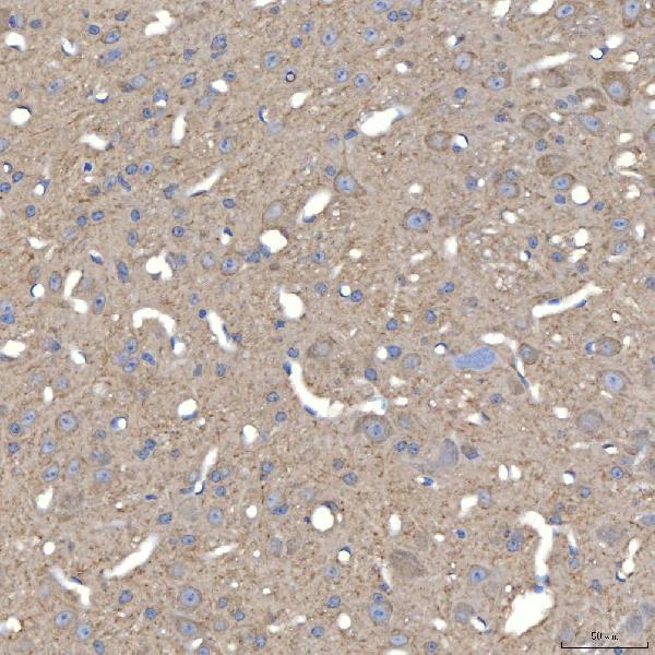
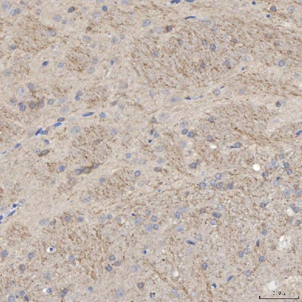
| Sample | Paraffin-embedded mouse brain tissue; fixative not specified (datasheet A08997-1) |
| Fixation | Image fixative and duration unreported (datasheet A08997-1); verify before use. |
| Sectioning | 4–5 µm sections on charged slides (standard) |
| Deparaffinisation | Xylene, graded ethanol series to water (standard) |
| Antigen retrieval | Heat retrieval: EDTA pH 8.0 (datasheet A08997-1); 20 min, 95–100 °C (standard) |
| Peroxidase block | 3% H2O2, 10 min, room temperature (standard) |
| Blocking | 10% goat serum (datasheet A08997-1) |
| Primary antibody | Rabbit anti-SLC25A23, 2-5 μg/ml (datasheet A08997-1) |
| Primary incubation | Overnight at 4 °C (datasheet A08997-1) |
| Detection | HRP-conjugated secondary, DAB chromogen (datasheet A08997-1) |
| Counterstain | Hematoxylin, blue, dehydrate and mount (standard) |
| Expected result | SLC25A23-positive staining in glandular cells of breast (HPA tissue IHC: Medium). HPA tissue profile: Granular cytoplasmic expression in CNS with additional cytoplasmic expression in several tissues. No signal in the no-primary control. |
SLC25A23 is an inner mitochondrial membrane protein with six transmembrane segments (UniProt Q9BV35 topology). In paraffin sections, expect granular cytoplasmic staining, particularly in CNS neuronal cells and several glandular or tubular cell populations (HPA tissue IHC: granular CNS staining; Medium in caudate and hippocampal neurons, breast glandular cells and kidney tubules). HPA rates tissue IHC as Approved but reports low consistency between antibody staining and RNA expression (HPA tissue IHC reliability).
| Granular cytoplasmic staining in hippocampal or caudate neurons, with an appropriate nuclear counterstain. | This fits the reported CNS pattern and Medium neuronal staining (HPA tissue IHC: hippocampus and caudate). Its cytoplasmic position is consistent with mitochondrial localization (UniProt Q9BV35; HPA subcellular ICC-IF). Chromogenic IHC alone does not resolve the inner mitochondrial membrane (general IHC practice). |
| Predominantly nuclear, cell-surface or extracellular staining, without a granular cytoplasmic component. | Those compartments conflict with the reported mitochondrial location (UniProt Q9BV35 topology; HPA subcellular ICC-IF). Treat the pattern as suspect and compare it with an expected-positive section and a detection control before assigning it to SLC25A23 (general IHC practice). |
| Strong signal in a cell population HPA lists as Not detected, such as adipocytes. | The result conflicts with the reported adipocyte observation (HPA tissue IHC: adipocytes Not detected). Possible explanations include antibody cross-reactivity or endogenous chromogenic detection activity (general IHC practice); the discordance alone cannot identify which cause applies, especially given HPA's low staining–RNA consistency (HPA tissue IHC reliability). |
| Diffuse color over cells and tissue spaces, obscuring cell boundaries and granules. | This is difficult to score as the reported granular cytoplasmic pattern (HPA tissue IHC profile). Widespread background can arise from detection chemistry, inadequate blocking or nonspecific antibody binding (general IHC practice); evaluate it with a primary-antibody omission control (general IHC practice). |
| No staining in hippocampal neurons or kidney tubule cells on a test section. | HPA reports Medium staining in these respective cell populations (HPA tissue IHC: hippocampus; kidney). Check tissue preservation, the antibody's IHC-P instructions and the detection system (general IHC practice). A blank section alone does not establish absent SLC25A23 expression (general IHC practice). |
| Subcellular location and topology | SLC25A23 has 6 membrane-spanning segments in the mitochondrial inner membrane (UniProt Q9BV35 topology). Interpret chromogenic granules at the cytoplasmic level; do not claim that light microscopy resolves membrane topology (general IHC practice). |
| Tissue and cell selection | HPA reports Medium staining in caudate and hippocampal neurons, breast glandular cells and kidney tubule cells, while adipocytes are Not detected (HPA tissue IHC). Score the named cell populations rather than treating every cell in a tissue as equivalent (general IHC practice). |
| Evidence strength | The tissue IHC assessment is Approved, with low consistency between antibody staining and RNA expression; GTEx data supports IHC (HPA tissue IHC reliability). The listed patterns are useful comparators, but a discordant section needs investigation rather than an automatic biological conclusion (general IHC practice). |
| IF/ICC question: should the IF result look identical to chromogenic IHC? | No exact visual match is implied: HPA reports supported mitochondrial localization by ICC-IF, while tissue IHC reports granular cytoplasmic staining (HPA subcellular ICC-IF; HPA tissue IHC). The modalities support a compartment-level comparison, not identical image texture (general IHC practice). |
| Situation | Likely cause | Next action |
|---|---|---|
| Expected-positive neuronal or tubular cells are blank. | An assay or detection failure is possible; those cells are reported at Medium intensity (HPA tissue IHC: hippocampus, caudate and kidney). | Check the antibody's IHC-P instructions, section integrity and chromogenic detection controls, then compare a separate reported-positive section (general IHC practice; HPA tissue IHC: listed Medium cells). |
| Signal is mainly nuclear or along cell surfaces. | That distribution is inconsistent with mitochondrial localization (UniProt Q9BV35; HPA subcellular ICC-IF). Nonspecific staining or interpretation of background is possible (general IHC practice). | Recheck morphology and the primary-antibody omission control; score only convincing cytoplasmic granules in the relevant cells (general IHC practice; HPA tissue IHC profile). |
| Broad brown background masks the expected granules. | Nonspecific binding or chromogenic detection background may obscure the reported granular cytoplasmic pattern (general IHC practice; HPA tissue IHC profile). | Inspect the omission control and review blocking, washing and detection conditions under the IHC-P workflow (general IHC practice). |
| Cells listed as Not detected appear strongly positive. | Cross-reactivity or endogenous chromogenic activity is possible (general IHC practice); the observation conflicts with the relevant HPA cell-level entry (HPA tissue IHC). | Check the omission control and inspect the specific cell type, then compare reported-positive tissue before interpreting the discrepancy (general IHC practice; HPA tissue IHC: listed Medium cells). |
| CNS regions give different staining results. | HPA lists Medium staining in caudate and hippocampal neurons but Not detected in cerebellar granular-layer cells (HPA tissue IHC); region and cell identity can therefore change the comparison. | Record the region and scored cell population explicitly, and compare each with its matching HPA entry (HPA tissue IHC). |
| A tissue result disagrees with an RNA-based expectation. | HPA reports low consistency between antibody staining and RNA expression, despite GTEx support for its IHC assessment (HPA tissue IHC reliability). | Document the observed cell-level pattern and assay controls; avoid converting RNA abundance alone into an expected IHC intensity (general IHC practice; HPA tissue IHC reliability). |


Comprehensive Human Protein Atlas IHC scoring per tissue (reliability: Approved — Low consistency between antibody staining and RNA expression data. GTEx data supports IHC.). Rows are taken directly from the HPA tissue chart — click any row's HPA link to view the source.
| Tissue | Cell type | Level | Evidence | Source |
|---|---|---|---|---|
| Breast | Glandular cells | Medium | Protein (IHC) | HPA → |
| Caudate | Neuronal cells | Medium | Protein (IHC) | HPA → |
| Gallbladder | Glandular cells | Medium | Protein (IHC) | HPA → |
| Hippocampus | Neuronal cells | Medium | Protein (IHC) | HPA → |
| Kidney | Cells in tubules | Medium | Protein (IHC) | HPA → |
| Tissue | Cell type | Level | Evidence | Source |
|---|---|---|---|---|
| Adipose tissue | Adipocytes | Not detected | Protein (IHC) | HPA → |
| Adrenal gland | Glandular cells | Not detected | Protein (IHC) | HPA → |
| Cerebellum | Cells in granular layer | Not detected | Protein (IHC) | HPA → |
| Cervix | Glandular cells | Not detected | Protein (IHC) | HPA → |
| Endometrium | Cells in endometrial stroma | Not detected | Protein (IHC) | HPA → |
Use the catalog antibody’s paraffin-section conditions as a starting point, then evaluate compartment, cell type and controls before scoring SLC25A23 staining.
The catalog antibody has paraffin-section IHC images from mouse and rat brain and human stomach (catalog image captions); no IF/ICC images are supplied (catalog payload).
A08997-1 has paraffin-section IHC images from mouse and rat brain and human stomach (catalog image captions). Its listed applications include IHC, while IF/ICC is unlisted and has no image evidence (catalog applications; catalog image captions).
Which to pick: Choose A08997-1 for paraffin-section tissue IHC: its captions document EDTA retrieval at pH 8.0 and 2 μg/ml primary antibody, but do not report the fixative (catalog image captions). For work across species, it lists human, mouse and rat reactivity, with IHC images for all three; clonality is unreported (catalog reactivity; catalog image captions; catalog payload). No SKU in this payload is validated for IF/ICC, so an IF/ICC choice cannot be supported here (catalog applications; catalog image captions).