SLC39A8 / Metal cation symporter ZIP8 · IHC design guide

Design Immunohistochemistry for SLC39A8

Plan paraffin-section SLC39A8 staining using kidney tubules and lung alveolar cells with high reported staining (HPA tissue IHC). Assess cytoplasmic and membranous signal (HPA tissue IHC), keeping the reported low agreement between antibody staining and RNA expression in view (HPA tissue IHC).

Evidence assembled Oct 2026 · For research use; verify linked source records and product datasheet before use
Immunohistochemistry protocol sheet for SLC39A8 (IHC for SLC39A8): expected localisation Cytoplasmic and membranous tissue staining (HPA tissue IHC), antibody A04007-1, validated IHC image, and IHC protocol steps
Printable SLC39A8 IHC protocol sheet — expected localisation Cytoplasmic and membranous tissue staining (HPA tissue IHC), antibody A04007-1, controls and protocol steps. Open the full SLC39A8 IHC guide →

SLC39A8 Immunohistochemistry Experimental Design Guide

Expected localisation, validated protocols, controls and antibodies — the at-a-glance facts below, then the full design guide.

Must know before staining
Expected localisation Cytoplasmic and membranous tissue staining (HPA tissue IHC)
Staining pattern Glandular and tubular cells show cytoplasmic/membranous staining (HPA tissue IHC)
Antigen retrieval EDTA pH 8.0 HIER, heat-mediated (datasheet A04007-1)
Positive control ⓘ Breast+4 more · see all
Negative control ⓘ None in HPA (detected in all 44 tissues); use no-primary + isotype controls
Important caveats
Reasons your staining may differ from the expected pattern.
Fixation Keep fixation conditions consistent across sections. (standard IHC practice; not target-specific)
Caveat Antibody staining has low consistency with RNA expression (HPA tissue IHC)
Regulation Expression regulation is unspecified (UniProt)
Isoform / epitope 3 isoforms; map the epitope to assess isoform coverage and membrane side (UniProt)
Section 1

Recommended SLC39A8 IHC & IF Protocols

The catalog antibody’s IHC-P protocol is paired with 4 published SLC39A8 paraffin-section IHC protocols (datasheet A04007-1; PMC8045709; PMC8804292; PMC13038716; PMC11869717).

Recommended immunohistochemistry (IHC-P) protocol parameters
SampleParaffin-embedded human endometrial cancer tissue; fixative not specified (datasheet A04007-1)
FixationImage fixative and duration unreported (datasheet A04007-1); verify before use.
Sectioning4–5 µm sections on charged slides (standard)
DeparaffinisationXylene, graded ethanol series to water (standard)
Antigen retrievalHeat retrieval: EDTA pH 8.0 (datasheet A04007-1); 20 min, 95–100 °C (standard)
Peroxidase block3% H2O2, 10 min, room temperature (standard)
Blocking10% goat serum (datasheet A04007-1)
Primary antibodyRabbit anti-SLC39A8, 2-5 μg/ml (datasheet A04007-1)
Primary incubationOvernight at 4 °C (datasheet A04007-1)
DetectionHRP-conjugated secondary, DAB chromogen (datasheet A04007-1)
CounterstainHematoxylin, blue, dehydrate and mount (standard)
Expected resultSLC39A8-positive staining in glandular cells of breast (HPA tissue IHC: High). HPA tissue profile: General cytoplasmic and membranous expression. No signal in the no-primary control.
💡Decision noteStart with heat-mediated EDTA pH 8.0 retrieval for the catalog antibody (datasheet A04007-1); the published excerpts do not specify a retrieval buffer (PMC13038716; PMC11869717).
Section 2

What Is the Expected SLC39A8 Staining Pattern?

SLC39A8 is a six-pass membrane protein found at the cell surface and lysosome membrane (UniProt Q9C0K1 topology and subcellular location). In paraffin-section IHC, expect cytoplasmic and membranous staining, including strong staining in kidney tubule cells and lung alveolar cells (HPA: general staining profile; High in both cell types). Treat intensity as a guide: HPA rates the tissue staining Approved but reports low consistency with RNA expression (HPA: reliability).

What am I looking at on my slide?
Membranous staining with some cytoplasmic signal in kidney tubule cells.This fits the reported distribution and a strong tissue example (HPA: general cytoplasmic and membranous profile; High in kidney tubule cells). Cytoplasmic signal can be compatible with intracellular SLC39A8 (UniProt Q9C0K1 subcellular location).
Staining confined to nuclei, with no convincing membrane or cytoplasmic pattern.Flag this as a possible artefact and review controls and morphology (UniProt Q9C0K1 subcellular location; HPA: general cytoplasmic and membranous profile). Neither source identifies the nucleus as an expected location.
Strong staining in cells outside the expected compartment or tissue pattern.Consider antibody cross-reactivity or endogenous detection activity, then compare with a no-primary control (general IHC practice). An unexpected cell type alone is inconclusive because expression is widespread and HPA reports low RNA–staining consistency (UniProt Q9C0K1 tissue specificity; HPA: reliability).
Diffuse staining across cells and surrounding tissue, obscuring boundaries.Treat the result as background until a control and a clearer membrane or cytoplasmic pattern support interpretation (general IHC practice; HPA: general staining profile). Diffuse color by itself cannot establish SLC39A8-positive cells.
No staining in kidney tubule cells despite intact tissue morphology.This conflicts with a reported strong IHC example (HPA: High in kidney tubule cells). Check the control, antibody dilution and general IHC workflow before calling the specimen negative (general IHC practice); HPA tissue staining is a reference, not a guarantee for every section (HPA: Approved; low consistency).
💡Expected SLC39A8 appearanceA convincing positive shows clearly assigned membranous and cytoplasmic staining in cells such as kidney tubule or lung alveolar cells, where HPA reports High staining; isolated nuclear color or diffuse tissue-wide color warrants control review (HPA: tissue IHC and general profile; UniProt Q9C0K1 subcellular location; general IHC practice).
How each factor affects the staining
Membrane topology and antibody epitopeSLC39A8 has six transmembrane segments and extracellular and cytoplasmic regions (UniProt Q9C0K1 topology). Epitope location could affect staining access, but the supplied sources do not identify this antibody’s epitope; topology alone cannot set retrieval or permeabilisation conditions.
Protein processing and glycosylationThe signal peptide spans residues 1–22, the annotated chain spans 23–460, and glycosylation sites occur at 40 and 88 (UniProt Q9C0K1 processing and glycosylation). These annotations do not establish epitope masking or a target-specific fixation effect.
IsoformsThree isoforms are listed (UniProt Q9C0K1 isoforms). Their existence does not establish which are recognized by an IHC antibody or how a particular section should stain; consult antibody-specific validation before interpreting isoform-dependent differences.
Strength of the tissue referenceHPA reports a general cytoplasmic and membranous pattern, while rating its IHC evidence Approved with low consistency against RNA expression (HPA: tissue IHC). The listed High cell types are useful reference examples, not an intensity threshold for diagnosis or scoring.
IF/ICC Q: Can this section define an IF/ICC pattern or protocol?A: HPA summarizes the subcellular location as Membrane but supplies no main location or ICC-IF image cell lines (HPA: subcellular). The listed antibodies have IHC Approved status and no ICC status (HPA: antibodies). Use the separate IF/ICC guide for that application.
Why is my staining missing, weak or wrong?
SituationLikely causeNext action
Kidney tubule control is blank.The result conflicts with reported High staining in those cells (HPA: tissue IHC); a failed staining run is possible (general IHC practice).Confirm tissue preservation and detection controls, then review the catalog antibody’s IHC-P instructions, dilution and retrieval steps (general IHC practice). Do not infer target-specific retrieval sensitivity from HPA.
Only nuclei show strong color.Nuclear localization is absent from the supplied location descriptions (UniProt Q9C0K1 subcellular location; HPA: general profile).Inspect the no-primary control and repeat with the validated IHC workflow if needed (general IHC practice); require a plausible membrane or cytoplasmic pattern before interpreting positivity.
Broad brown color persists when primary antibody is omitted.Primary-independent signal can arise from endogenous detection activity or nonspecific background (general chromogenic IHC practice).Review the detection system’s blocking steps, including endogenous peroxidase control when using peroxidase detection, and reassess staining with the no-primary control (general IHC practice).
Signal covers tissue without clear cell boundaries.Diffuse background can obscure the reported cellular pattern (HPA: general cytoplasmic and membranous profile; general IHC practice).Check blocking, washes and antibody dilution against the validated IHC instructions; compare the no-primary control and score only identifiable cells (general IHC practice).
Unexpected cell types appear stronger than the reference cells.Cross-reactivity or background is possible, but SLC39A8 is broadly expressed and HPA reports low RNA–staining consistency (UniProt Q9C0K1 tissue specificity; HPA: reliability).Compare morphology, no-primary staining and a reported High reference tissue before attributing the color to SLC39A8 (HPA: tissue IHC; general IHC practice).
Cytoplasmic staining appears without a sharp surface outline.Intracellular SLC39A8 is plausible, and HPA describes cytoplasmic as well as membranous staining (UniProt Q9C0K1 subcellular location; HPA: general profile).Evaluate whether the signal is cell-associated and reproducible against controls; record cytoplasmic and membranous components separately when scoring (general IHC practice).

Sample controls for SLC39A8 IHC & IF

🧪Run kidney first and assess staining in tubular cells (HPA: High in cells in tubules). HPA detects SLC39A8 in all 44 scored tissues, so no negative tissue is established; use no-primary and isotype controls, and treat any unstained cells on the positive slide as a local background reference rather than confirmed target-negative cells (HPA: no negative rows; standard IHC practice).
Positive control tissue: Breast (Glandular cells, HPA High)
Negative control tissue: None in HPA: SLC39A8 is detected in all 44 scored tissues. Use a no-primary (secondary-only) and an isotype control instead.
ICC-IF cell lines (HPA subcellular resource): HPA carries no ICC-IF cell line for SLC39A8; derive a cell-line control from the positive tissue's cell type (Glandular cells) and confirm it by RNA or western blot first.
Technical controls: Include a no-primary, secondary-only slide and a matched rabbit isotype control for the rabbit catalog antibody; a SLC39A8 knockout sample or validated immunizing-peptide block can provide a biological specificity control (selected-SKU caption: rabbit primary; standard IHC practice). For kidney HRP–DAB sections, block endogenous peroxidase and inspect control sections for background staining (HPA: High in kidney tubular cells; selected-SKU caption: HRP–DAB; standard IHC practice).
⚠️Feasibility: A target-specific fixation window or fixation effect is unreported, and the selected-SKU paraffin-section caption does not state the fixative (selected-SKU caption: fixative not stated). The caption uses heat retrieval in EDTA at pH 8.0, but does not establish whether retrieval is required or how frozen sections compare (selected-SKU caption: EDTA retrieval). IF/ICC feasibility relative to IHC is unestablished from the supplied evidence; for kidney chromogenic IHC, assess endogenous peroxidase background with the controls (HPA: kidney tubular cells High; selected-SKU caption: HRP–DAB; standard IHC practice).

HPA tissue IHC evidence for SLC39A8

Comprehensive Human Protein Atlas IHC scoring per tissue (reliability: Approved — Low consistency between antibody staining and RNA expression data.). Rows are taken directly from the HPA tissue chart — click any row's HPA link to view the source.

Positive expression · recommended positive controls

TissueCell typeLevelEvidenceSource
Breast Glandular cells High Protein (IHC) HPA →
Duodenum Glandular cells High Protein (IHC) HPA →
Endometrium Glandular cells Medium Protein (IHC) HPA →
Kidney Cells in tubules High Protein (IHC) HPA →
Lung Alveolar cells High Protein (IHC) HPA →

Undetected expression · recommended negative controls

TissueCell typeLevelEvidenceSource
None in HPA: SLC39A8 is detected in all 44 scored tissues. Use a no-primary (secondary-only) and an isotype control instead.
Section 3

Advanced SLC39A8 IHC Tips

Troubleshooting guidance for chromogenic SLC39A8 IHC in paraffin sections, with one entry on IF assay design.

What should I change when SLC39A8 staining is weak after retrieval?
Start with heat-mediated retrieval in EDTA at pH 8.0 for this paraffin-section assay (datasheet A04007-1). The product image used 2 μg/ml primary antibody overnight at 4°C, so check those conditions before changing retrieval (datasheet A04007-1). Keep heating and cooling consistent across sections, then compare staining in a tissue with documented expression, such as kidney tubules (standard IHC practice; HPA: High in kidney tubule cells). If staining remains weak, test an alternative retrieval condition on adjacent sections as a fallback and assess tissue damage alongside signal (standard IHC practice). Record the condition selected for every sample in a scoring set (standard IHC practice).
Could fixation explain weak or uneven SLC39A8 staining?
Target-specific fixation sensitivity is unknown: the selected product caption describes a paraffin section but does not state its fixative (datasheet A04007-1). For new specimens, document the fixative and fixation duration, then process comparison sections consistently before attributing a staining difference to SLC39A8 (standard IHC practice). Check morphology and compare internal cell populations on the same section; poor preservation or uneven processing can make a stain difficult to interpret (standard IHC practice). Keep EDTA at pH 8.0 and the primary antibody at 2 μg/ml constant during the fixation comparison (datasheet A04007-1). Use a documented positive-control section in each run to distinguish a processing problem from a staining-run problem (standard IHC practice).
How should I assess cytoplasmic staining from a membrane transporter?
Score membrane-associated and intracellular staining separately: SLC39A8 is annotated at cell, apical, basolateral and lysosomal membranes (UniProt Q9C0K1 localisation). Intracellular signal can be plausible because the record describes a substantial intracellular fraction in blood–brain barrier microvascular endothelial cells (UniProt Q9C0K1 localisation). HPA also reports general cytoplasmic and membranous tissue staining, though its staining-to-RNA consistency is low (HPA: tissue IHC profile and reliability description). Examine whether the signal follows cell boundaries or a reproducible intracellular pattern in intact cells, using the same chromogen development across sections (standard IHC practice). Treat diffuse staining over damaged tissue or section edges as a technical concern requiring control comparison (standard IHC practice).
Can this stain distinguish SLC39A8 isoforms or epitope accessibility?
Do not assign an isoform from this IHC stain alone: the record lists 3 isoforms without giving this antibody's epitope or isoform coverage (UniProt Q9C0K1 isoforms; datasheet A04007-1). The annotated protein has 6 transmembrane segments, an extracellular region at residues 23–132 and a cytoplasmic region at 213–365 (UniProt Q9C0K1 topology). Glycosylation sites at residues 40 and 88 lie in the annotated extracellular region, but their effect on this antibody's staining is unknown (UniProt Q9C0K1 topology and glycosylation). Check the antibody's mapped immunogen or epitope before interpreting retrieval changes as epitope effects (standard IHC practice). If isoform identity matters, use an independently validated isoform-specific method alongside the tissue stain (standard IHC practice).
How can I investigate SLC39A8 staining with multiplex IF?
Use IF as a separately optimised assay; the supplied product evidence documents chromogenic staining in a paraffin section, while HPA provides no cell-line ICC/IF images (datasheet A04007-1; HPA: subcellular record). Multiplex SLC39A8 with a macrophage marker such as CD68 when studying that population, because macrophage protein expression is documented (UniProt Q9C0K1 tissue specificity; standard IF practice). Choose a far-red fluorophore and inspect single-channel controls to reduce confusion with tissue autofluorescence or spectral bleed-through (standard IF practice). If the antibody epitope is cytoplasmic, optimise permeabilisation; if it is extracellular, compare staining without permeabilisation first (UniProt Q9C0K1 topology; standard IF practice). Confirm the epitope location before treating either condition as definitive for this antibody (datasheet A04007-1: epitope not specified).
How can I separate SLC39A8 signal from chromogenic background?
Start by comparing stained sections with a no-primary control and checking whether DAB deposits follow intact cells rather than folds, edges or damaged areas (standard IHC practice). The selected product image used 10% goat serum blocking, 2 μg/ml primary antibody and overnight incubation at 4°C (datasheet A04007-1). Its detection used a peroxidase-conjugated secondary and DAB; include a peroxidase block when building the workflow, treating that block as general IHC practice (datasheet A04007-1; standard IHC practice). If background persists, adjust blocking, washing and primary concentration systematically while keeping retrieval at EDTA pH 8.0 for the initial comparison (standard IHC practice; datasheet A04007-1). Review a documented positive-control tissue alongside the no-primary control before changing the interpretation threshold (standard IHC practice).
What is a defensible way to score SLC39A8 IHC across samples? ⚠ ANSWER MARKED FOR VERIFICATION
Define the cell population and compartment before scoring, because the annotated protein can appear at cell and lysosomal membranes (UniProt Q9C0K1 localisation; standard IHC practice). For epithelial or tubular populations, report percentage positive cells and an H-score using intensity grades 0–3, giving a possible range of 0–300 (standard IHC practice). If counting scattered positive cells, report density per mm² of evaluable tissue and specify the counted cell population (standard IHC practice). Normalise counts to intact tissue area or the number of eligible cells, and keep chromogen development and threshold settings consistent (standard IHC practice). Report membrane and cytoplasmic scores separately rather than merging distinct staining patterns (UniProt Q9C0K1 localisation; standard IHC practice).
When should an apparent SLC39A8-positive cell be considered artefactual?
Assess whether staining is in an expected compartment and cell population: SLC39A8 is annotated at cell and lysosomal membranes and expressed by macrophages (UniProt Q9C0K1 localisation and tissue specificity). HPA reports high staining in kidney tubule cells and lung alveolar cells, but also notes low consistency between antibody staining and RNA expression (HPA: tissue IHC and reliability description). Be cautious with signal confined to tissue edges, necrotic areas or damaged cells, and compare a no-primary control for nonspecific DAB or endogenous enzyme signal (standard IHC practice). A plausible pattern still needs reproducibility across intact fields and an appropriate positive control (standard IHC practice). Avoid inferring metal-transport activity from stain intensity alone; the assay detects antibody binding in tissue (standard IHC practice).
Boster reagents

Best SLC39A8 / Metal cation symporter ZIP8 IHC Antibodies

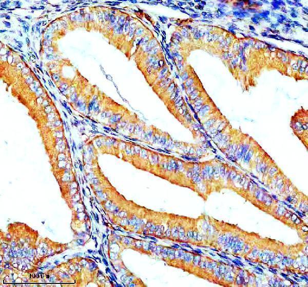
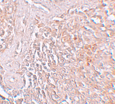
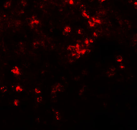
The catalog shows human spleen IHC and IF images for A04007 (image captions) and human endometrial cancer paraffin-section IHC for A04007-1 (IHC caption).

Real IHC data IHC analysis of ZIP8/SLC39A8 using anti-ZIP8/SLC39A8 antibody (A04007-1). ZIP8/SLC39A8 was detected in a paraffin-embedded section of human endometrial cancer tissue. Heat mediated antigen retrieval was performed in EDTA buffer (pH 8.0, epitope retrieval solution). The tissue section was blocked with 10% goat serum. The tissue section was then incubated with 2 μg/ml rabbit anti-ZIP8/SLC39A8 Antibody (A04007-1) overnight at 4°C. Peroxidase Conjugated Goat Anti-rabbit IgG was used as secondary antibody and incubated for 30 minutes at 37°C. The tissue section was developed using HRP Conjugated Rabbit IgG Super Vision Assay Kit (Catalog # SV0002) with DAB as the chromogen.
Anti-ZIP8/SLC39A8 Antibody ®
Cat # A04007-1
Real IHC data Immunohistochemistry of ZIP8 in human spleen tissue with ZIP8 antibody at 5 μg/mL.
Anti-ZIP8 SLC39A8 Antibody
Cat # A04007

A04007 has human spleen IHC and IF images (A04007 image captions). A04007-1 has a human endometrial cancer paraffin-section IHC image; its listed reactivity is human, mouse and rat (A04007-1 IHC caption; catalog: reactivity).

Which to pick: For paraffin-section IHC, choose A04007-1 at 2–5 μg/mL (A04007-1 datasheet: IHC dilution; IHC caption: paraffin section). For human tissue IF, choose A04007 at 20 μg/mL (A04007 datasheet: IF dilution; IF caption: human spleen); ICC validation is unreported (A04007 application list). For mouse or rat work, A04007-1 lists those species as reactive, but its IHC image shows human tissue only; the fixative is unreported (A04007-1 catalog: reactivity; IHC caption).

Each figure is that product's own IHC / IF validation image from its datasheet.

References

  1. UniProt Consortium. UniProt entry Q9C0K1 (S39A8_HUMAN, Metal cation symporter ZIP8).
  2. Human Protein Atlas. SLC39A8 tissue IHC expression (reliability: Approved).
  3. Human Protein Atlas. SLC39A8 subcellular location (ICC-IF): Membrane.
  4. Human Protein Atlas. SLC39A8 antibody validation summary (2 antibodies).
  5. SLC39A8/Zinc Suppresses the Progression of Clear Cell Renal Cell Carcinoma. Frontiers in oncology 2021 — PMC8045709.
  6. An Iron Metabolism-Related Gene Signature for the Prognosis of Colon Cancer. Frontiers in cell and developmental biology 2021 — PMC8804292.
  7. Unveiling the prognostic and therapeutic landscape of the zinc transporter protein SLC39A family in colorectal cancer through multi-omics and machine learning approaches. Clinical and experimental medicine 2026 — PMC13038716.
  8. PCR array analysis reveals a novel expression profile of ferroptosis-related genes in idiopathic pulmonary fibrosis. BMC pulmonary medicine 2025 — PMC11869717.
  9. PubMed PMID:12504855 — UniProt-cited evidence.
  10. PubMed PMID:14702039 — UniProt-cited evidence.
  11. PubMed PMID:15498874 — UniProt-cited evidence.