This website uses cookies to ensure you get the best experience on our website.


- Table of Contents
Plan chromogenic SLFN11 IHC on paraffin sections using the catalog antibody at 2–5 μg/mL (datasheet A08259). Assess cytoplasmic tissue staining (HPA tissue IHC) alongside the protein’s nuclear and chromosomal localisation (UniProt), and interpret the tissue pattern with its uncertain reliability in mind (HPA tissue IHC).

Expected localisation, validated protocols, controls and antibodies — the at-a-glance facts below, then the full design guide.
| Expected localisation | Cytoplasmic tissue staining (HPA tissue IHC); nuclear/chromosomal localisation (UniProt) | |
| Staining pattern | Cytoplasmic staining across several tissue cell types (HPA tissue IHC) | |
| Antigen retrieval | EDTA pH 8.0 HIER, heat-mediated (datasheet A08259) | |
| Positive control | Adipose tissue+4 more · see all | |
| Negative control | Appendix+4 more · see all |
| Fixation | Keep paraffin-section fixation consistent (standard IHC practice; not target-specific) | |
| Caveat | Tissue staining reliability is uncertain (HPA tissue IHC) | |
| Regulation | Interferon-induced expression (UniProt) | |
| Isoform / epitope | No annotated isoforms; single chain, residues 1–901 (UniProt) |
The catalog antibody’s IHC-P protocol is accompanied by published SLFN11 staining conditions for soft tissue sarcoma, ovarian serous carcinoma, and bladder cancer (PMC13008703; PMC13576481; PMC8819307).
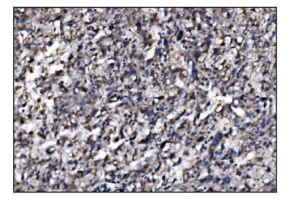
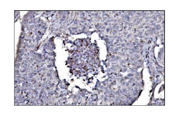
| Sample | Paraffin-embedded human liver cancer tissue; fixative not specified (datasheet A08259) |
| Fixation | Image fixative and duration unreported (datasheet A08259); verify before use. |
| Sectioning | 4–5 µm sections on charged slides (standard) |
| Deparaffinisation | Xylene, graded ethanol series to water (standard) |
| Antigen retrieval | Heat retrieval: EDTA pH 8.0 (datasheet A08259); 20 min, 95–100 °C (standard) |
| Peroxidase block | 3% H2O2, 10 min, room temperature (standard) |
| Blocking | 10% goat serum (datasheet A08259) |
| Primary antibody | Rabbit anti-SLFN11, 2-5μg/ml (datasheet A08259) |
| Primary incubation | Overnight at 4 °C (datasheet A08259) |
| Detection | Streptavidin-biotin complex (SABC), DAB chromogen (datasheet A08259) |
| Counterstain | Hematoxylin, blue, dehydrate and mount (standard) |
| Expected result | SLFN11-positive staining in adipocytes of adipose tissue (HPA tissue IHC: Medium). HPA tissue profile: Cytoplasmic expression in several different tissue types. No signal in the no-primary control. |
SLFN11 is assigned to the nucleus and chromosomes and has no transmembrane segment (UniProt Q7Z7L1). Nuclear staining in expressing cells is therefore biologically plausible. HPA instead reports cytoplasmic tissue IHC staining across several tissues, with uncertain reliability; its ICC-IF data support nucleoplasmic localisation (HPA tissue IHC; HPA subcellular). Interpret compartment and cell type together, and treat either IHC pattern as requiring antibody and control review.
| Nuclear signal in cells of endometrial stroma, with limited background. | Nuclear localisation agrees with UniProt and supported HPA ICC-IF nucleoplasmic localisation (UniProt Q7Z7L1; HPA subcellular). HPA rates endometrial stromal tissue staining medium, but its tissue IHC reliability is uncertain; this slide alone does not validate the IHC antibody (HPA tissue IHC). |
| Predominantly cytoplasmic staining in adipocytes or adrenal glandular cells. | HPA reports medium staining in those cells and describes its tissue IHC profile as cytoplasmic (HPA tissue IHC). This differs from UniProt nuclear localisation and supported HPA ICC-IF nucleoplasmic localisation (UniProt Q7Z7L1; HPA subcellular). Record it as an observed, uncertain IHC pattern rather than proof of cytoplasmic SLFN11. |
| Strong staining confined to cell membranes, with little nuclear or cytoplasmic signal. | A membrane-only pattern conflicts with UniProt nuclear localisation and the absence of a transmembrane segment (UniProt Q7Z7L1). Check antibody specificity and detection background before assigning it to SLFN11. HPA's uncertain cytoplasmic tissue IHC pattern does not support a membrane-only interpretation (HPA tissue IHC). |
| Prominent signal in appendix glandular cells or bronchial respiratory epithelium. | HPA lists those cell types as not detected (HPA tissue IHC). If signal is convincing, assess cross-reactivity or endogenous detection activity with appropriate controls (general IHC practice). Because HPA rates its tissue IHC evidence uncertain, a discrepant result warrants investigation rather than an automatic false-positive label (HPA tissue IHC). |
| No signal in adipocytes despite intact tissue and a working counterstain. | HPA reports medium adipocyte staining, making them a useful comparison within this evidence set, although the tissue IHC profile remains uncertain (HPA tissue IHC). Check the antibody and detection controls, retrieval conditions and reagent dilution (general IHC practice). A negative slide alone cannot establish absent SLFN11 expression. |
| Compartment evidence | UniProt assigns SLFN11 to the nucleus and chromosomes; HPA ICC-IF supports nucleoplasm, whereas HPA tissue IHC describes cytoplasmic staining with uncertain reliability (UniProt Q7Z7L1; HPA subcellular; HPA tissue IHC). Keep the assay-specific discrepancy visible when scoring. |
| Tissue and cell selection | HPA reports medium staining in adipocytes and endometrial stromal cells, low staining in glial cells, and no detection in appendix glandular cells (HPA tissue IHC). Compare the named cell populations, rather than treating an entire section as uniformly positive or negative. |
| Antibody evidence | HPA023030 is rated uncertain for IHC and supported for ICC; HPA062062 has no listed IHC status and is supported for ICC (HPA antibodies). An ICC rating does not establish performance in paraffin-section IHC. HPA also rates its tissue IHC profile uncertain (HPA tissue IHC). |
| Protein topology and processing | UniProt lists no transmembrane segment, signal peptide, propeptide or annotated glycosylation site for SLFN11 (UniProt Q7Z7L1). These annotations support caution with a membrane-only pattern; they do not predict how fixation or antigen retrieval affects the IHC signal. |
| IF/ICC Q: what pattern should be expected? | A: Mainly nucleoplasmic signal is supported; additional cytosolic localisation is uncertain (HPA subcellular). That ICC-IF observation helps interpret localisation, but it cannot substitute for validation of chromogenic paraffin-section IHC (HPA subcellular; HPA antibodies). |
| Situation | Likely cause | Next action |
|---|---|---|
| Nuclear signal is absent but cytoplasmic colour is present. | HPA describes uncertain cytoplasmic tissue IHC, while UniProt and supported ICC-IF favour nuclear localisation (HPA tissue IHC; UniProt Q7Z7L1; HPA subcellular). | Record both compartment and named cell type; compare antibody and detection controls before interpreting the cytoplasmic colour as SLFN11 (general IHC practice). |
| Signal appears only along membranes. | Membrane-only localisation is unsupported by UniProt's nuclear assignment and lack of a transmembrane segment (UniProt Q7Z7L1). | Review antibody specificity and a no-primary control, then reassess localisation in intact cells (general IHC practice). |
| Unexpected epithelial cells stain strongly. | Appendix glandular and bronchial respiratory epithelial cells are listed as not detected by HPA, although its tissue IHC evidence is uncertain (HPA tissue IHC). | Confirm the cell identity and compare a no-primary control; investigate cross-reactivity or endogenous detection activity before scoring a new positive pattern (general IHC practice). |
| Colour is diffuse across tissue or appears in cell-free areas. | Diffuse colour prevents a reliable cell-level or compartment-level assignment (general IHC practice). | Compare a no-primary control and review blocking, washes and chromogen development; score only distinguishable cellular signal (general IHC practice). |
| No staining appears in adipocytes or endometrial stromal cells. | Both are reported at medium staining by HPA, but the overall tissue IHC profile has uncertain reliability (HPA tissue IHC). | Verify section integrity, antibody dilution, retrieval and detection controls; document the negative finding without assuming biological absence (general IHC practice). |
| An ICC-IF result is nucleoplasmic while paraffin IHC is cytoplasmic. | That difference mirrors supported HPA ICC-IF localisation and its uncertain cytoplasmic tissue IHC profile (HPA subcellular; HPA tissue IHC). | Report assay and compartment separately; assess the IHC antibody and controls within paraffin sections before reconciling the results (HPA antibodies; general IHC practice). |


Comprehensive Human Protein Atlas IHC scoring per tissue (reliability: Uncertain — Medium consistency between antibody staining and RNA expression data. Pending external verification.). Rows are taken directly from the HPA tissue chart — click any row's HPA link to view the source.
| Tissue | Cell type | Level | Evidence | Source |
|---|---|---|---|---|
| Adipose tissue | Adipocytes | Medium | Protein (IHC) | HPA → |
| Adrenal gland | Glandular cells | Medium | Protein (IHC) | HPA → |
| Caudate | Neuronal cells | Medium | Protein (IHC) | HPA → |
| Endometrium | Cells in endometrial stroma | Medium | Protein (IHC) | HPA → |
| Epididymis | Glandular cells | Medium | Protein (IHC) | HPA → |
| Tissue | Cell type | Level | Evidence | Source |
|---|---|---|---|---|
| Appendix | Glandular cells | Not detected | Protein (IHC) | HPA → |
| Bronchus | Respiratory epithelial cells | Not detected | Protein (IHC) | HPA → |
| Colon | Endothelial cells | Not detected | Protein (IHC) | HPA → |
| Duodenum | Glandular cells | Not detected | Protein (IHC) | HPA → |
| Esophagus | Squamous epithelial cells | Not detected | Protein (IHC) | HPA → |
Troubleshoot SLFN11 staining in paraffin-section chromogenic IHC by checking retrieval, nuclear localisation, controls and cell-specific scoring (datasheet A08259; UniProt Q7Z7L1).
A human-reactive anti-SLFN11 antibody has real IHC data from paraffin sections of human liver and lung cancer tissue (catalog: A08259 reactivity and IHC image captions).
A08259 is listed for human IHC and has chromogenic images from paraffin sections of human liver and lung cancer tissue (catalog: A08259 applications, reactivity and IHC image captions). IF/ICC is absent from its application list, and no IF image is supplied (catalog: A08259 applications and IF image alts).
Which to pick: Choose A08259 for human paraffin-section IHC: the rabbit antibody is listed for IHC, and its own captions document EDTA retrieval at pH 8.0 and DAB detection (catalog: A08259 host, applications and IHC image captions). The clone and fixative are unreported; the captions establish paraffin embedding, not a specific fixation method (catalog: A08259 clone and IHC image captions). No listed SKU has IF/ICC or nonhuman validation: A08259 lists human reactivity and no IF application or IF image (catalog: A08259 applications, reactivity and IF image alts).