This website uses cookies to ensure you get the best experience on our website.


- Table of Contents
Plan chromogenic SLITRK1 IHC on paraffin sections with the catalog antibody, starting at 2.5 μg/mL (datasheet). Use cortical glia as a strong staining reference and interpret cytoplasmic CNS staining with the reported secreted variant in mind (HPA tissue IHC).

Expected localisation, validated protocols, controls and antibodies — the at-a-glance facts below, then the full design guide.
| Expected localisation | Cytoplasmic staining in CNS cells (HPA tissue IHC) | |
| Staining pattern | Selective cytoplasmic CNS staining, strongest in cortical glia (HPA tissue IHC) | |
| Antigen retrieval | Citrate pH 6.0 HIER, 95–98 °C, 20 min (rule: cytoplasmic / membrane antigen) | |
| Positive control | Cerebral cortex+2 more · see all | |
| Negative control | Adipose tissue+4 more · see all |
| Fixation | Keep fixation consistent across paraffin sections. (standard IHC practice; not target-specific) | |
| Caveat | A secreted variant may shift protein from RNA-positive cells (HPA tissue IHC) | |
| Regulation | Predominant expression in frontal cortex (UniProt) | |
| Isoform / epitope | No listed isoforms; extracellular versus cytoplasmic epitope matters (UniProt) |
The catalog antibody has an IHC-P protocol (datasheet); one published brain-section DAB protocol provides a comparison (PMC3292218).
| Sample | Tissue sections; selected-image fixative not specified (standard IHC workflow) |
| Fixation | Image fixative and duration unreported (datasheet A07507); verify before use. |
| Sectioning | 4–5 µm sections on charged slides (standard) |
| Deparaffinisation | Xylene, graded ethanol series to water (standard) |
| Antigen retrieval | Heat-induced epitope retrieval in citrate buffer, pH 6.0, 20 min at 95–98 °C (standard rule: cytoplasmic / membrane antigen) |
| Peroxidase block | 3% H2O2, 10 min, room temperature (standard) |
| Blocking | 10% normal serum of the secondary host, 30 min, room temperature (standard) |
| Primary antibody | Rabbit anti-SLITRK1, 2.5 μg/mL (datasheet A07507) |
| Primary incubation | Overnight at 4 °C (standard) |
| Detection | HRP-polymer secondary, DAB chromogen 5–10 min (standard) |
| Counterstain | Hematoxylin, blue, dehydrate and mount (standard) |
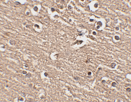
| Expected result | SLITRK1-positive staining in glial cells of cerebral cortex (HPA tissue IHC: High). HPA tissue profile: Selective cytoplasmic expression in CNS. No signal in the no-primary control. |
SLITRK1 is annotated at the membrane, synapse and in secreted form, with an extracellular region at residues 18–622, a transmembrane segment at 623–643 and a cytoplasmic tail at 644–696 (UniProt Q96PX8 topology). In tissue IHC, expect selective cytoplasmic CNS staining: high in cerebral-cortex glia and medium in cerebellar Purkinje cells and hippocampal neurons (HPA tissue IHC). HPA rates the tissue pattern Enhanced, while reporting medium RNA–protein consistency (HPA tissue IHC).
| Cerebral-cortex glia show strong cytoplasmic chromogen; cerebellar Purkinje cells and hippocampal neurons show weaker staining. | This matches the reported high cortical-glial and medium Purkinje-cell and hippocampal-neuronal levels (HPA tissue IHC). Compare the named cells within each region; a uniformly dark section does not establish this selective pattern (general IHC interpretation). |
| Predominantly nuclear staining appears in the expected positive cells. | A nuclear-only pattern does not match HPA’s selective cytoplasmic tissue profile or UniProt’s membrane, synapse and secreted annotations (HPA tissue IHC; UniProt Q96PX8). Check nuclear counterstain overlap and detection controls before interpreting it as SLITRK1 (general IHC practice). |
| Strong signal appears in an unexpected cell type, or in adipocytes used as a negative comparison. | HPA reports adipocytes as not detected in adipose tissue and breast, while its named CNS positives are specific cell classes (HPA tissue IHC). Unexpected staining may reflect cross-reactivity or endogenous detection activity; morphology alone cannot distinguish these causes (general IHC interpretation). |
| Chromogen spreads across tissue compartments and obscures cell boundaries. | Diffuse staining prevents a reliable call on HPA’s selective cytoplasmic pattern (HPA tissue IHC). Review background in a no-primary control and inspect the detection chemistry before scoring cells (general IHC practice). A diffuse result alone does not demonstrate secreted SLITRK1 (UniProt Q96PX8). |
| No signal is visible in the expected cells of a cerebral-cortex section. | That conflicts with HPA’s high cortical-glial result, but one negative slide cannot establish absent protein (HPA tissue IHC; general IHC interpretation). Verify that glia are present, the positive control and detection reagents work, and the IHC-validated antibody was used under its documented conditions (general IHC practice). |
| Epitope location | SLITRK1 spans an extracellular region, one transmembrane segment and a cytoplasmic tail (UniProt Q96PX8 topology). The supplied evidence does not locate the IHC antibody’s epitope, so topology alone cannot predict which compartment that antibody will highlight. |
| Secreted annotation | UniProt lists secreted SLITRK1, and HPA notes that at least one protein variant is secreted; RNA and tissue protein locations may therefore differ (UniProt Q96PX8; HPA tissue IHC). Do not treat diffuse extracellular color by itself as proof of secretion (general IHC interpretation). |
| Strength of the tissue reference | HPA labels the tissue IHC result Enhanced and notes external characterization, medium RNA–protein consistency and presumed off-target binding that was disregarded (HPA tissue IHC). Use the named cell and region pattern with controls when judging a new stain (general IHC practice). |
| Antigen retrieval | Target-specific fixation effects are not established by the supplied assay evidence. Verify with a matched IHC source before attributing a result to fixation. |
| IF/ICC Q&A: Is a cell-line staining pattern established? | No ICC-IF image-bearing cell line or main subcellular location is supplied by HPA (HPA subcellular). Its membrane/secreted summary and UniProt’s topology provide context, but do not validate an IF/ICC pattern or a protocol option (HPA subcellular; UniProt Q96PX8). |
| Situation | Likely cause | Next action |
|---|---|---|
| Cortical glia are unstained. | A failed detection step, unsuitable antibody conditions or a section lacking the expected cells can mimic a negative result (general IHC practice); HPA reports high cortical-glial staining (HPA tissue IHC). | Confirm glial morphology and section quality, run a documented positive section, and check antibody use and detection reagents before changing retrieval (general IHC practice). No SLITRK1-specific retrieval condition is supplied (HPA tissue IHC; UniProt Q96PX8). |
| The whole section has brown haze. | Background from detection reagents or inadequate blocking can obscure selective cytoplasmic staining (general IHC practice; HPA tissue IHC). | Compare a no-primary section, adjust blocking or washes, and inspect the chromogen reaction according to the detection system (general IHC practice). Score SLITRK1 only where cells and compartments remain distinguishable (HPA tissue IHC). |
| Nuclei are the dominant positive compartment. | The pattern is discordant with reported cytoplasmic tissue staining and annotated membrane/synapse localization; counterstain or nonspecific detection may confuse interpretation (HPA tissue IHC; UniProt Q96PX8; general IHC practice). | Review an unstained or no-primary control and examine cells at higher magnification before calling nuclear positivity (general IHC practice). |
| Adipocytes stain as strongly as cortical glia. | HPA reports adipocytes as not detected in adipose tissue and breast, versus high cortical-glial staining (HPA tissue IHC). Cross-reactivity or detection background is possible, not proven (general IHC interpretation). | Compare the tissues in the same staining run and review no-primary controls; reassess antibody specificity if the unexpected cellular signal persists (general IHC practice). |
| Cerebellum or hippocampus looks weaker than cortex. | Medium Purkinje-cell and hippocampal-neuronal staining versus high cortical-glial staining is the reported relationship (HPA tissue IHC). | Judge the named cell populations against their own background before increasing signal development; stronger cortex alone does not imply failure in the other regions (HPA tissue IHC; general IHC practice). |
| A negative comparison shows a few faint cells. | Duodenal and small-intestinal enterocytes are reported at low levels, so they are unsuitable as absolute negatives; adipose-tissue adipocytes are reported not detected (HPA tissue IHC). | Choose a reported not-detected cell population for the negative comparison, confirm its morphology, and interpret faint staining alongside a no-primary control (HPA tissue IHC; general IHC practice). |


Comprehensive Human Protein Atlas IHC scoring per tissue (reliability: Enhanced — Medium consistency between antibody staining and RNA expression data. At least one protein variant secreted, tissue location of RNA and protein might differ and correlation is complex. Presumed off target binding observed and disregarded. External characterization data supports antibody staining.). Rows are taken directly from the HPA tissue chart — click any row's HPA link to view the source.
| Tissue | Cell type | Level | Evidence | Source |
|---|---|---|---|---|
| Cerebral cortex | Glial cells | High | Protein (IHC) | HPA → |
| Cerebellum | Purkinje cells | Medium | Protein (IHC) | HPA → |
| Hippocampus | Neuronal cells | Medium | Protein (IHC) | HPA → |
| Tissue | Cell type | Level | Evidence | Source |
|---|---|---|---|---|
| Adipose tissue | Adipocytes | Not detected | Protein (IHC) | HPA → |
| Adrenal gland | Glandular cells | Not detected | Protein (IHC) | HPA → |
| Appendix | Endocrine cells | Not detected | Protein (IHC) | HPA → |
| Bone marrow | Hematopoietic cells | Not detected | Protein (IHC) | HPA → |
| Breast | Adipocytes | Not detected | Protein (IHC) | HPA → |
Troubleshoot SLITRK1 staining in paraffin sections by checking retrieval, cell identity and signal location against the available tissue evidence.
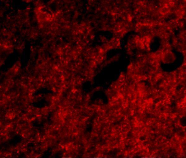
A07507 has IHC and IF images from human brain tissue (catalog image captions). Its listed applications include IHC-P and IF, with stated reactivity to human, mouse and rat (catalog applications and reactivity).
A07507 will render with a human brain tissue IHC image at 2.5 μg/mL (catalog IHC image caption). Its IF image also shows human brain tissue at 20 μg/mL; the catalog lists human, mouse and rat reactivity (catalog IF image caption; catalog reactivity).
Which to pick: Choose A07507 for tissue IHC because IHC-P is listed, though its IHC caption identifies human brain tissue without reporting the fixative (catalog applications; catalog IHC image caption). For IF, choose A07507 based on its human brain image; confirm performance for ICC separately because the catalog lists IF but provides no ICC image (catalog IF image caption; catalog applications). A07507 is the listed cross-species option for human, mouse and rat, but the supplied images show human tissue only; clonality is unreported (catalog reactivity; catalog image captions; catalog clone field).