SLK / STE20-like serine/threonine-protein kinase · IHC design guide

Design Immunohistochemistry for SLK

Plan chromogenic SLK IHC in paraffin sections using the catalog antibody at 2–5 μg/ml (datasheet A01614-2). Score cytoplasmic staining and include kidney glomerular cells, which show high staining (HPA tissue IHC).

Evidence assembled Oct 2026 · For research use; verify linked source records and product datasheet before use
Immunohistochemistry protocol sheet for SLK (IHC for SLK): expected localisation Cytoplasmic tissue staining (HPA tissue IHC), antibody A01614-2, validated IHC image, and IHC protocol steps
Printable SLK IHC protocol sheet — expected localisation Cytoplasmic tissue staining (HPA tissue IHC), antibody A01614-2, controls and protocol steps. Open the full SLK IHC guide →

SLK Immunohistochemistry Experimental Design Guide

Expected localisation, validated protocols, controls and antibodies — the at-a-glance facts below, then the full design guide.

Must know before staining
Expected localisation Cytoplasmic tissue staining (HPA tissue IHC)
Staining pattern Cytoplasmic staining; high in glomerular cells (HPA tissue IHC)
Antigen retrieval EDTA pH 8.0 HIER, heat-mediated (datasheet A01614-2)
Positive control ⓘ Kidney+4 more · see all
Negative control ⓘ None in HPA (detected in all 45 tissues); use no-primary + isotype controls
Important caveats
Reasons your staining may differ from the expected pattern.
Fixation Matched tissue-IHC evidence does not establish the fixation claim. Validate the specimen-specific method before use. (selected-SKU IHC image A01614-2)
Caveat Staining has medium agreement with RNA data (HPA tissue IHC)
Regulation No stimulus-dependent regulation annotated (UniProt)
Isoform / epitope 2 isoforms; epitope coverage unspecified (UniProt)
Section 1

Recommended SLK IHC & IF Protocols

The catalog antibody uses heat-mediated EDTA pH 8.0 retrieval (datasheet: A01614-2). The published mouse IHC protocol below uses citrate pH 6.0 retrieval (PMC12423296: Methods).

Recommended immunohistochemistry (IHC-P) protocol parameters
SampleParaffin-embedded human colon tissue; fixative not specified (datasheet A01614-2)
FixationImage fixative and duration unreported (datasheet A01614-2); verify before use.
Sectioning4–5 µm sections on charged slides (standard)
DeparaffinisationXylene, graded ethanol series to water (standard)
Antigen retrievalHeat retrieval: EDTA pH 8.0 (datasheet A01614-2); 20 min, 95–100 °C (standard)
Peroxidase block3% H2O2, 10 min, room temperature (standard)
Blocking10% goat serum (datasheet A01614-2)
Primary antibodyRabbit anti-SLK, 2-5 μg/ml (datasheet A01614-2)
Primary incubationOvernight at 4 °C (datasheet A01614-2)
DetectionHRP-conjugated secondary, DAB chromogen (datasheet A01614-2)
CounterstainHematoxylin, blue, dehydrate and mount (standard)
Expected resultSLK-positive staining in cells in glomeruli of kidney (HPA tissue IHC: High). HPA tissue profile: General cytoplasmic expression. No signal in the no-primary control.
💡Decision noteStart with EDTA pH 8.0 for the catalog antibody (datasheet: A01614-2); use citrate pH 6.0 when reproducing the published method (PMC12423296: Methods).
Section 2

What Is the Expected SLK Staining Pattern?

SLK is a cytoplasmic protein with no transmembrane segment (UniProt Q9H2G2). In paraffin sections, expect general cytoplasmic staining, with high signal in kidney glomerular cells and medium signal in several listed epithelial and glandular cell groups (HPA tissue IHC). HPA rates the tissue staining Approved, with medium consistency between antibody staining and RNA expression; interpret the pattern with that qualification (HPA tissue IHC).

What am I looking at on my slide?
Glomerular cells show clear cytoplasmic staining, stronger than nearby weakly stained areas.This fits the high level reported for kidney glomerular cells (HPA tissue IHC) and SLK's cytoplasmic location (UniProt Q9H2G2). Compare the signal within identified cells; a dark glomerular region alone does not establish which cells contain it (standard IHC interpretation).
Glandular or epithelial cells show moderate cytoplasmic staining in an HPA-listed tissue.Medium staining is reported in adrenal gland, breast, duodenum, endometrium and gallbladder glandular cells, and bronchial respiratory epithelial cells (HPA tissue IHC). Cerebellar Purkinje cells are also reported at medium level (HPA tissue IHC). Identify the sampled tissue and cell population before scoring.
The dominant signal is nuclear, with little cytoplasmic staining.A nuclear-dominant pattern conflicts with UniProt's cytoplasmic location (UniProt Q9H2G2) and HPA's general cytoplasmic tissue profile (HPA tissue IHC). Treat it as suspect and check controls and localization before assigning it to SLK (standard IHC interpretation). Plasma membrane signal alone needs care because HPA also reports it in ICC-IF (HPA subcellular).
The strongest staining falls in a cell population other than the one expected for that tissue.For example, lung macrophages are reported at low level, whereas kidney glomerular cells are high (HPA tissue IHC). An unexpectedly strong population may reflect cross-reactivity or endogenous detection activity (standard IHC interpretation); it is not proof of either, and HPA reports no negative tissue group (HPA tissue IHC).
Color covers tissue broadly without readable cell boundaries, or a known positive field has no signal.Broad haze prevents a localization call and can arise from background detection or inadequate washing (standard IHC practice). Absence of signal in kidney glomerular cells conflicts with their reported high staining (HPA tissue IHC), but first check tissue identity, controls and whether the staining run worked (standard IHC practice).
💡Expected SLK appearanceCall a result consistent when kidney glomerular cells show high cytoplasmic staining (HPA tissue IHC; UniProt Q9H2G2); dominant nuclear color or diffuse color without cell localization is suspect (HPA tissue IHC; standard IHC interpretation).
How each factor affects the staining
Tissue and cell selectionHPA reports high staining in kidney glomerular cells, medium staining in specified glandular, epithelial and Purkinje cells, and low staining in several other populations (HPA tissue IHC). Its low tissue specificity designation and UniProt's ubiquitous expression argue against treating an unlisted tissue as a validated negative (HPA tissue IHC; UniProt Q9H2G2).
Antibody evidenceHPA015757 is Approved for IHC; HPA056556 has ICC approval but no listed IHC status (HPA antibodies). HPA calls tissue staining Approved with medium RNA-to-staining consistency (HPA tissue IHC). Use the IHC-validated antibody's own controls when judging a section; the stated evidence does not establish agreement between two IHC antibodies.
Isoform and epitope coverageUniProt lists 2 SLK isoforms, but the supplied record gives no antibody epitope or isoform-specific staining result (UniProt Q9H2G2; HPA antibodies). If staining differs between samples, check the catalog antibody's epitope information before attributing that difference to an isoform (standard antibody interpretation).
What should IF/ICC show?Mainly cytosol, with an additional plasma membrane location, is HPA's approved ICC-IF pattern (HPA subcellular). This supports a cytoplasmic IHC expectation but does not establish how prominent membrane staining should be in a paraffin section (HPA subcellular; HPA tissue IHC).
Detection and retrieval checksBlocking, antigen retrieval, detection reagents and counterstaining can affect readability in routine paraffin IHC (standard IHC practice). The supplied UniProt and HPA records give no SLK-specific retrieval condition or fixation sensitivity; evaluate these steps with run controls without claiming a target-specific effect.
Why is my staining missing, weak or wrong?
SituationLikely causeNext action
No staining in kidney glomerular cells.A failed staining step, unsuitable retrieval or an uninformative section is possible (standard IHC practice); HPA reports these cells as high (HPA tissue IHC).Confirm glomerular cells are present, inspect the run's positive control, and review the catalog antibody's IHC-P conditions before adjusting one routine staining step at a time (standard IHC practice).
Strong nuclear staining dominates the section.This disagrees with cytoplasmic SLK localization (UniProt Q9H2G2; HPA tissue IHC); nonspecific signal or detection background is possible (standard IHC interpretation).Check whether cytoplasmic signal is also present in an expected cell group, then compare appropriate negative and positive staining controls (standard IHC practice). Do not score nuclear color alone as the expected SLK pattern.
Diffuse chromogen obscures tissue structure.Background from blocking, washing or the detection system can obscure cells (standard IHC practice). HPA's general cytoplasmic profile requires a discernible cellular pattern for comparison (HPA tissue IHC).Review reagent-only and other run controls, blocking and wash steps, and whether the counterstain permits cell identification (standard IHC practice). Reassess localization after background is controlled.
An unexpected cell population stains more strongly than the HPA-listed population.Cross-reactivity or endogenous detection activity is possible (standard IHC interpretation); HPA levels describe observations, not an exhaustive map of SLK-negative cells (HPA tissue IHC).Verify cell identity and compare staining controls in the same tissue (standard IHC practice). Report the discrepancy with the tissue and cell type instead of labeling the population positive solely by color intensity.
A reported low-level population appears unstained.Low staining may fall below what this run resolves; HPA lists low staining in lung macrophages and several neuronal, endothelial or epithelial populations (HPA tissue IHC).Judge run performance using a reported high-level population, such as kidney glomerular cells (HPA tissue IHC), then document the low-level result as undetected under the tested conditions (standard IHC interpretation).
IHC and IF appear to disagree on membrane localization.HPA reports plasma membrane as an additional approved ICC-IF location, while tissue IHC has a general cytoplasmic profile (HPA subcellular; HPA tissue IHC).Score the paraffin section against its tissue IHC cell pattern, and describe the IF observation separately (HPA tissue IHC; HPA subcellular). Check each assay's controls before treating the difference as biological (standard IHC/IF interpretation).

Sample controls for SLK IHC & IF

🧪Run kidney first and look for staining in cells of the glomeruli (HPA: High in kidney glomerular cells). HPA detects SLK in all 45 scored tissues, so there is no supported negative tissue; use no-primary and isotype controls, and treat cells outside glomeruli on the same slide as background comparators rather than proven SLK-negative cells (HPA: no negative tissues).
Positive control tissue: Kidney (Cells in glomeruli, HPA High)
Negative control tissue: None in HPA: SLK is detected in all 45 scored tissues. Use a no-primary (secondary-only) and an isotype control instead.
ICC-IF cell lines (HPA subcellular resource): HPA ICC-IF images show SLK in A-431, U-251MG, U2OS, ASC52telo, RPTEC/TERT1, hTERT-RPE1 (serum starved), with annotated localisation: Cytosol (approved) (HPA subcellular).
Technical controls: Include a no-primary, secondary-only section, a concentration-matched rabbit isotype control, and an SLK knockout sample processed alongside the test section (selected-SKU caption: rabbit primary antibody). Block endogenous peroxidase and inspect kidney sections for pigment or other background before interpreting DAB staining (selected-SKU caption: HRP/DAB detection).
⚠️Feasibility: A target-specific fixation window or fixation effect is unreported, and the selected-SKU paraffin-section caption does not state the fixative (selected-SKU caption: fixative not stated). The reported kidney-independent starting condition is heat retrieval in EDTA at pH 8.0, followed by 2 μg/ml primary antibody overnight at 4°C; its necessity for kidney sections is unreported (selected-SKU caption: human colon IHC). The supplied evidence does not establish that frozen sections or IF are easier; kidney glomeruli can carry background that should be checked against the controls (HPA: High in kidney glomerular cells; selected-SKU caption: HRP/DAB detection).

HPA tissue IHC evidence for SLK

Comprehensive Human Protein Atlas IHC scoring per tissue (reliability: Approved — Medium consistency between antibody staining and RNA expression data.). Rows are taken directly from the HPA tissue chart — click any row's HPA link to view the source.

Positive expression · recommended positive controls

TissueCell typeLevelEvidenceSource
Kidney Cells in glomeruli High Protein (IHC) HPA →
Adrenal gland Glandular cells Medium Protein (IHC) HPA →
Breast Glandular cells Medium Protein (IHC) HPA →
Bronchus Respiratory epithelial cells Medium Protein (IHC) HPA →
Cerebellum Purkinje cells Medium Protein (IHC) HPA →

Undetected expression · recommended negative controls

TissueCell typeLevelEvidenceSource
None in HPA: SLK is detected in all 45 scored tissues. Use a no-primary (secondary-only) and an isotype control instead.
Section 3

Advanced SLK IHC Tips

Use the paraffin-section protocol for SLK as the starting point, then evaluate cytoplasmic staining against tissue morphology and appropriate controls (caption A01614-2; HPA tissue IHC).

How should I optimize retrieval when SLK staining is weak?
Start with heat-mediated antigen retrieval in EDTA at pH 8.0 for this paraffin-section assay (datasheet A01614-2). The selected image used that retrieval before 10% goat-serum blocking and 2 μg/ml primary antibody overnight at 4°C (caption A01614-2). If staining is weak, vary heating duration across matched sections while holding antibody concentration and DAB development constant (standard IHC practice). Compare staining in kidney glomerular cells with a no-primary control, and inspect the sections for retrieval-related tissue damage (HPA: High in glomerular cells; standard IHC practice). Record heating time and buffer conditions for each slide so the comparison remains interpretable (standard IHC practice).
Could fixation explain variable SLK staining between paraffin blocks?
The selected SLK image documents a paraffin section but does not state its fixative or fixation duration (caption A01614-2). Target-specific sensitivity to fixation chemistry or duration is therefore unknown from the supplied evidence (caption A01614-2). For a new specimen series, record fixation conditions and process comparison sections together using EDTA at pH 8.0 for retrieval (datasheet A01614-2; standard IHC practice). Include a no-primary control and assess tissue preservation alongside DAB signal before changing antibody concentration (standard IHC practice). If blocks differ, report their processing histories with the staining results; a difference in signal alone cannot establish a fixation effect (standard IHC practice).
Where should convincing SLK staining appear in a tissue section?
Expect predominantly cytoplasmic staining: UniProt places SLK in the cytoplasm, and HPA describes general cytoplasmic tissue expression (UniProt Q9H2G2; HPA tissue IHC). HPA also reports cytosol as the main location and plasma membrane as an additional location in its subcellular data (HPA subcellular). SLK has no annotated transmembrane segment, so a membrane-associated pattern does not establish membrane insertion (UniProt Q9H2G2 topology). Assess cytoplasmic signal within intact cells and compare its distribution with section morphology and a no-primary control (standard IHC practice). Strong isolated nuclear or extracellular DAB deposits warrant further specificity checks before being scored as SLK (UniProt Q9H2G2; standard IHC practice).
Can this stain distinguish SLK isoforms or phosphorylation states?
SLK has 2 annotated isoforms, while the supplied IHC caption does not identify the antibody epitope (UniProt Q9H2G2; caption A01614-2). Its kinase domain spans residues 34–292, and annotated modified residues include phosphothreonine 183 and phosphoserine 189 (UniProt Q9H2G2). Without an epitope map or phospho-specific validation, treat DAB intensity as staining by this antibody, not as an isoform or phosphorylation-state measurement (caption A01614-2; standard IHC interpretation). If that distinction matters, obtain epitope information and test an independently validated isoform- or modification-specific reagent on matched sections (standard IHC practice). Keep retrieval and development conditions matched during that comparison (standard IHC practice).
How can IF help check the cellular pattern seen by chromogenic IHC?
Use IF as a separate spatial check of the cell populations and compartments scored by chromogenic IHC (standard IF practice). Multiplex SLK with a validated marker for the expected cell type, such as glomerular cells when examining the HPA-positive kidney compartment (HPA: High in glomerular cells; standard IF practice). Select spectrally separated fluorophores and favor a far-red channel where tissue autofluorescence obscures shorter-wavelength signal (standard IF practice). Because SLK is mainly cytosolic with an additional plasma-membrane location, titrate permeabilisation to expose a cytosolic epitope while preserving membrane-associated signal; the antibody's epitope is unspecified here (HPA subcellular; caption A01614-2; standard IF practice). Evaluate overlap against single-label and no-primary controls (standard IF practice).
How do I separate SLK signal from diffuse DAB background?
The selected IHC image used a peroxidase-linked secondary antibody and DAB development after 10% goat-serum blocking (caption A01614-2). Add an endogenous-peroxidase block and a no-primary control to identify tissue enzyme activity and detection-reagent background (standard chromogenic IHC practice). If diffuse color persists, compare matched sections with shorter DAB development and review wash and blocking conditions before interpreting faint staining (standard IHC practice). SLK is broadly expressed, so widespread cytoplasmic staining alone is insufficient evidence of nonspecific binding (UniProt Q9H2G2 tissue specificity; HPA tissue IHC). Score signal within preserved cells and document background separately for each staining run (standard IHC practice).
What is a defensible way to quantify SLK IHC across specimens? ⚠ ANSWER MARKED FOR VERIFICATION
Define the tissue compartment and cell population before scoring because HPA reports cell-specific staining levels, including High staining in kidney glomerular cells (HPA tissue IHC; standard IHC practice). For intact cells, record percent positive and staining intensity, then calculate an H-score from 0–300 using one documented scoring rule (standard IHC practice). For spatial counts, report positive cells per mm² of viable, evaluable tissue rather than raw counts per image (standard IHC practice). Normalize comparisons to the same cell population, section area, retrieval conditions, and DAB development run (standard IHC practice). Include control-slide results and avoid interpreting DAB intensity as kinase activity (standard IHC practice; UniProt Q9H2G2 function).
Which staining patterns should make me question an apparent SLK-positive result?
A convincing result should show cellular staining consistent with SLK's cytoplasmic annotation and HPA's mainly cytosolic pattern (UniProt Q9H2G2; HPA subcellular). Check the stained cell population against tissue morphology; HPA reports High staining in kidney glomerular cells and Low staining in lung macrophages (HPA tissue IHC). Isolated nuclear deposits, sharp section-edge color, or staining confined to necrotic material call for review with a no-primary control (UniProt Q9H2G2 localisation; standard IHC practice). Endogenous peroxidase can also produce DAB color, so assess the peroxidase-blocked control before assigning that signal to SLK (standard chromogenic IHC practice). Report uncertain areas separately from scored viable cells (standard IHC practice).
Boster reagents

Best SLK / STE20-like serine/threonine-protein kinase IHC Antibodies

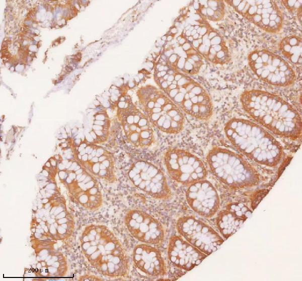
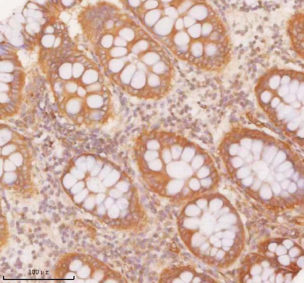
One anti-SLK antibody has real IHC data from paraffin sections of human colon and colon adenocarcinoma (A01614-2 image captions); no IF image is provided (catalog IF image list).

Real IHC data IHC analysis of SLK using anti-SLK antibody (A01614-2). SLK was detected in a paraffin-embedded section of human colon tissue. Heat mediated antigen retrieval was performed in EDTA buffer (pH 8.0, epitope retrieval solution). The tissue section was blocked with 10% goat serum. The tissue section was then incubated with 2 μg/ml rabbit anti-SLK Antibody (A01614-2) overnight at 4°C. Peroxidase Conjugated Goat Anti-rabbit IgG was used as secondary antibody and incubated for 30 minutes at 37°C. The tissue section was developed using HRP Conjugated Rabbit IgG Super Vision Assay Kit (Catalog # SV0002) with DAB as the chromogen.
Anti-SLK Antibody ®
Cat # A01614-2

A01614-2 is listed for IHC and reacts with human and mouse samples (catalog applications and reactivity). Its IHC images show human colon and colon adenocarcinoma paraffin sections (A01614-2 image captions).

Which to pick: Choose A01614-2 for chromogenic IHC on paraffin sections; its caption describes EDTA pH 8.0 retrieval and DAB detection, but does not report the fixative (A01614-2 IHC image caption). No IF/ICC-validated SKU is provided (catalog applications and IF image list). A01614-2 lists mouse reactivity, but its IHC images show human tissue only; it is a rabbit antibody with no clone specified (catalog reactivity, image captions, host and clone fields).

Each figure is that product's own IHC / IF validation image from its datasheet.

References

  1. UniProt Consortium. UniProt entry Q9H2G2 (SLK_HUMAN, STE20-like serine/threonine-protein kinase).
  2. Human Protein Atlas. SLK tissue IHC expression (reliability: Approved).
  3. Human Protein Atlas. SLK subcellular location (ICC-IF): Mainly localized to the cytosol. In addition localized to the plasma membrane..
  4. Human Protein Atlas. SLK antibody validation summary (2 antibodies).
  5. Estrogen Promotes Endometrial Cancer Development by Modulating ZNF626, SLK, and RFWD3 Gene Expression and Inducing Immune Inflammatory Changes. Biomedicines 2025 — PMC11853163.
  6. Dysregulated RNA splicing impairs regeneration in alcohol-associated liver disease. Nature communications 2025 — PMC12423296.
  7. Circuit-selective cell-autonomous regulation of inhibition in pyramidal neurons by Ste20-like kinase. Cell reports 2022 — PMC9756112.
  8. Deletion of the Ste20-like kinase SLK in skeletal muscle results in a progressive myopathy and muscle weakness. Skeletal muscle 2017 — PMC5288853.
  9. PubMed PMID:10699464 — UniProt-cited evidence.
  10. PubMed PMID:11149944 — UniProt-cited evidence.
  11. PubMed PMID:9039502 — UniProt-cited evidence.