This website uses cookies to ensure you get the best experience on our website.


- Table of Contents
Use A01554-1 at 0.5–1 μg/mL for chromogenic IHC-P (datasheet A01554-1). Score nuclear and cytoplasmic staining, using seminiferous duct cells as a positive comparison and adipocytes as a negative comparison; tissue staining reliability is uncertain (HPA tissue IHC).

Expected localisation, validated protocols, controls and antibodies — the at-a-glance facts below, then the full design guide.
| Expected localisation | Observed nuclear and cytoplasmic staining (HPA tissue IHC) | |
| Staining pattern | Nuclear and cytoplasmic staining in most tissues (HPA tissue IHC) | |
| Antigen retrieval | Citrate pH 6 HIER, heat-mediated (datasheet A01554-1) | |
| Positive control | Gallbladder+1 more · see all | |
| Negative control | Adipose tissue+4 more · see all |
| Fixation | Keep fixation consistent across sections (standard IHC practice; not target-specific); Matched tissue-IHC evidence does not establish the fixation claim. Validate the specimen-specific method before use. (selected-SKU IHC image A01554-1) | |
| Caveat | Tissue staining reliability is uncertain (HPA tissue IHC) | |
| Regulation | Strong in testis; widely expressed (UniProt) | |
| Isoform / epitope | 2 isoforms; epitope coverage unreported (UniProt) |
The catalog antibody’s IHC-P protocol (datasheet: A01554-1) is followed by two published SMC6 IHC protocols (PMC3763431; PMC12826375).
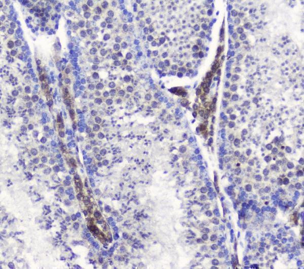
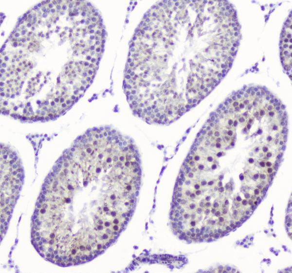
| Sample | Paraffin-embedded mouse testis tissue; fixative not specified (datasheet A01554-1) |
| Fixation | Image fixative and duration unreported (datasheet A01554-1); verify before use. |
| Sectioning | 4–5 µm sections on charged slides (standard) |
| Deparaffinisation | Xylene, graded ethanol series to water (standard) |
| Antigen retrieval | Heat retrieval: Citrate pH 6, 20 min (datasheet A01554-1) |
| Peroxidase block | 3% H2O2, 10 min, room temperature (standard) |
| Blocking | 10% goat serum (datasheet A01554-1) |
| Primary antibody | Rabbit anti-SMC6, 0.5-1μg/ml (datasheet A01554-1) |
| Primary incubation | Overnight at 4 °C (datasheet A01554-1) |
| Detection | Streptavidin-biotin complex (SABC), DAB chromogen (datasheet A01554-1) |
| Counterstain | Hematoxylin, blue, dehydrate and mount (standard) |
| Expected result | SMC6-positive staining in glandular cells of gallbladder (HPA tissue IHC: Medium). HPA tissue profile: Cytoplasmic and nuclear expression in most tissues. No signal in the no-primary control. |
SMC6 is a nuclear protein without a transmembrane segment (UniProt Q96SB8: localisation and topology). In paraffin sections, expect staining in gallbladder glandular cells and cells in testis seminiferous ducts, both reported at medium intensity (HPA tissue IHC). HPA also reports cytoplasmic and nuclear staining across most tissues, but rates its tissue IHC profile Uncertain because antibody staining and RNA expression have only medium consistency (HPA tissue IHC).
| Distinct nuclear staining in gallbladder glandular cells or cells in testis seminiferous ducts. | This fits the reported medium staining in those cells (HPA tissue IHC) and SMC6’s nuclear localisation (UniProt Q96SB8). Score the named cell population and its nuclear signal separately from staining elsewhere; the HPA tissue IHC reliability is Uncertain (HPA tissue IHC). |
| Strong cytoplasmic staining with little or no nuclear signal in the expected cells. | Treat a cytoplasmic-only result as a compartment mismatch that needs an artefact check, given SMC6’s nuclear localisation (UniProt Q96SB8) and supported nucleoplasmic ICC-IF location (HPA subcellular). Some cytoplasmic tissue staining is reported, so cytoplasmic signal alone does not prove artefact (HPA tissue IHC). |
| Staining dominates an HPA-listed, not-detected cell population while the expected cells are weak. | Check antibody specificity or detection-system activity before assigning that signal to SMC6. HPA reports adipocytes and lymph-node germinal center cells as not detected, among other cell populations (HPA tissue IHC). Those observations are comparison points, not proof that every cell in either tissue lacks SMC6 (HPA tissue IHC: Uncertain). |
| Colour spreads across the section, including areas without a clear cellular pattern. | Diffuse staining is hard to score as SMC6; background from the detection workflow is a possible explanation (general IHC practice). Compare a no-primary control and review blocking, washing and detection before interpreting cell-specific signal. HPA’s medium staining calls refer to named cell populations, not diffuse section-wide colour (HPA tissue IHC). |
| No staining appears in gallbladder glandular cells or cells in testis seminiferous ducts. | A blank result misses both HPA-reported medium-positive populations (HPA tissue IHC). Review the catalog antibody’s IHC-P procedure, retrieval and detection controls before calling the sample negative (general IHC practice). An absent signal does not resolve SMC6 expression on its own, especially with Uncertain tissue IHC reliability (HPA tissue IHC). |
| Tissue and cell choice | Gallbladder glandular cells and cells in testis seminiferous ducts are reported at medium staining; adipocytes and several specified brain, lymphoid and other cell populations are listed as not detected (HPA tissue IHC). Compare like cell types when judging a section; HPA rates the tissue profile Uncertain (HPA tissue IHC). |
| Compartment | SMC6 is assigned to the nucleus, including speckles, chromosomes and PML bodies (UniProt Q96SB8). Tissue IHC reports nuclear and cytoplasmic expression, whereas ICC-IF supports nucleoplasm (HPA tissue IHC; HPA subcellular). These observations support a nuclear expectation but do not make every cytoplasmic IHC signal specific. |
| Antibody validation | The HPA tissue-IHC antibodies HPA042733 and CAB037186 are each rated Uncertain for IHC; HPA035057 is Supported for ICC, with no IHC rating given (HPA antibodies). Keep those application-specific ratings separate when weighing a paraffin-section result. |
| Isoforms and processing | UniProt lists 2 isoforms, no signal peptide or propeptide, and one annotated chain spanning residues 1–1091 (UniProt Q96SB8). No supplied epitope map shows whether a given IHC antibody distinguishes the isoforms; avoid assigning a staining difference to either isoform. |
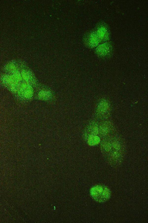
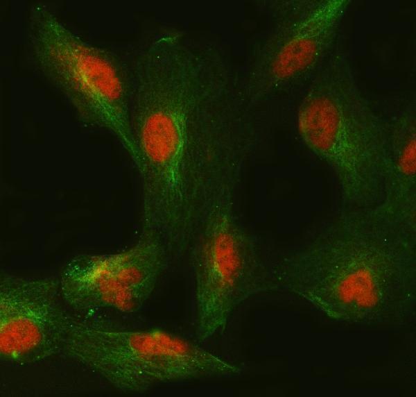
| IF/ICC Q&A: what location is supported? | HPA reports supported nucleoplasmic localisation in ICC-IF and lists images from A-431, Rh30 and U2OS (HPA subcellular). This answers the localisation question for IF/ICC; it does not provide an IF protocol or upgrade an IHC antibody’s validation rating (HPA antibodies). |
| Situation | Likely cause | Next action |
|---|---|---|
| Expected cell population is blank in IHC-P. | Retrieval or detection may be underperforming (general IHC practice); HPA reports medium staining in gallbladder glandular cells and cells in testis seminiferous ducts (HPA tissue IHC). | Check the catalog antibody’s IHC-P instructions, the retrieval and detection steps, and a positive control containing a reported cell population before scoring the specimen as negative (general IHC practice; HPA tissue IHC). |
| Signal appears mainly cytoplasmic. | The localisation differs from UniProt’s nuclear assignment and HPA’s supported nucleoplasmic ICC-IF result, although HPA tissue IHC reports some cytoplasmic staining (UniProt Q96SB8; HPA subcellular; HPA tissue IHC). | Score nuclear and cytoplasmic staining separately. Compare the named positive cells with a no-primary control and assess whether cytoplasmic colour persists as background (general IHC practice). |
| A reported not-detected cell type stains strongly. | Cross-reactivity or endogenous detection activity is possible (general IHC practice); HPA lists, for example, adipocytes as not detected (HPA tissue IHC). | Check a no-primary control and the relevant endogenous-activity blocking step, then compare the result with the reported positive cell populations (general IHC practice; HPA tissue IHC). |
| Diffuse colour obscures cell boundaries. | Nonspecific background can arise within an IHC detection workflow (general IHC practice); a diffuse field cannot establish the cell-specific HPA pattern (HPA tissue IHC). | Review blocking, washes, detection exposure and the no-primary control; score only interpretable cellular staining after the background is resolved (general IHC practice). |
| Two antibodies give different tissue patterns. | Both HPA-listed IHC antibodies have Uncertain IHC validation, and HPA calls overall tissue reliability Uncertain (HPA antibodies; HPA tissue IHC). | Keep antibody results separate, compare the same cell populations and compartments, and avoid treating agreement in one region as validation of every stained cell type (HPA tissue IHC; general IHC practice). |
| Nucleoplasmic IF/ICC signal is clear, but IHC-P is weak. | HPA supports the nucleoplasmic ICC-IF location while rating its listed IHC antibodies Uncertain; the application-specific evidence does not establish identical performance (HPA subcellular; HPA antibodies). | Interpret each application against its own controls and validation status. For IHC-P, review the catalog antibody’s IHC-P procedure and a reported positive cell population (general IHC practice; HPA tissue IHC). |


Comprehensive Human Protein Atlas IHC scoring per tissue (reliability: Uncertain — Medium consistency between antibody staining and RNA expression data.). Rows are taken directly from the HPA tissue chart — click any row's HPA link to view the source.
| Tissue | Cell type | Level | Evidence | Source |
|---|---|---|---|---|
| Gallbladder | Glandular cells | Medium | Protein (IHC) | HPA → |
| Testis | Cells in seminiferous ducts | Medium | Protein (IHC) | HPA → |
| Tissue | Cell type | Level | Evidence | Source |
|---|---|---|---|---|
| Adipose tissue | Adipocytes | Not detected | Protein (IHC) | HPA → |
| Caudate | Glial cells | Not detected | Protein (IHC) | HPA → |
| Cerebellum | Cells in granular layer | Not detected | Protein (IHC) | HPA → |
| Cerebral cortex | Endothelial cells | Not detected | Protein (IHC) | HPA → |
| Hippocampus | Glial cells | Not detected | Protein (IHC) | HPA → |
Troubleshoot SMC6 staining in paraffin sections by checking retrieval, nuclear localisation, controls and cell level scoring before interpreting the chromogenic signal.
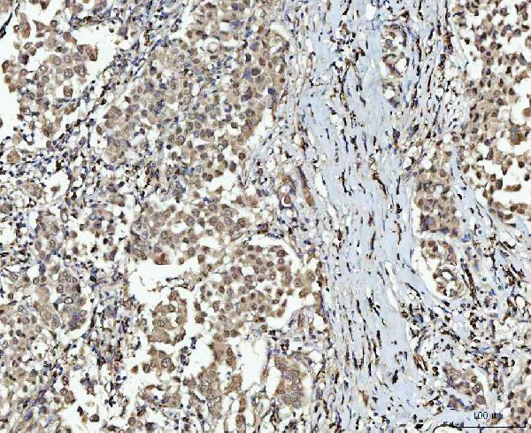
The catalog shows SMC6 IHC images from paraffin sections of human, mouse and rat tissue, plus IF/ICC images from human cell lines (catalog IHC and IF captions).
A01554-1 will render with a mouse testis paraffin-section IHC image; its catalog also shows human and rat testis IHC images and A431 cell IF/ICC (A01554-1 image captions). A01554-2 will render with a human breast cancer paraffin-section IHC image; its catalog also shows A549 cell IF/ICC and human breast cancer tissue IF (A01554-2 image captions).
Which to pick: For paraffin-section IHC, choose A01554-1 for testis across human, mouse and rat (A01554-1 IHC captions: citrate retrieval, pH 6), or A01554-2 for human breast cancer tissue (A01554-2 IHC caption: EDTA retrieval, pH 8.0). For IF/ICC, both have cell images: A01554-1 in A431 cells and A01554-2 in A549 cells (catalog IF captions). A01554-1 has broader listed species reactivity (catalog: Human, Mouse, Rat; A01554-2: Human, Rat); clonality and the fixative used for either paraffin-section IHC image are unreported (catalog clone fields and IHC captions).