This website uses cookies to ensure you get the best experience on our website.


- Table of Contents
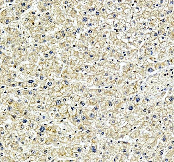
Plan SPRED2 IHC in paraffin sections using the variable cytoplasmic tissue pattern as a reference (HPA tissue IHC). The catalog antibody has a human liver IHC example and a 2–5 μg/ml IHC dilution range (datasheet A06368-1).

Expected localisation, validated protocols, controls and antibodies — the at-a-glance facts below, then the full design guide.
| Expected localisation | Cytoplasmic staining is observed in tissue (HPA tissue IHC) | |
| Staining pattern | Variable cytoplasmic staining across tissue cell types (HPA tissue IHC) | |
| Antigen retrieval | EDTA pH 8.0 HIER, heat-mediated (datasheet A06368-1) | |
| Positive control | Adrenal gland+4 more · see all | |
| Negative control | None in HPA (detected in all 45 tissues); use no-primary + isotype controls |
| Fixation | Keep fixation conditions consistent across sections. (standard IHC practice; not target-specific) | |
| Caveat | Staining and RNA show medium consistency (HPA tissue IHC) | |
| Regulation | Expression regulator not specified (UniProt) | |
| Isoform / epitope | 2 isoforms; epitope differences unspecified (UniProt) |
The catalog antibody uses EDTA pH 8.0 retrieval (datasheet A06368-1). Three published SPRED2 IHC protocols below use citric acid retrieval (PMC8612556; PMC10003366; PMC12563662).
| Sample | Paraffin-embedded human liver tissue; fixative not specified (datasheet A06368-1) |
| Fixation | Image fixative and duration unreported (datasheet A06368-1); verify before use. |
| Sectioning | 4–5 µm sections on charged slides (standard) |
| Deparaffinisation | Xylene, graded ethanol series to water (standard) |
| Antigen retrieval | Heat retrieval: EDTA pH 8.0 (datasheet A06368-1); 20 min, 95–100 °C (standard) |
| Peroxidase block | 3% H2O2, 10 min, room temperature (standard) |
| Blocking | 10% goat serum (datasheet A06368-1) |
| Primary antibody | Rabbit anti-SPRED2, 2-5 μg/ml (datasheet A06368-1) |
| Primary incubation | Overnight at 4 °C (datasheet A06368-1) |
| Detection | HRP-conjugated secondary, DAB chromogen (datasheet A06368-1) |
| Counterstain | Hematoxylin, blue, dehydrate and mount (standard) |
| Expected result | SPRED2-positive staining in glandular cells of adrenal gland (HPA tissue IHC: High). HPA tissue profile: General cytoplasmic expression of varying levels. No signal in the no-primary control. |
In paraffin sections, expect predominantly cytoplasmic SPRED2 staining, potentially including vesicle-associated and membrane-adjacent signal (UniProt Q7Z698 localisation; no transmembrane segment). HPA reports general cytoplasmic expression of varying levels, with high staining in several glandular cell types, cardiomyocytes and kidney tubule cells (HPA: tissue IHC). Its tissue IHC assessment is Approved, with medium consistency between antibody staining and RNA expression (HPA: reliability).
| Cytoplasmic chromogen in adrenal or salivary glandular cells, cardiomyocytes, or kidney tubule cells. | This matches the reported compartment and examples of high-staining cells (HPA: tissue IHC). Compare the cells with their neighbors and score both the proportion stained and intensity; a positive result does not require every cell to stain equally, because HPA describes varying cytoplasmic levels (HPA: tissue IHC). |
| Predominantly nuclear chromogen in a paraffin section, with little convincing cytoplasmic staining. | Treat this as a compartment mismatch requiring investigation, rather than confirmed SPRED2 staining: HPA tissue IHC describes general cytoplasmic expression (HPA: tissue IHC). HPA reports uncertain nucleoplasmic localisation in ICC-IF, alongside supported cytosol localisation; that separate observation does not establish a nuclear IHC pattern (HPA: subcellular ICC-IF). |
| Strong staining chiefly in cells outside the expected positive cell population. | Check cell identity and the detection control before assigning a biological pattern. HPA identifies specific high-staining populations, including pancreatic exocrine glandular cells and kidney tubule cells (HPA: tissue IHC). Unanticipated staining may reflect nonspecific binding or endogenous detection activity (standard IHC practice); the HPA list alone cannot prove cross-reactivity. |
| Uniform haze across cells and surrounding tissue, obscuring cytoplasmic boundaries. | This is background until a cell-associated pattern can be resolved (standard IHC practice). Compare an antibody-omission control and review blocking, washes, detection reagent and chromogen development (standard IHC practice). A broadly expressed target can complicate visual comparison, since HPA reports low tissue specificity rather than a reliably negative tissue (HPA: RNA specificity). |
| No convincing stain in an expected high-staining control section. | First check section integrity and the staining run, including retrieval, antibody incubation and detection controls (standard IHC practice). HPA reports high staining in duodenal glandular cells and kidney tubule cells, among other populations (HPA: tissue IHC). A blank control section warrants troubleshooting but does not, by itself, establish a target-specific fixation effect. |
| Tissue and cell selection | HPA reports high staining in adrenal, duodenal, gallbladder, rectal and salivary glandular cells, cardiomyocytes, kidney tubule cells, and pancreatic exocrine glandular cells (HPA: tissue IHC). Select an identifiable reported population for a positive comparison; HPA supplies no negative tissue list (HPA: tissue IHC). |
| Compartment and topology | Cytoplasm, secretory vesicle membrane and cell membrane are UniProt localisations; SPRED2 has no transmembrane segment (UniProt Q7Z698 localisation and topology). Interpret a membrane-adjacent accent within a broader cellular pattern cautiously: this record gives no IHC criterion requiring a crisp membrane outline. |
| Evidence strength | The HPA tissue IHC assessment is Approved but has medium consistency with RNA expression (HPA: reliability). HPA064394 is IHC Approved; the supplied record gives no IHC status for HPA038256 (HPA: antibody validation). Agreement with a positive control and expected compartment is therefore useful when interpreting an individual run. |
| Isoforms and epitope coverage | UniProt lists two SPRED2 isoforms and the WH1, KBD and SPR domains (UniProt Q7Z698). The supplied sources do not locate the catalog antibody's epitope or establish whether it detects both isoforms; avoid treating a difference between samples as an isoform difference without separate evidence. |
| IF/ICC Q&A: should nuclear fluorescence define the IHC result? | No. HPA calls nucleoplasm uncertain and cytosol supported in ICC-IF (HPA: subcellular ICC-IF); its tissue IHC summary is cytoplasmic (HPA: tissue IHC). Assess IF/ICC on its separate guide page rather than transferring an ICC-IF nuclear call into a paraffin-section IHC score. |
| Situation | Likely cause | Next action |
|---|---|---|
| Expected positive cells stain weakly or remain blank. | A staining-run failure is possible; the supplied sources do not identify a SPRED2-specific retrieval condition or fixation sensitivity. | Confirm an identifiable HPA high-staining population, then review retrieval, antibody incubation and detection performance using routine run controls (HPA: tissue IHC; standard IHC practice). |
| Predominantly nuclear stain appears in IHC. | The observed compartment conflicts with the general cytoplasmic tissue pattern (HPA: tissue IHC); nuclear ICC-IF localisation is uncertain (HPA: subcellular ICC-IF). | Recheck morphology and counterstain, compare a positive control, and assess antibody-omission staining before scoring the nuclear signal (standard IHC practice). |
| Diffuse chromogen obscures cell boundaries. | Nonspecific staining or excessive detection signal may obscure a cellular pattern (standard IHC practice). | Check the antibody-omission control; review blocking, washes and chromogen development, then reassess whether staining resolves to cytoplasm (standard IHC practice; HPA: tissue IHC). |
| Unexpected cells stain more strongly than the reported positive population. | Cell identification, nonspecific antibody binding or endogenous detection activity may explain the contrast (standard IHC practice); HPA reports variable cytoplasmic levels (HPA: tissue IHC). | Verify the cell population on the counterstained section and inspect detection controls before calling cross-reactivity (standard IHC practice). |
| A low-staining tissue shows faint cytoplasmic signal. | Low does not mean absent: HPA reports low staining in parathyroid glandular cells and smooth muscle cells, among others (HPA: tissue IHC). | Record the cell type and intensity, and compare them with a reported high-staining control; do not use these low-staining tissues as proven negatives (HPA: tissue IHC). |
| Two assays give different nuclear and cytoplasmic impressions. | HPA's ICC-IF summary includes uncertain nucleoplasm and supported cytosol, while its tissue IHC summary is generally cytoplasmic (HPA: subcellular ICC-IF; tissue IHC). | Interpret each assay against its own evidence and controls; use the separate IF/ICC guide for fluorescence-specific decisions (HPA: subcellular ICC-IF; standard IHC practice). |


Comprehensive Human Protein Atlas IHC scoring per tissue (reliability: Approved — Medium consistency between antibody staining and RNA expression data.). Rows are taken directly from the HPA tissue chart — click any row's HPA link to view the source.
| Tissue | Cell type | Level | Evidence | Source |
|---|---|---|---|---|
| Adrenal gland | Glandular cells | High | Protein (IHC) | HPA → |
| Duodenum | Glandular cells | High | Protein (IHC) | HPA → |
| Gallbladder | Glandular cells | High | Protein (IHC) | HPA → |
| Heart muscle | Cardiomyocytes | High | Protein (IHC) | HPA → |
| Kidney | Cells in tubules | High | Protein (IHC) | HPA → |
| Tissue | Cell type | Level | Evidence | Source |
|---|---|---|---|---|
| None in HPA: SPRED2 is detected in all 45 scored tissues. Use a no-primary (secondary-only) and an isotype control instead. | ||||
Troubleshoot SPRED2 staining in paraffin sections using the catalog antibody’s liver IHC conditions, then interpret results against tissue and subcellular evidence.
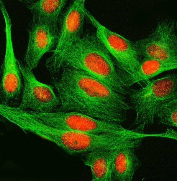
A06368-1 has IHC data from human paraffin-embedded liver and IF data from U20S cells (catalog IHC/IF captions). Its listed reactivity covers human, mouse and rat (catalog reactivity).
A06368-1 will render with human liver paraffin-section IHC using EDTA retrieval at pH 8.0 and DAB detection (A06368-1 IHC caption). The same SKU has an IF example in U20S cells and lists both IF and ICC applications (A06368-1 IF caption; catalog applications).
Which to pick: Choose A06368-1 for human tissue IHC: its own image documents a paraffin-embedded liver section, but the fixative is unreported (A06368-1 IHC caption). For IF/ICC, the same SKU has a U20S cell IF example and lists both applications (A06368-1 IF caption; catalog applications). For mouse or rat samples, A06368-1 lists reactivity with both species, while its supplied IHC image documents human tissue (catalog reactivity; A06368-1 IHC caption).