This website uses cookies to ensure you get the best experience on our website.


- Table of Contents
Plan paraffin STAMBP IHC around nuclear and cytoplasmic staining (HPA tissue IHC). Breast and colon glandular cells show high staining (HPA tissue IHC); the catalog antibody lists 2–5 μg/ml for IHC (datasheet A05964-1).

Expected localisation, validated protocols, controls and antibodies — the at-a-glance facts below, then the full design guide.
| Expected localisation | Nuclear and cytoplasmic tissue staining (HPA tissue IHC) | |
| Staining pattern | General nuclear and cytoplasmic staining (HPA tissue IHC) | |
| Antigen retrieval | EDTA pH 8.0 HIER, heat-mediated (datasheet A05964-1) | |
| Positive control | Adrenal gland+4 more · see all | |
| Negative control | None in HPA (detected in all 45 tissues); use no-primary + isotype controls |
| Fixation | Keep fixation consistent across sections (standard IHC practice; not target-specific) | |
| Caveat | IHC staining has uncertain reliability versus RNA data (HPA tissue IHC) | |
| Regulation | Ubiquitously expressed; regulation unreported (UniProt) | |
| Isoform / epitope | 2 isoforms; epitope impact unresolved (UniProt) |
Compare the catalog antibody’s IHC-P protocol (datasheet: A05964-1) with published STAMBP methods for human tissue and subcutaneous tumors (PMC11379802) and human LUAD tissue microarrays (PMC8190130).
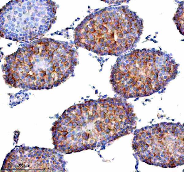
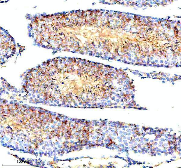
| Sample | Paraffin-embedded rat testis tissue; fixative not specified (datasheet A05964-1) |
| Fixation | Image fixative and duration unreported (datasheet A05964-1); verify before use. |
| Sectioning | 4–5 µm sections on charged slides (standard) |
| Deparaffinisation | Xylene, graded ethanol series to water (standard) |
| Antigen retrieval | Heat retrieval: EDTA pH 8.0 (datasheet A05964-1); 20 min, 95–100 °C (standard) |
| Peroxidase block | 3% H2O2, 10 min, room temperature (standard) |
| Blocking | 10% goat serum (datasheet A05964-1) |
| Primary antibody | Rabbit anti-STAMBP, 2-5 μg/ml (datasheet A05964-1) |
| Primary incubation | Overnight at 4 °C (datasheet A05964-1) |
| Detection | HRP-conjugated secondary, DAB chromogen (datasheet A05964-1) |
| Counterstain | Hematoxylin, blue, dehydrate and mount (standard) |
| Expected result | STAMBP-positive staining in glandular cells of adrenal gland (HPA tissue IHC: High). HPA tissue profile: General nuclear and cytoplasmic expression. No signal in the no-primary control. |
For paraffin-section IHC, expect nuclear and cytoplasmic staining in several cell types, including glandular cells in adrenal gland, appendix, breast, colon, duodenum, fallopian tube, and gallbladder, and glial cells in cerebral cortex (HPA tissue IHC: High; general nuclear and cytoplasmic expression). Treat this as a provisional pattern because tissue IHC reliability is Uncertain (HPA tissue IHC). STAMBP also associates with membranes and early endosomes but has no transmembrane segment (UniProt O95630: localization and topology).
| Nuclear and cytoplasmic signal in glandular cells of a listed high-staining tissue. | This fits the reported tissue pattern, especially when the signal is stronger than nearby background (HPA tissue IHC: general nuclear and cytoplasmic expression; High in glandular cells). Interpret intensity within the same run: the HPA label describes its observed staining, while its overall IHC reliability remains Uncertain (HPA tissue IHC). |
| Predominantly sharp staining in an unexpected compartment, with little nuclear or cytoplasmic signal. | Question the result before assigning a new IHC localization: HPA reports general nuclear and cytoplasmic tissue staining (HPA tissue IHC), while UniProt also lists membrane and early endosome localization (UniProt O95630). Check morphology and controls; an unusual pattern alone cannot establish either artifact or a genuine compartment-specific pool. |
| Strong staining in a cell population expected to stain weakly, such as esophageal squamous cells or adipocytes. | Investigate possible cross-reactivity or endogenous detection activity before calling it STAMBP: those cell populations are reported Low, while STAMBP is broadly expressed (HPA tissue IHC: Low in esophageal squamous cells and adipocytes; UniProt O95630: ubiquitous expression). Low is a comparison point, not a required absence of signal. |
| Diffuse colour across cells, extracellular spaces, and the section edge. | Treat a pattern without clear cell boundaries as background until controls support a cellular signal (general IHC practice). Compare a reagent-control section and inspect uneven counterstain or detection deposits (general IHC practice). The reported STAMBP pattern is cellular and generally nuclear and cytoplasmic (HPA tissue IHC). |
| No convincing signal in glandular cells of a listed high-staining tissue. | Review the run before interpreting the sample as STAMBP-negative: these cells were reported High by HPA, although the tissue IHC assessment is Uncertain (HPA tissue IHC). Check section quality, retrieval, antibody dilution, and detection controls as general IHC troubleshooting; no STAMBP-specific retrieval or fixation sensitivity is supplied. |
| Tissue IHC confidence | HPA calls its tissue staining Uncertain because antibody staining and RNA expression have medium consistency (HPA tissue IHC); interpret a match as support for the pattern, not independent proof of specificity. |
| Antibody-specific validation | HPA035800 is Uncertain for IHC and Supported for ICC; HPA035801 has Supported ICC status but no listed IHC status (HPA antibodies). Do not transfer an ICC validation label to paraffin-section IHC. |
| Cell type and comparison tissue | HPA reports High staining in specified glandular cells and cerebral-cortex glia, but Low staining in esophageal squamous cells and adipocytes (HPA tissue IHC). Score the stated cell population rather than a whole-section average. |
| Subcellular context | UniProt lists nucleus, cytoplasm, membrane, and early endosome, with no transmembrane segment (UniProt O95630: localization and topology). HPA tissue IHC reports general nuclear and cytoplasmic staining; its finer nucleoplasm, plasma-membrane, and cytosol assignments come from ICC-IF (HPA tissue IHC; HPA subcellular). |
| Isoforms and epitope scope | Two isoforms are listed (UniProt O95630: isoforms 1 and 2). Their presence alone does not establish that an antibody recognizes both; use the antibody's documented immunogen or epitope information when available (general IHC practice). No epitope location is supplied here. |
| Situation | Likely cause | Next action |
|---|---|---|
| A listed high-staining tissue appears blank. | The run may lack usable antigen or detection signal; the HPA High label is observational and its IHC reliability is Uncertain (HPA tissue IHC). | Check tissue preservation, retrieval, antibody dilution, and detection with routine controls (general IHC practice). No target-specific fixation or retrieval effect is established here. |
| Nuclear signal is absent but cytoplasmic signal remains. | The observed compartment balance may differ across cells or runs; HPA describes general nuclear and cytoplasmic tissue expression (HPA tissue IHC). | Compare matched cells and control sections, then document each compartment separately (general IHC practice); avoid declaring a new STAMBP localization from this slide alone. |
| Low-reference cells stain as strongly as the selected high-reference cells. | Cross-reactivity, endogenous chromogenic activity, or a run-wide background problem is possible (general IHC practice); HPA reports those cell groups as Low (HPA tissue IHC). | Compare reagent controls and inspect whether colour follows cellular structures (general IHC practice). Treat Low as relative staining, not a mandatory negative result (HPA tissue IHC). |
| Colour is diffuse or concentrated at section edges. | Uneven reagent coverage, nonspecific binding, or detection deposits can produce this appearance (general IHC practice). | Review blocking, washes, and detection controls, and score only interpretable cells (general IHC practice). The reported tissue pattern is cellular and generally nuclear and cytoplasmic (HPA tissue IHC). |
| Two antibodies give different IHC patterns. | Their validation information differs: HPA035800 has Uncertain IHC status, and no IHC status is listed for HPA035801 (HPA antibodies). | Keep antibody identities separate in the record and compare each result with cell type, compartment, and controls (HPA antibodies; HPA tissue IHC; general IHC practice). |
| IF/ICC: what localization should be checked? | HPA's ICC-IF summary places STAMBP mainly in the nucleoplasm, with additional plasma-membrane and cytosol localization (HPA subcellular: Supported). | Use the separate IF/ICC guide for that application; record nuclear, cytosolic, and membrane signal separately (HPA subcellular; general IF practice). |
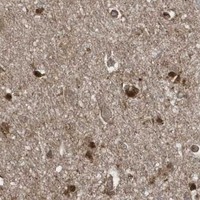


Comprehensive Human Protein Atlas IHC scoring per tissue (reliability: Uncertain — Medium consistency between antibody staining and RNA expression data.). Rows are taken directly from the HPA tissue chart — click any row's HPA link to view the source.
| Tissue | Cell type | Level | Evidence | Source |
|---|---|---|---|---|
| Adrenal gland | Glandular cells | High | Protein (IHC) | HPA → |
| Appendix | Glandular cells | High | Protein (IHC) | HPA → |
| Breast | Glandular cells | High | Protein (IHC) | HPA → |
| Cerebral cortex | Glial cells | High | Protein (IHC) | HPA → |
| Colon | Glandular cells | High | Protein (IHC) | HPA → |
| Tissue | Cell type | Level | Evidence | Source |
|---|---|---|---|---|
| None in HPA: STAMBP is detected in all 45 scored tissues. Use a no-primary (secondary-only) and an isotype control instead. | ||||
Troubleshoot STAMBP staining in paraffin sections by checking retrieval, compartment pattern, cell identity and scoring against the available tissue evidence.
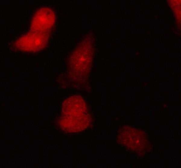
A05964-1 has IHC images from human, mouse and rat paraffin sections and an IF image from PC3 cells (catalog: IHC and IF image captions).
A05964-1 is shown in IHC on rat and mouse testis, human ovarian cancer tissue and human spleen (catalog: IHC image captions). The same SKU is shown in IF on PC3 cells (catalog: IF image caption).
Which to pick: For tissue IHC, choose A05964-1 for paraffin sections; its images use heat retrieval in EDTA at pH 8.0 and primary antibody at 2 μg/ml (catalog: IHC image captions). For IF/ICC, A05964-1 is listed at 5 μg/ml and shown on PC3 cells (catalog: applications, IF dilution and IF image caption). For cross-species work, A05964-1 lists human, monkey, mouse and rat reactivity, with IHC images for human, mouse and rat; it is rabbit hosted, while clonality and the paraffin sections’ fixative are unreported (catalog: reactivity, host and clone; IHC image captions).