STAT6 / Signal transducer and activator of transcription 6 · IHC design guide

Design Immunohistochemistry for STAT6

Plan chromogenic STAT6 IHC in paraffin sections using the catalog antibody’s 2–5 μg/ml range (datasheet A00523-4). Compare staining with high signal in tonsil germinal center cells and undetected signal in adipocytes (HPA tissue IHC).

Evidence assembled Oct 2026 · For research use; verify linked source records and product datasheet before use
Immunohistochemistry protocol sheet for STAT6 (IHC for STAT6): expected localisation Cytoplasmic and nuclear staining (HPA tissue IHC), antibody A00523-4, validated IHC image, and IHC protocol steps
Printable STAT6 IHC protocol sheet — expected localisation Cytoplasmic and nuclear staining (HPA tissue IHC), antibody A00523-4, controls and protocol steps. Open the full STAT6 IHC guide →

STAT6 Immunohistochemistry Experimental Design Guide

Expected localisation, validated protocols, controls and antibodies — the at-a-glance facts below, then the full design guide.

Must know before staining
Expected localisation Cytoplasmic and nuclear staining (HPA tissue IHC)
Staining pattern Cells in several tissues: cytoplasmic and nuclear staining (HPA tissue IHC)
Antigen retrieval EDTA pH 8.0 HIER, heat-mediated (datasheet A00523-4)
Positive control ⓘ Appendix+4 more · see all
Negative control ⓘ Adipose tissue+4 more · see all
Important caveats
Reasons your staining may differ from the expected pattern.
Fixation Keep fixation consistent across paraffin sections. (standard IHC practice; not target-specific)
Caveat Nuclear staining may vary with phosphorylation (UniProt)
Regulation Phosphorylation drives nuclear entry (UniProt)
Isoform / epitope 3 isoforms; check antibody epitope coverage (UniProt)
Section 1

Recommended STAT6 IHC & IF Protocols

The catalog antibody’s IHC-P protocol (datasheet A00523-4) is followed by one published STAT6 protocol using mesenchymal tumor sections (PMC4505928: Methods).

Recommended immunohistochemistry (IHC-P) protocol parameters
SampleParaffin-embedded human placenta tissue; fixative not specified (datasheet A00523-4)
FixationImage fixative and duration unreported (datasheet A00523-4); verify before use.
Sectioning4–5 µm sections on charged slides (standard)
DeparaffinisationXylene, graded ethanol series to water (standard)
Antigen retrievalHeat retrieval: EDTA pH 8.0 (datasheet A00523-4); 20 min, 95–100 °C (standard)
Peroxidase block3% H2O2, 10 min, room temperature (standard)
Blocking10% goat serum (datasheet A00523-4)
Primary antibodyRabbit anti-STAT6, 2-5 μg/ml (datasheet A00523-4)
Primary incubationOvernight at 4 °C (datasheet A00523-4)
DetectionHRP-conjugated secondary, DAB chromogen (datasheet A00523-4)
CounterstainHematoxylin, blue, dehydrate and mount (standard)
Expected resultSTAT6-positive staining in lymphoid tissue of appendix (HPA tissue IHC: High). HPA tissue profile: Cytoplasmic and nuclear expression in several tissues. No signal in the no-primary control.
💡Decision noteStart with heat-mediated EDTA pH 8.0 retrieval for the catalog antibody (datasheet A00523-4); the published protocol does not specify retrieval conditions (PMC4505928: Methods).
Section 2

What Is the Expected STAT6 Staining Pattern?

STAT6 staining may be cytoplasmic, nuclear, or both; phosphorylation can drive nuclear translocation (UniProt P42226: subcellular location). For IHC-P, inspect the relevant cell population: germinal center cells in lymph node and tonsil, red pulp cells in spleen, and urothelial cells in bladder are reported at high levels (HPA: tissue IHC). STAT6 has no transmembrane segment (UniProt P42226: topology). HPA rates its tissue IHC profile Approved, with medium consistency against RNA data (HPA: reliability).

What am I looking at on my slide?
Nuclear and/or cytoplasmic staining in the expected cells, with discernible cell boundaries.This fits the reported tissue profile (HPA: cytoplasmic and nuclear expression). Nuclear enrichment can fit activated STAT6 because phosphorylation promotes nuclear translocation (UniProt P42226: subcellular location); localization alone does not prove pathway activation.
Signal is confined to the plasma membrane or extracellular material.Reassess specificity and slide preparation: STAT6 is reported in cytoplasm and nucleus and lacks a transmembrane segment (UniProt P42226: subcellular location and topology). An isolated edge deposit is not the expected intracellular pattern.
Strong staining appears in a cell type listed as not detected by HPA.For example, HPA reports adipocytes and cardiomyocytes as not detected (HPA: tissue IHC). Check cell identity and compare controls; cross-reactivity or endogenous chromogen activity are possibilities, not diagnoses. HPA’s Approved rating has medium RNA agreement (HPA: reliability).
Brown color spreads across tissue, stroma, or the whole section without clear cellular localization.Treat this as background until a clean, cell-resolved pattern is established. Review antibody concentration, blocking, washes, and endogenous enzyme controls (general chromogenic IHC practice); diffuse color cannot establish STAT6 localization.
No signal appears in an expected positive control, such as tonsil germinal center cells.A failed control makes a negative test section uninterpretable; tonsil germinal center cells are reported High (HPA: tissue IHC). Check retrieval, primary and detection reagents, and development using the catalog antibody’s IHC-P instructions (general IHC troubleshooting).
💡Expected STAT6 appearanceCall positive when clear cytoplasmic and/or nuclear signal occurs in an expected cell population, such as High-level tonsil germinal center cells (HPA: tissue IHC); isolated membrane color or diffuse, cell-free deposit is suspect (UniProt P42226: topology; general IHC interpretation).
How each factor affects the staining
Compartment and signaling stateSTAT6 occupies cytoplasm and nucleus; phosphorylation can shift it into the nucleus (UniProt P42226: subcellular location). Score each compartment separately rather than requiring one fixed distribution.
Tissue and cell selectionHPA reports low tissue specificity at the RNA level, while IHC levels vary by cell population (HPA: tissue IHC). Use a documented High population for a positive control and identify cells before scoring.
Antibody evidenceThe listed antibody, HPA001861, is Approved for IHC, while the tissue profile has medium agreement with RNA data (HPA: antibody validation; HPA: reliability). Interpret unexpected staining with controls.
Isoforms and modificationThree isoforms and phosphorylation at Tyr641 are annotated (UniProt P42226: isoforms and modified residues). Without an epitope or phospho-specificity record, total staining cannot distinguish isoforms or phosphorylation states.
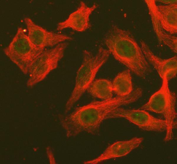
IF/ICC: where should fluorescence appear?HPA reports supported nucleoplasm and cytosol localization, plus an approved connecting-piece location in ICC-IF images (HPA: subcellular). This localization evidence does not define an IHC-P staining requirement.
Why is my staining missing, weak or wrong?
SituationLikely causeNext action
Expected positive tissue is blank.The assay may have failed, or the sampled section may lack the expected cell population (general IHC practice).Confirm the cell population against HPA’s High examples, then check the catalog antibody’s IHC-P procedure, retrieval, detection reagents, and development with a positive control (HPA: tissue IHC; general IHC practice).
A test tissue is negative but its positive control stains correctly.The sampled cell type may have low or undetected staining; HPA reports substantial variation across cell types (HPA: tissue IHC).Identify and score the relevant cells, document the working control, and report the result for that cell population rather than labeling an entire organ negative (general IHC interpretation).
The section has widespread brown haze.Excess primary or detection signal, incomplete washing, or endogenous peroxidase can obscure localization (general chromogenic IHC practice).Inspect a primary-omission control, check peroxidase blocking and wash steps, and adjust antibody conditions according to the catalog IHC-P procedure (general IHC practice).
Staining is exclusively membranous.The distribution conflicts with the reported cytoplasmic and nuclear location and absence of a transmembrane segment (UniProt P42226: subcellular location and topology).Compare with a known positive section and appropriate negative controls; verify that apparent edges are intracellular before assigning STAT6 staining (general IHC interpretation).
Only nuclei stain, or nuclear staining varies between fields.Nuclear translocation can follow phosphorylation, but a total STAT6 stain cannot establish phosphorylation or explain field variation by itself (UniProt P42226: subcellular location).Score nuclear and cytoplasmic compartments separately, compare equivalent cell types and controls, and avoid calling pathway activation from localization alone (general IHC interpretation).
A supposedly negative cell type stains strongly.The HPA reference may report that cell type as not detected, or nonspecific signal and endogenous detection activity may contribute (HPA: tissue IHC; general IHC practice).Verify cell identity, inspect omission and enzyme-block controls, and seek agreement with an independent antibody before treating the unexpected pattern as STAT6 (general IHC practice).

Sample controls for STAT6 IHC & IF

🧪Run tonsil first and confirm staining in germinal center cells (HPA: High in tonsil germinal center cells). Use heart muscle cardiomyocytes as a negative tissue comparator (HPA: Not detected in cardiomyocytes); on the tonsil slide, cells without specific signal should show counterstain alone, but the HPA row does not establish every cell outside the germinal center as STAT6-negative (HPA: tonsil germinal center cells).
Positive control tissue: Appendix (Lymphoid tissue, HPA High)
Negative control tissue: Adipose tissue (HPA Not detected)
ICC-IF cell lines (HPA subcellular resource): HPA ICC-IF images show STAT6 in A-431, U2OS, Sperm, with annotated localisation: Nucleoplasm (supported), Cytosol (supported) (HPA subcellular).
Technical controls: Include a no-primary, secondary-only control and a concentration-matched rabbit IgG isotype control (selected-SKU caption: rabbit primary antibody and anti-rabbit secondary); use STAT6 knockout material as a biological specificity control where available (standard IHC practice). Quench endogenous peroxidase and check blood-rich regions of tonsil for nonspecific DAB signal (standard chromogenic IHC practice).
⚠️Feasibility: A target-specific fixation window or fixation effect is unreported, and the selected-SKU paraffin-section caption does not state the fixative (selected-SKU tissue-IHC caption: fixative not stated). The demonstrated IHC conditions use heat retrieval in EDTA at pH 8.0, followed by 2 μg/ml primary antibody overnight at 4°C; these conditions establish a starting protocol, not a measured retrieval dependency (selected-SKU tissue-IHC caption). The supplied evidence does not establish that frozen sections or tissue IF are easier; control peroxidase-related background in tonsil when interpreting DAB staining (HPA: ICC-IF images in A-431, U2OS and sperm; standard chromogenic IHC practice).

HPA tissue IHC evidence for STAT6

Comprehensive Human Protein Atlas IHC scoring per tissue (reliability: Approved — Medium consistency between antibody staining and RNA expression data.). Rows are taken directly from the HPA tissue chart — click any row's HPA link to view the source.

Positive expression · recommended positive controls

TissueCell typeLevelEvidenceSource
Appendix Lymphoid tissue High Protein (IHC) HPA →
Fallopian tube Glandular cells High Protein (IHC) HPA →
Lymph node Germinal center cells High Protein (IHC) HPA →
Spleen Cells in red pulp High Protein (IHC) HPA →
Tonsil Germinal center cells High Protein (IHC) HPA →

Undetected expression · recommended negative controls

TissueCell typeLevelEvidenceSource
Adipose tissue Adipocytes Not detected Protein (IHC) HPA →
Caudate Glial cells Not detected Protein (IHC) HPA →
Heart muscle Cardiomyocytes Not detected Protein (IHC) HPA →
Hippocampus Glial cells Not detected Protein (IHC) HPA →
Liver Cholangiocytes Not detected Protein (IHC) HPA →
Section 3

Advanced STAT6 IHC Tips

Troubleshoot STAT6 staining in paraffin sections by checking retrieval, compartment, controls, and cell-specific scoring before interpreting nuclear signal.

What retrieval should I try first if STAT6 staining is weak?
Start with heat-mediated antigen retrieval in EDTA at pH 8.0 for paraffin sections (datasheet A00523-4). The documented tissue image used 2 μg/ml primary antibody overnight at 4°C, so compare retrieval changes under those stated primary incubation conditions (datasheet A00523-4). If staining remains weak, adjust heating time or cooling consistently across sections and check tissue preservation before changing buffer (standard IHC practice). Use a control containing cells expected to stain, such as tonsil germinal center cells, to distinguish failed retrieval from a low-expression specimen (HPA: High in tonsil germinal center cells; standard IHC practice).
Could fixation explain loss of STAT6 staining?
Target-specific fixation sensitivity is unknown because the selected paraffin-section caption does not report a fixative (datasheet A00523-4). Record the fixative, fixation duration, and processing history for each specimen, then compare sections processed alike when troubleshooting weak staining (standard IHC practice). Excessive or inconsistent fixation can reduce access to epitopes in IHC generally, but the supplied evidence does not establish a STAT6-specific fixation effect (standard IHC practice; datasheet A00523-4). Repeat the documented EDTA pH 8.0 retrieval on a suitable positive control before attributing a negative section to fixation (datasheet A00523-4; standard IHC practice).
How should I assess cytoplasmic versus nuclear STAT6 staining?
Score nuclear and cytoplasmic staining separately because tissue IHC shows both compartments, while subcellular evidence supports nucleoplasm and cytosol localisation (HPA tissue IHC: cytoplasmic and nuclear expression; HPA subcellular: supported nucleoplasm and cytosol). STAT6 can move into the nucleus following phosphorylation, including JAK-mediated phosphorylation at Tyr641 (UniProt P42226). Nuclear staining alone does not prove pathway activation unless the reagent and experimental controls establish what molecular state is detected (UniProt P42226; standard IHC interpretation). Compare staining in intact cells with expected positive tissue compartments, and exclude poorly preserved regions from compartment scoring (HPA tissue IHC; standard IHC practice).
Can this antibody distinguish STAT6 isoforms or phosphorylated STAT6?
STAT6 has 3 annotated isoforms, but the supplied product caption gives no epitope sequence or isoform coverage (UniProt P42226; datasheet A00523-4). Its SH2 domain spans residues 517–632, and a JAK phosphorylation site is Tyr641; neither feature establishes where this antibody binds (UniProt P42226). Treat the chromogenic signal as STAT6 immunoreactivity, without assigning it to an isoform or phosphorylation state from staining pattern alone (datasheet A00523-4; standard IHC interpretation). For a state-specific conclusion, check documented epitope specificity and compare appropriate stimulated and unstimulated controls under the same processing conditions (standard IHC practice).
How can IF help resolve an ambiguous IHC localisation pattern?
Use IF as a separate localisation check and multiplex STAT6 with a validated marker for the cell population being assessed, such as germinal center cells in tonsil (HPA: High in tonsil germinal center cells; standard IF practice). Choose a spectrally separated fluorophore, favouring a longer-wavelength channel when tissue autofluorescence obscures the STAT6 channel (standard IF practice). STAT6 has no transmembrane segment and occupies cytosol and nucleoplasm, so permeabilisation should allow access to intracellular epitopes while preserving compartment boundaries (UniProt P42226 topology; HPA subcellular; standard IF practice). Confirm the observed channels with single-stain and no-primary controls before comparing IF localisation with chromogenic IHC (standard IF practice).
What should I check when STAT6 DAB staining is diffuse?
Check whether diffuse colour persists in a no-primary control; persistence points toward detection-system or tissue background rather than primary-dependent staining (standard IHC practice). The documented section used 10% goat serum blocking, a peroxidase-conjugated secondary, and DAB development (datasheet A00523-4). Include a peroxidase-blocking step and inspect DAB development time because endogenous enzyme activity and overdevelopment can obscure cellular boundaries (standard chromogenic IHC practice). Compare intact cells with the expected cytoplasmic or nuclear pattern, and review a negative control beside the same staining run before changing primary concentration (HPA tissue IHC; standard IHC practice).
How should I quantify STAT6 staining across paraffin sections? ⚠ ANSWER MARKED FOR VERIFICATION
Define the cell population and compartment before scoring because STAT6 can appear in both cytoplasm and nucleus (HPA tissue IHC; UniProt P42226). Report the percentage of positive cells and an intensity-weighted H-score for each compartment, using the same threshold and scoring rules across sections (standard IHC practice). If cell abundance varies, report positive-cell density per mm² of viable tissue and normalise counts to the relevant cell population or analysed area (standard IHC practice). Exclude folds, section edges, and necrotic regions, and include a consistently processed positive control to monitor run-to-run staining variation (standard IHC practice).
How do I distinguish a true STAT6 positive from staining artefact?
A credible result shows cellular cytoplasmic or nuclear staining in preserved tissue, consistent with reported STAT6 compartments (HPA tissue IHC; HPA subcellular). Check the cell identity: germinal center cells in tonsil are reported High, whereas cardiomyocytes in heart muscle are reported Not detected (HPA tissue IHC). Staining restricted to section edges, necrotic areas, or a no-primary control warrants investigation before calling cells positive (standard IHC practice). Because STAT6 can translocate after phosphorylation, a nuclear pattern is biologically plausible, but this antibody’s supplied caption does not establish phosphorylation specificity or pathway activation (UniProt P42226; datasheet A00523-4).
Boster reagents

Best STAT6 / Signal transducer and activator of transcription 6 IHC Antibodies

Anti-STAT6 antibodies have IHC images from human, mouse and rat tissues and IF images from HeLa cells (catalog image captions); listed reactivity varies by SKU (catalog reactivity).

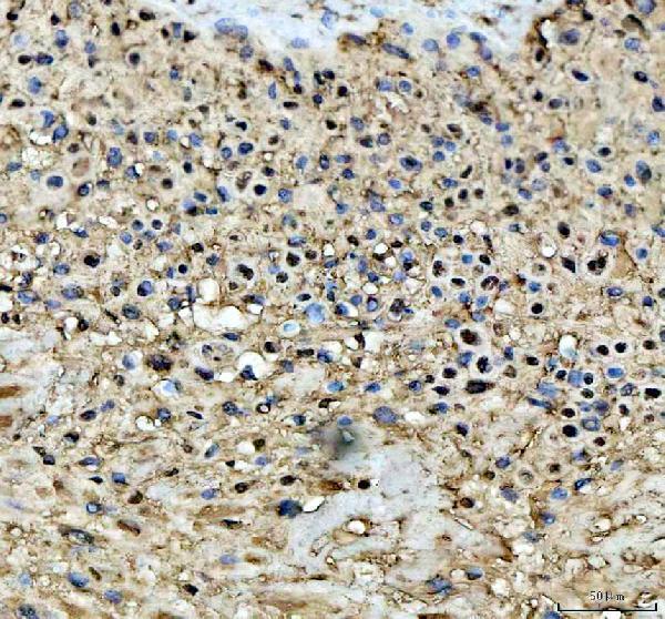
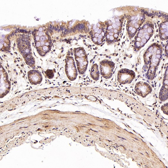
Real IHC data IHC analysis of STAT6 using anti-STAT6 antibody (A00523-4). STAT6 was detected in a paraffin-embedded section of human placenta tissue. Heat mediated antigen retrieval was performed in EDTA buffer (pH 8.0, epitope retrieval solution). The tissue section was blocked with 10% goat serum. The tissue section was then incubated with 2 μg/ml rabbit anti-STAT6 Antibody (A00523-4) overnight at 4°C. Peroxidase Conjugated Goat Anti-rabbit IgG was used as secondary antibody and incubated for 30 minutes at 37°C. The tissue section was developed using HRP Conjugated Rabbit IgG Super Vision Assay Kit (Catalog # SV0002) with DAB as the chromogen.
Anti-STAT6 Antibody ®
Cat # A00523-4
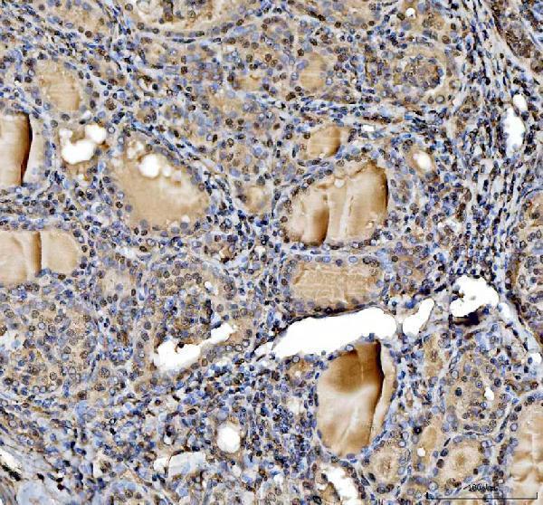
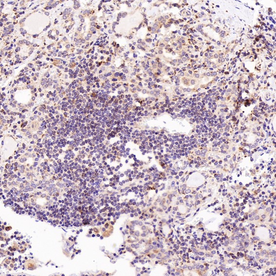
Real IHC data IHC analysis of STAT6 using anti-STAT6 antibody (PA1691). STAT6 was detected in a paraffin-embedded section of human lung cancer tissue. Heat mediated antigen retrieval was performed in EDTA buffer (pH 8.0, epitope retrieval solution). The tissue section was blocked with 10% goat serum. The tissue section was then incubated with 2 μg/ml rabbit anti-STAT6 Antibody (PA1691) overnight at 4°C. Peroxidase Conjugated Goat Anti-rabbit IgG was used as secondary antibody and incubated for 30 minutes at 37°C. The tissue section was developed using HRP Conjugated Rabbit IgG Super Vision Assay Kit (Catalog # SV0002) with DAB as the chromogen.
Anti-STAT6 Antibody ®
Cat # PA1691
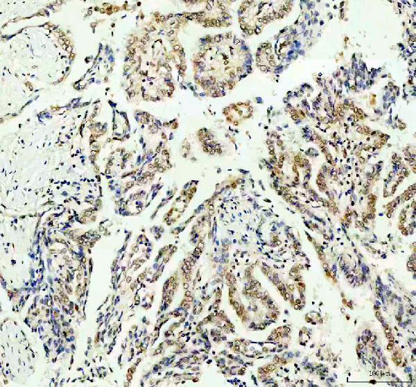
Real IHC data Immunohistochemical analysis of paraffin-embedded Rat stomach, using the Antibody at 1:100 dilution.
Anti-STAT6 Rabbit Monoclonal Antibody
Cat # M00523
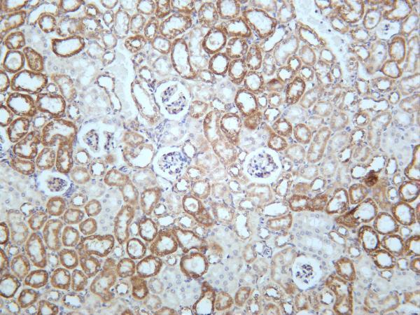
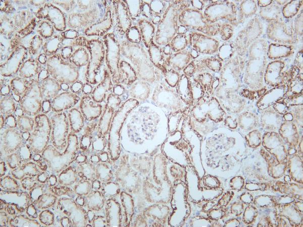
Real IHC data Mouse kidney was stained with anti-STAT6 rabbit antibody
Anti-STAT6 Rabbit Monoclonal Antibody
Cat # M00523-3
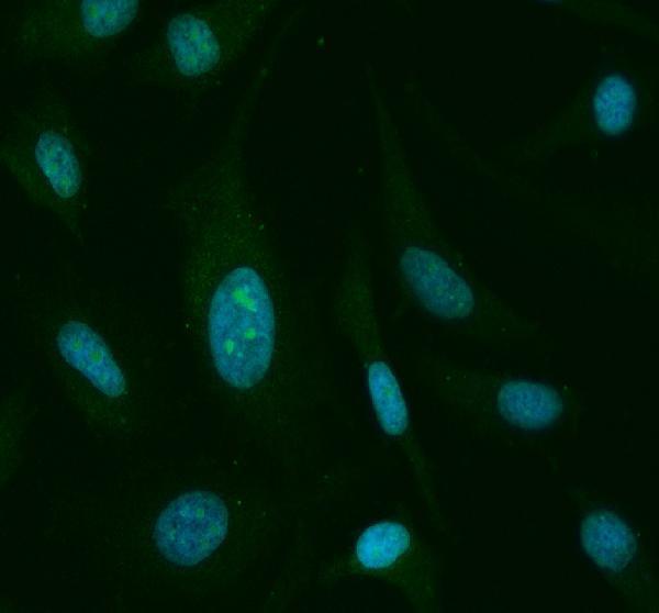
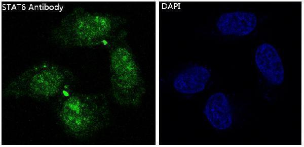
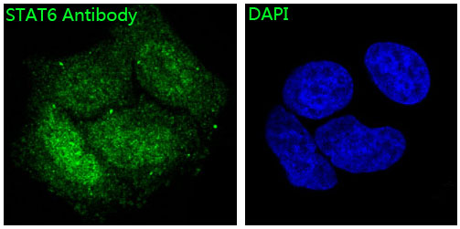
Real IF data Immunofluorescent analysis of Hela cells, using STAT6 Antibody .
Anti-STAT6 Rabbit Monoclonal Antibody
Cat # M00523-1

A00523-4 shows human placenta IHC, PA1691 shows human lung cancer IHC, and M00523 shows rat stomach IHC, although M00523 lists only human and mouse reactivity (respective IHC captions; catalog reactivity). M00523-3 shows mouse kidney IHC, while M00523-1 shows HeLa cell IF and has no listed IHC application (respective image captions; catalog applications).

Which to pick: For human paraffin-section IHC, A00523-4 and PA1691 each have a caption documenting EDTA retrieval at pH 8.0, primary antibody at 2 μg/ml, and DAB detection (respective IHC captions); M00523 offers a rabbit monoclonal option with a human thyroid cancer paraffin-section image (catalog clone; M00523 IHC caption). For IF/ICC, choose M00523-1 when an IF image in HeLa cells and listed human reactivity meet the need (M00523-1 IF caption; catalog applications and reactivity). For cross-species IHC, M00523-3 lists human, mouse and rat reactivity and has kidney IHC captions for all three (catalog reactivity; M00523-3 IHC captions); the cited paraffin-section captions do not report the fixative (catalog IHC captions).

Each figure is that product's own IHC / IF validation image from its datasheet.