This website uses cookies to ensure you get the best experience on our website.
- Table of Contents
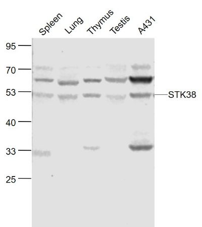
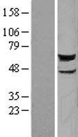
Facts about Serine/threonine-protein kinase 38.
.
Human | |
---|---|
Gene Name: | STK38 |
Uniprot: | Q15208 |
Entrez: | 11329 |
Belongs to: |
---|
protein kinase superfamily |
EC 2.7.11; EC 2.7.11.1; Ndr Ser/Thr kinase-like protein; NDR1 protein kinase; NDR1; NDRnuclear Dbf2-related 1; Nuclear Dbf2-related kinase 1; serine threonine protein kinase; serine/threonine kinase 38; serine/threonine-protein kinase 38
Mass (kDA):
54.19 kDA
Human | |
---|---|
Location: | 6p21.31 |
Sequence: | 6; NC_000006.12 (36493889..36547479, complement) |
Ubiquitously expressed with highest levels observed in peripheral blood leukocytes.
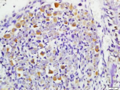
Nucleus. Cytoplasm.
Boster bio Anti-STK38 marker has many options to detect Western Blot. If you are unsure which antibody you should use, you can read more about the different detection options, including DAB and chemiluminescent detection. Boster Bio products are highly respected and validated by the research world, regardless of which antibody is used.
Boster Bio Anti-STK38 Marker is for human antibodies that react with STK38L. This enzyme is involved in the regulation structural processes in neuronal neurons. The product has been validated for Western Blotting, Immunohistochemistry, and ELISA. This antibody is safe for human use and should not cause adverse reactions.
A clear and accurate Western blot begins with sample preparation. A good choice of protein extraction methods can make the difference between a boring blot or one that is stunning. Boster offers several different lysis buffers and specialized protein extraction kits, and its guide can help you choose the right one for your experiment. Different types of transfer have different methods, equipment requirements, and speeds. It is important that you choose carefully.
ELISA, or enzyme linked immunosorbent system, is a method that detects specific antibodies with high accuracy and sensitive. ELISAs are highly sensitive and require high-affinity antibody coating. Boster Bio, a leader in this sector, is the preferred choice among scientists worldwide. PicoKine(tm), the company's proprietary platform, uses a patented technology that delivers high-sensitivity ELISA kit kits.
Two common methods to detect protein levels in biological samples are fluorescence and chemiluminescence. These methods produce a temporary burst of light which must be detected using a digital camera. As a result, a good quality blocking buffer must minimize nonspecific binding of antibodies and reduce background levels. Nonfat dry milk diluted into TBST is the most popular blocking buffer.
Dual labeling with primary and secondary antibodies allows for robust answers and more context than a single Western blot. This method is particularly useful for identifying the presence of a specific protein in a sample. The primary antibody detects the epitope of the target protein, while the secondary antibody binds to it. This method is recommended to analyze proteins with epitope-specific gene expression.
Chemiluminescent Western Blotting uses a primary antibody that recognizes target protein and a secondary antibody labeled using an enzyme such as horseradish Peroxidase or a luminol based HRP. The enzyme releases photons which can be measured using an xray film or CCD imaging device.
The ECL chemiluminescent detection device uses Luminol, which emits a weak beam of light that makes it difficult to measure the protein content on a membrane. The ECL method is enhanced by the addition of enhancers that boost the luminol's emission to a higher level. This enzymatic reaction, known as enhanced chemiluminescence, allows researchers to detect targets containing very low levels of protein.
Amersham ECL Prime is a great reagent that emits stable signal emission. This reagent allows multiple exposures as well as multiple blots to all be processed in one run. Hybond ECL membranes, 0.45 micron pores, offer excellent sensitivity with low background. They also provide excellent sensitivity, dynamic range, and excellent sensitivity. Hybond ECL membranes, which are unsupported and cooled, come with a well plate that can be used for quick and efficient processing.
Boster Bio's ECL chemi-blotting system is equipped with a microplate reader that allows researchers to see the amount of protein on a blot. The system allows multiple exposures to optimize signal/noise ratios. It is possible to rescan the blots once the enzyme or substratum have been removed. Because the signal is reproducible, many protein labs are quickly adopting chemiluminescent detection.
A high salt wash buffer is required for the ECL chemiluminescent detection process. This buffer is 10 mM Tris pH 7.4 at 22 degC. The buffer contains 0.2% SDS (w/v) and 0.1% Tween-20 (w/v). For best results, rinse the samples with buffer within the first 3 days. If the background persists, repeat the process for another three days to eliminate any traces of residual protein.
Boster Bio DAB Chromogenic Discovery System uses DAB chromogenic Substrat Kit for staining tissues or cells. The solution should be clear, colorless, and precipitate-free. Boster DAB is a chromogenic detection system. You can also use the Boster 44% Paraformaldehyde to prepare the sample. Mix the sample with the Boster DAB development reagents. For example, Boster's 4X Dual Color Protein loading buffer.
The STK38 protein is a key marker in the regulation of structural changes in neuronal cells. It is found in the membrane and is associated with actin cytoskeleton. STK38 can help scientists identify and detect neuronal cell types. STK38 markers can be used by scientists to study the structure, function, and function of different neuronal types. This protein can be detected with antibodies developed by Boster Bio.
PMID: 7761441 by Millward T.A., et al. Molecular cloning and characterization of a conserved nuclear serine/threonine protein kinase.
PMID: 12493777 by Tamaskovic R., et al. Mechanism of Ca2+-mediated regulation of NDR protein kinase through autophosphorylation and phosphorylation by an upstream kinase.