This website uses cookies to ensure you get the best experience on our website.


- Table of Contents
Plan chromogenic STX7 IHC in paraffin sections using placental Hofbauer cells or tonsil germinal center cells as positive controls (HPA tissue IHC). Start the catalog antibody at 1:50 (datasheet A07855-1) and assess granular cytoplasmic staining (HPA tissue IHC).

Expected localisation, validated protocols, controls and antibodies — the at-a-glance facts below, then the full design guide.
| Expected localisation | Tissue: granular cytoplasm (HPA tissue IHC); protein: early endosome membrane (UniProt) | |
| Staining pattern | Granular cytoplasm, most abundant in lymphoid tissues (HPA tissue IHC) | |
| Antigen retrieval | EDTA pH 8.0 HIER, heat-mediated (datasheet A07855-1) | |
| Positive control | Appendix+4 more · see all | |
| Negative control | Adipose tissue+4 more · see all |
| Fixation | Keep fixation consistent across paraffin sections. (standard IHC practice; not target-specific) | |
| Caveat | In placenta, assess Hofbauer cells specifically (HPA tissue IHC) | |
| Regulation | Expression regulation is unspecified (UniProt) | |
| Isoform / epitope | 2 isoforms; check epitope against cytoplasmic residues 2–238 (UniProt) |
The catalog antibody’s IHC-P protocol is paired with one published STX7 IHC protocol for ovarian tissues (PMC10525231).
| Sample | Paraffin-embedded human ovarian cancer tissue; fixative not specified (datasheet A07855-1) |
| Fixation | Image fixative and duration unreported (datasheet A07855-1); verify before use. |
| Sectioning | 4–5 µm sections on charged slides (standard) |
| Deparaffinisation | Xylene, graded ethanol series to water (standard) |
| Antigen retrieval | Heat retrieval: EDTA pH 8.0 (datasheet A07855-1); 20 min, 95–100 °C (standard) |
| Peroxidase block | 3% H2O2, 10 min, room temperature (standard) |
| Blocking | 10% goat serum (datasheet A07855-1) |
| Primary antibody | Rabbit anti-STX7, 1:50 recommended; image 2 μg/ml (datasheet A07855-1) |
| Primary incubation | Overnight at 4 °C (datasheet A07855-1) |
| Detection | HRP-conjugated secondary, DAB chromogen (datasheet A07855-1) |
| Counterstain | Hematoxylin, blue, dehydrate and mount (standard) |
| Expected result | STX7-positive staining in lymphoid tissue of appendix (HPA tissue IHC: High). HPA tissue profile: Granular cytoplasmic expression in several tissues, most abundant in lymphoid tissues. No signal in the no-primary control. |
STX7 is an endosomal membrane protein with a cytoplasmic domain spanning residues 2–238 (UniProt O15400 topology). In paraffin-section IHC, expect granular cytoplasmic staining in selected cells, especially lymphoid cells and placental Hofbauer cells (HPA tissue IHC). HPA rates its tissue staining reliability Enhanced, while reporting medium consistency with RNA expression (HPA tissue IHC).
| Distinct cytoplasmic granules in tonsil or lymph-node germinal center cells, or placental Hofbauer cells. | This matches HPA's High staining in those cell populations (HPA tissue IHC). Granules fit an intracellular vesicular distribution; IHC alone cannot identify the stained organelle (UniProt O15400; HPA subcellular ICC-IF). |
| Predominantly nuclear staining or a smooth, continuous outline of the plasma membrane. | These patterns do not fit the reported granular cytoplasmic tissue pattern (HPA tissue IHC). Check whether the signal also appears in the negative control before assigning it to STX7 (standard IHC practice). |
| Strong staining in adipocytes or liver cholangiocytes while expected positive cells stain weakly. | HPA reports STX7 as Not detected in those cell types (HPA tissue IHC). Consider antibody cross-reactivity or endogenous chromogenic activity, then compare the control and positive tissue (standard IHC practice). |
| A diffuse haze covers cells and extracellular areas, obscuring individual granules. | A broad haze is harder to reconcile with HPA's granular cytoplasmic pattern (HPA tissue IHC). Review background in the negative control and the distribution across compartments (standard IHC practice). |
| No staining in a section containing identifiable germinal center cells or Hofbauer cells. | Those cells are reported as High by HPA, so absent signal warrants a technical check (HPA tissue IHC). Confirm their presence on the section before interpreting a negative result (standard IHC practice). |
| Cell population and tissue context | HPA reports High staining in germinal center cells and Hofbauer cells, but Not detected in adipocytes and cholangiocytes (HPA tissue IHC). Score the named cell population, not the whole organ. |
| Subcellular location | UniProt places STX7 at the early endosome membrane, while HPA ICC-IF supports lysosomal localization (UniProt O15400; HPA subcellular ICC-IF). Granular cytoplasmic IHC is compatible with both; chromogenic IHC does not resolve them. |
| Topology and antibody epitope | STX7 has one transmembrane segment at residues 239–259 and a cytoplasmic region at 2–238 (UniProt O15400 topology). Antibody epitope location is not supplied, so the staining pattern cannot establish which region is recognized. |
| Isoforms and modifications | UniProt lists 2 isoforms and several modified residues (UniProt O15400). Their effects on this antibody's IHC staining are unreported; do not use them to explain a weak or absent slide result. |
| Strength of HPA evidence | HPA labels tissue IHC Enhanced and reports medium agreement between staining and RNA expression (HPA tissue IHC). Use its cell-specific observations as a reference while checking each new specimen and control. |
| IF/ICC comparison | For the IF/ICC question, HPA reports supported lysosomal localization and images from A-431, U-251MG and U2OS (HPA subcellular ICC-IF). That evidence informs compartment interpretation; IF/ICC experimental conditions belong in its separate guide. |
| Situation | Likely cause | Next action |
|---|---|---|
| Expected positive cells are present, but the IHC slide has no detectable signal. | The assay may have failed, or the sampled cells may be difficult to identify; HPA reports High staining in germinal center and Hofbauer cells (HPA tissue IHC). | Confirm cell identity on the counterstained section; review the catalog antibody's IHC-P procedure, antigen retrieval, detection reagents and positive control (standard IHC practice). |
| Signal appears mainly in nuclei or as a continuous cell-surface rim. | The compartment differs from HPA's granular cytoplasmic tissue pattern (HPA tissue IHC). | Compare a positive tissue and the negative control; reassess staining specificity before scoring these cells as STX7 positive (standard IHC practice). |
| Adipocytes or cholangiocytes stain strongly. | HPA reports STX7 Not detected in those cells (HPA tissue IHC); cross-reactivity or endogenous detection activity is possible (standard IHC practice). | Check the negative control and any tissue pigment; apply appropriate endogenous-activity controls for the chromogenic detection system (standard IHC practice). |
| Granules are visible, but diffuse background makes their boundaries unclear. | Background can obscure the reported granular cytoplasmic distribution (HPA tissue IHC; standard IHC practice). | Compare background in the negative control, then review blocking, washing, antibody dilution and chromogen development against the validated IHC procedure (standard IHC practice). |
| A tissue looks negative despite a positive result reported elsewhere in the same organ. | HPA's levels refer to specified cells: for example, kidney proximal tubules (microvilli) are High, while liver cholangiocytes are Not detected (HPA tissue IHC). | Identify and score the relevant cell population on the section before comparing results across tissues or regions (standard IHC practice). |
| IHC granules are interpreted as proof of lysosomal localization. | HPA supports lysosomes in ICC-IF, while UniProt lists the early endosome membrane; chromogenic IHC has limited compartment resolution (HPA subcellular ICC-IF; UniProt O15400; standard IHC practice). | Report the IHC observation as granular cytoplasmic staining. Use the separate IF/ICC guide when organelle-level localization is the experimental question (standard IHC practice). |
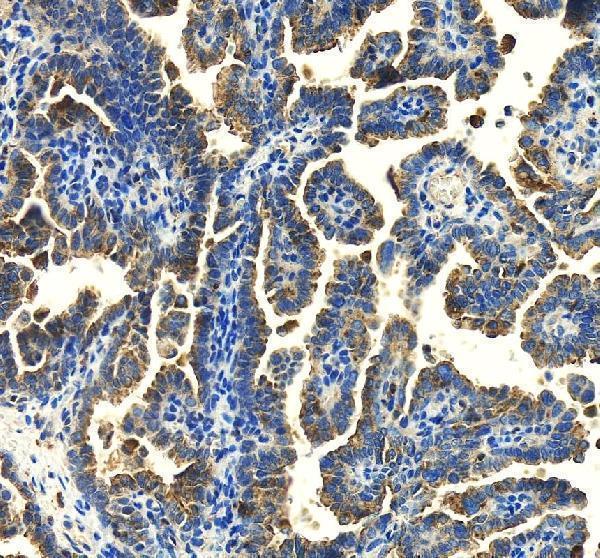
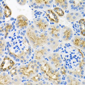
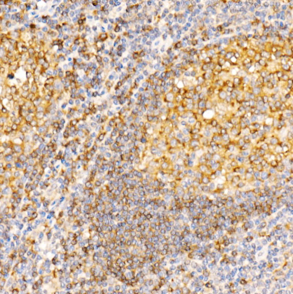


Comprehensive Human Protein Atlas IHC scoring per tissue (reliability: Enhanced — Medium consistency between antibody staining and RNA expression data.). Rows are taken directly from the HPA tissue chart — click any row's HPA link to view the source.
| Tissue | Cell type | Level | Evidence | Source |
|---|---|---|---|---|
| Appendix | Lymphoid tissue | High | Protein (IHC) | HPA → |
| Kidney | Proximal tubules (microvilli) | High | Protein (IHC) | HPA → |
| Lymph node | Germinal center cells | High | Protein (IHC) | HPA → |
| Placenta | Hofbauer cells | High | Protein (IHC) | HPA → |
| Skin | Melanocytes | High | Protein (IHC) | HPA → |
| Tissue | Cell type | Level | Evidence | Source |
|---|---|---|---|---|
| Adipose tissue | Adipocytes | Not detected | Protein (IHC) | HPA → |
| Breast | Adipocytes | Not detected | Protein (IHC) | HPA → |
| Cerebellum | Cells in granular layer | Not detected | Protein (IHC) | HPA → |
| Duodenum | Glandular cells | Not detected | Protein (IHC) | HPA → |
| Fallopian tube | Glandular cells | Not detected | Protein (IHC) | HPA → |
Troubleshoot STX7 staining in paraffin sections by checking retrieval, cell type, subcellular pattern and controls before interpreting intensity.
Two anti-STX7 antibodies have real IHC images from human ovarian cancer, placenta and tonsil, and mouse spleen paraffin sections (catalog IHC captions); A07855-1 also lists IF/ICC (catalog applications).
A07855-1 will render with IHC from a human ovarian cancer paraffin section (A07855-1 IHC caption); it lists IF/ICC, but has no IF image in the payload (catalog applications and image alts). A07855 will render with IHC from human placenta (A07855 card caption); additional captions show human tonsil and mouse spleen paraffin sections (A07855 IHC captions).
Which to pick: For tissue IHC, choose A07855 for its 1:50–1:100 range and human and mouse paraffin-section examples (A07855 catalog dilution and IHC captions), or A07855-1 for its 1:50 starting dilution and human ovarian cancer paraffin-section example (A07855-1 catalog dilution and IHC caption). For IF/ICC, choose A07855-1, a rabbit polyclonal antibody with IF and ICC listed at 1:50, without an IF image in the payload (A07855-1 catalog applications, dilution and image alts). Both list human, mouse and rat reactivity, but neither has a rat IHC image in the payload (catalog reactivity and IHC captions); their captions establish paraffin sections, while the fixative is unreported (catalog IHC captions).