This website uses cookies to ensure you get the best experience on our website.


- Table of Contents
Plan SYNE2 chromogenic IHC-P around nuclear-membranous staining in several cell types (HPA tissue IHC). Start the catalog antibody at 2–5 μg/ml (datasheet A02818-1) and interpret staining with isoform-dependent localization in mind (UniProt).

Expected localisation, validated protocols, controls and antibodies — the at-a-glance facts below, then the full design guide.
| Expected localisation | Nuclear membrane in several cell types (HPA tissue IHC) | |
| Staining pattern | Nuclear-membranous staining across several cell types (HPA tissue IHC) | |
| Antigen retrieval | EDTA pH 8.0 HIER, heat-mediated (datasheet A02818-1) | |
| Positive control | Colon+4 more · see all | |
| Negative control | Adipose tissue+4 more · see all |
| Fixation | Matched tissue-IHC evidence does not establish the fixation claim. Validate the specimen-specific method before use. (selected-SKU IHC image A02818-1) | |
| Caveat | Isoform-dependent localization may change the pattern (UniProt) | |
| Regulation | Expression varies by isoform (UniProt) | |
| Isoform / epitope | 13 isoforms; cytoplasmic and C-terminal epitopes may differ (UniProt) |
The catalog antibody’s paraffin IHC protocol (datasheet A02818-1) is accompanied by a published mouse embryo staining protocol (PMC6244730).
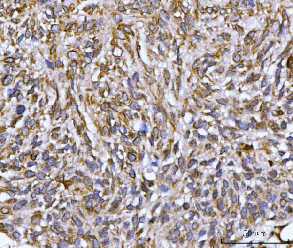
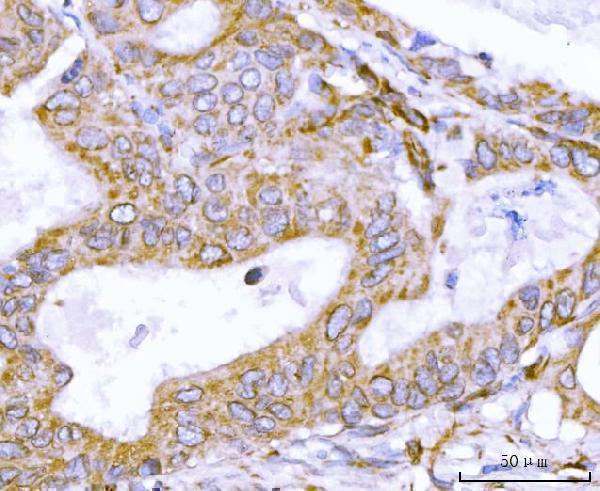
| Sample | Paraffin-embedded human penis squamous cell carcinoma tissue; fixative not specified (datasheet A02818-1) |
| Fixation | Image fixative and duration unreported (datasheet A02818-1); verify before use. |
| Sectioning | 4–5 µm sections on charged slides (standard) |
| Deparaffinisation | Xylene, graded ethanol series to water (standard) |
| Antigen retrieval | Heat retrieval: EDTA pH 8.0 (datasheet A02818-1); 20 min, 95–100 °C (standard) |
| Peroxidase block | 3% H2O2, 10 min, room temperature (standard) |
| Blocking | 10% goat serum (datasheet A02818-1) |
| Primary antibody | Rabbit anti-SYNE2, 2-5 μg/ml (datasheet A02818-1) |
| Primary incubation | Overnight at 4 °C (datasheet A02818-1) |
| Detection | HRP-conjugated secondary, DAB chromogen (datasheet A02818-1) |
| Counterstain | Hematoxylin, blue, dehydrate and mount (standard) |
| Expected result | SYNE2-positive staining in glandular cells of colon (HPA tissue IHC: High). HPA tissue profile: Nuclear membranous expression in several different cell types. No signal in the no-primary control. |
SYNE2 should appear chiefly as a rim around nuclei in paraffin-section IHC: HPA reports nuclear membranous staining across several cell types (HPA tissue IHC: Enhanced; medium consistency with RNA data). Expect clear staining in colon glandular cells, kidney tubular cells, placental cytotrophoblasts, nasopharyngeal respiratory epithelium and basal skin cells (HPA tissue IHC: High). Its C-terminal membrane span anchors an isoform at the outer nuclear membrane, with most of that protein facing the cytoplasm (UniProt Q8WXH0 topology and subcellular location).
| A defined brown rim outlines nuclei in kidney tubules or colon glands (HPA tissue IHC: High in both cell types). | This matches the reported nuclear membranous pattern (HPA tissue IHC). Assess the rim in the specified cells, rather than scoring all nearby cells together; HPA's intensity calls apply to named cell types (HPA tissue IHC). |
| Cytoplasm stains strongly while nuclear outlines are indistinct (HPA tissue IHC: nuclear membranous profile). | This does not reproduce the principal tissue-IHC pattern (HPA tissue IHC). Cytoplasmic signal is biologically possible for shorter soluble isoforms, so compartment alone cannot prove artefact; compare morphology, antibody validation and controls (UniProt Q8WXH0 isoforms and subcellular location; standard IHC practice). |
| Strong staining appears mainly in adipocytes or bone-marrow hematopoietic cells (HPA tissue IHC: Not detected). | That distribution conflicts with the sampled HPA observations and warrants a specificity check, including detection controls (HPA tissue IHC; standard IHC practice). An HPA 'Not detected' call is an observation for those cells, rather than proof that every specimen must be negative (HPA tissue IHC). |
| A broad brown haze covers tissue and obscures cell borders or nuclear rims (standard IHC practice). | The section is difficult to interpret for SYNE2 localisation (HPA tissue IHC: nuclear membranous profile). Examine a no-primary control for detection background and review blocking, washing and chromogen development (standard IHC practice). |
| Kidney tubular cells and colon glandular cells show no visible rim under the same run (HPA tissue IHC: High). | These are useful positive reference cell populations, but their absence does not by itself identify the failure step (HPA tissue IHC; standard IHC practice). Check tissue preservation, antibody and detection performance, and the retrieval conditions used for that assay (standard IHC practice). |
| Membrane topology (UniProt Q8WXH0 topology). | The C-terminal segment spans residues 6835–6855; residues 1–6834 face the cytoplasm and 6856–6885 the perinuclear space (UniProt Q8WXH0 topology). A nuclear outline is consistent with this arrangement, but the record does not locate the antibody epitope (UniProt Q8WXH0 topology; supplied HPA antibody data). |
| Isoform distribution (UniProt Q8WXH0 subcellular location). | The record lists 13 isoforms and reports distinct compartments: membrane-bearing forms can occupy the nuclear envelope or other membranes, while shorter soluble forms occur in cytoplasm or nucleus (UniProt Q8WXH0 isoforms and subcellular location). Interpret secondary staining with the antibody's epitope coverage in mind (standard IHC practice). |
| Strength of tissue evidence (HPA tissue IHC; HPA antibodies). | HPA calls the tissue-IHC profile Enhanced but reports only medium consistency between antibody staining and RNA expression; both listed antibodies have Enhanced IHC status (HPA tissue IHC; HPA antibodies: HPA003435, HPA050204). Use the reported cell-level pattern as a reference, not an absolute cutoff for every specimen (HPA tissue IHC). |
| Retrieval and detection (standard IHC practice). | Optimize antigen retrieval and primary-antibody concentration against a positive reference section, then inspect a no-primary control for endogenous detection activity (standard IHC practice). The supplied HPA and UniProt records provide no SYNE2-specific retrieval condition or fixation-sensitivity result (supplied HPA and UniProt records). |
| IF/ICC Q&A: should the IHC rim appear in fluorescence images? (HPA subcellular ICC-IF). | HPA reports nuclear membrane localisation as uncertain, intermediate filaments as supported, and primary cilium localisation as uncertain in ICC-IF (HPA subcellular ICC-IF). Those cell-image observations can differ from the tissue-IHC profile; evaluate IF/ICC on its own guide page (HPA subcellular ICC-IF; HPA tissue IHC). |
| Situation | Likely cause | Next action |
|---|---|---|
| No stain in kidney tubular cells or colon glandular cells (HPA tissue IHC: High). | The assay may have missed a visible signal, or the selected section may not reproduce the reference observation (standard IHC practice; HPA tissue IHC). | Verify that the named cells are present; review antibody dilution, retrieval, incubation and detection with a positive reference section (standard IHC practice). No target-specific retrieval setting is supplied (supplied HPA and UniProt records). |
| Predominantly cytoplasmic stain with no clear nuclear rim (HPA tissue IHC: nuclear membranous profile). | Possible nonspecific staining or detection background; shorter soluble isoforms also occur in cytoplasm (standard IHC practice; UniProt Q8WXH0 subcellular location). | Compare with a no-primary control and the HPA cell-level pattern; assess any cytoplasmic signal separately rather than treating it as proof of the expected tissue-IHC result (standard IHC practice; HPA tissue IHC). |
| Brown signal in adipocytes or bone-marrow hematopoietic cells (HPA tissue IHC: Not detected). | The observed distribution differs from HPA's sampled pattern; nonspecific binding or endogenous detection activity is possible (HPA tissue IHC; standard IHC practice). | Check a no-primary control, blocking and detection chemistry; compare staining in a named HPA-high population on the same run (standard IHC practice; HPA tissue IHC). |
| Diffuse brown background obscures nuclear boundaries (standard IHC practice). | Excess primary or detection signal, inadequate washing, or endogenous activity can obscure compartment calls (standard IHC practice). | Review titration, washing and chromogen development; require a readable nuclear outline before scoring SYNE2 localisation (standard IHC practice; HPA tissue IHC: nuclear membranous profile). |
| Some cells show a rim, but intensity varies across tissue (HPA tissue IHC: several cell types). | HPA reports different levels by cell type, including High in kidney tubules and Medium in bronchial respiratory epithelium; its antibody–RNA consistency is medium (HPA tissue IHC). | Record cell identity and intensity separately. Compare like cells across sections and use the HPA levels as reference observations, not universal pass–fail thresholds (HPA tissue IHC; standard IHC practice). |

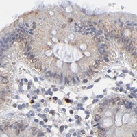


Comprehensive Human Protein Atlas IHC scoring per tissue (reliability: Enhanced — Medium consistency between antibody staining and RNA expression data.). Rows are taken directly from the HPA tissue chart — click any row's HPA link to view the source.
| Tissue | Cell type | Level | Evidence | Source |
|---|---|---|---|---|
| Colon | Glandular cells | High | Protein (IHC) | HPA → |
| Kidney | Cells in tubules | High | Protein (IHC) | HPA → |
| Nasopharynx | Respiratory epithelial cells | High | Protein (IHC) | HPA → |
| Placenta | Cytotrophoblasts | High | Protein (IHC) | HPA → |
| Skin | Cells in basal layer | High | Protein (IHC) | HPA → |
| Tissue | Cell type | Level | Evidence | Source |
|---|---|---|---|---|
| Adipose tissue | Adipocytes | Not detected | Protein (IHC) | HPA → |
| Adrenal gland | Glandular cells | Not detected | Protein (IHC) | HPA → |
| Bone marrow | Hematopoietic cells | Not detected | Protein (IHC) | HPA → |
| Caudate | Glial cells | Not detected | Protein (IHC) | HPA → |
| Cerebellum | Cells in granular layer | Not detected | Protein (IHC) | HPA → |
Troubleshoot SYNE2 chromogenic IHC in paraffin sections by checking retrieval, staining compartment, controls and scoring; IF/ICC considerations appear in one entry.
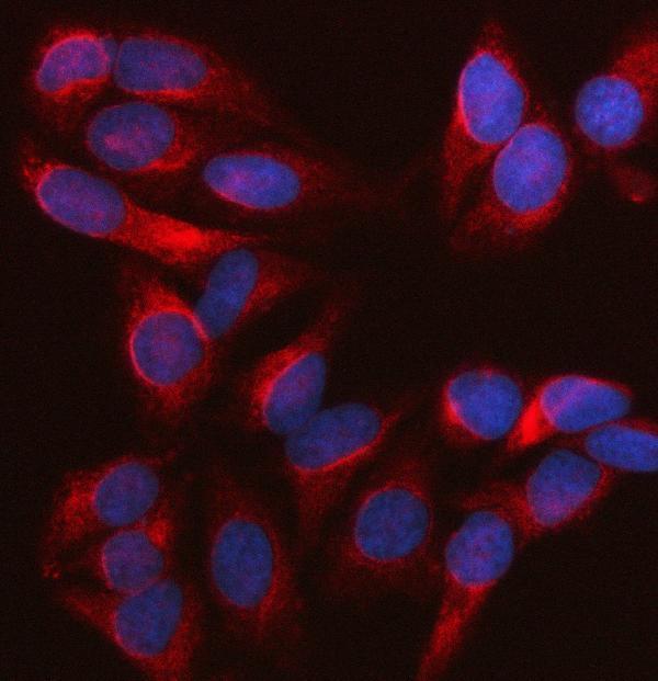
Catalog antibody A02818-1 has IHC data from human paraffin sections and IF data from HeLa cells (catalog image captions); its listed reactivity is human and monkey (catalog: reactivity).
A02818-1 will render with its own IHC figure from a human penis squamous cell carcinoma paraffin section (catalog: IHC image caption). The same SKU lists IHC, IF and ICC applications and has IF data from HeLa cells (catalog: applications; IF image caption).
Which to pick: Choose A02818-1 for paraffin-section IHC: its own figure documents EDTA pH 8.0 retrieval and 2 μg/ml primary antibody in a human tissue section; the fixative is unreported (catalog: IHC image caption). For IF/ICC, A02818-1 is also listed for both applications, with an IF image from HeLa cells at 5 μg/ml (catalog: applications; IF image caption). For monkey samples, A02818-1 lists monkey reactivity, but its supplied IHC and IF images show human samples (catalog: reactivity; IHC and IF image captions).