This website uses cookies to ensure you get the best experience on our website.


- Table of Contents
Plan SYNE3 chromogenic IHC on paraffin sections with the catalog antibody's 2–5 μg/ml range (datasheet A13164-2). Compare staining with the reported nuclear pattern and high signal in testis spermatogonia (HPA tissue IHC).

Expected localisation, validated protocols, controls and antibodies — the at-a-glance facts below, then the full design guide.
| Expected localisation | Nuclear staining in tissue (HPA tissue IHC); outer nuclear membrane location (UniProt) | |
| Staining pattern | Nuclear staining in several tissues; high in spermatogonia (HPA tissue IHC) | |
| Antigen retrieval | EDTA pH 8.0 HIER, heat-mediated (datasheet A13164-2) | |
| Positive control | Testis | |
| Negative control | Adipose tissue+4 more · see all |
| Fixation | Matched tissue-IHC evidence does not establish the fixation claim. Validate the specimen-specific method before use. (selected-SKU IHC image A13164-2) | |
| Caveat | Bone marrow RNA is enhanced, but hematopoietic IHC is undetected (HPA tissue IHC) | |
| Regulation | Tissue-enhanced RNA in bone marrow (HPA tissue RNA) | |
| Isoform / epitope | 3 isoforms; epitope differences unspecified (UniProt) |
Compare the catalog antibody’s IHC-P protocol (datasheet A13164-2) with a published SYNE3 IHC protocol for mouse and human tissues (PMC7501639).
| Sample | Paraffin-embedded human liver cancer tissue; fixative not specified (datasheet A13164-2) |
| Fixation | Image fixative and duration unreported (datasheet A13164-2); verify before use. |
| Sectioning | 4–5 µm sections on charged slides (standard) |
| Deparaffinisation | Xylene, graded ethanol series to water (standard) |
| Antigen retrieval | Heat retrieval: EDTA pH 8.0 (datasheet A13164-2); 20 min, 95–100 °C (standard) |
| Peroxidase block | 3% H2O2, 10 min, room temperature (standard) |
| Blocking | 10% goat serum (datasheet A13164-2) |
| Primary antibody | Rabbit anti-SYNE3, 2-5 μg/ml (datasheet A13164-2) |
| Primary incubation | Overnight at 4 °C (datasheet A13164-2) |
| Detection | HRP-conjugated secondary, DAB chromogen (datasheet A13164-2) |
| Counterstain | Hematoxylin, blue, dehydrate and mount (standard) |
| Expected result | SYNE3-positive staining in spermatogonia cells of testis (HPA tissue IHC: High). HPA tissue profile: Nuclear expression in several tissues. No signal in the no-primary control. |
SYNE3 is an outer nuclear membrane protein with a cytoplasmic region spanning residues 1–925 (UniProt Q6ZMZ3 topology). In paraffin-section IHC, expect nuclear-associated staining, especially in spermatogonia, where HPA reports High staining (HPA: Testis, spermatogonia High). HPA describes nuclear expression in several tissues, but rates its tissue IHC profile Approved with low consistency between antibody staining and RNA expression (HPA: tissue IHC reliability). Interpret additional positive cell populations cautiously.
| Strong staining outlines nuclei in spermatogonia within an otherwise interpretable testis section. | This fits the reported High spermatogonial staining (HPA: Testis, spermatogonia High) and nuclear membrane location (UniProt Q6ZMZ3; HPA: ICC-IF supported nuclear membrane). On chromogenic sections, assess nuclear association across multiple cells; a thin rim may be hard to resolve by light microscopy (general IHC practice). |
| Signal is predominantly diffuse cytoplasmic or uniformly fills nuclei, with no nuclear-associated pattern. | That distribution warrants caution because SYNE3 is assigned to the outer nuclear membrane and rough endoplasmic reticulum (UniProt Q6ZMZ3 subcellular location), while HPA supports a nuclear membrane location in ICC-IF (HPA: subcellular). Compare with controls and tissue structure before calling the stain specific (general IHC practice). |
| Strong staining appears in adipocytes or bone marrow hematopoietic cells. | HPA reports SYNE3 as Not detected in those cell populations (HPA: adipose adipocytes; bone marrow hematopoietic cells). Consider nonspecific antibody binding or endogenous chromogenic activity, then check the negative control and blocking steps (general IHC practice). A single unexpected positive result cannot establish expression. |
| Color covers broad tissue areas or appears equally in unrelated cell populations. | This is difficult to reconcile with a cell-associated, nuclear pattern (HPA: tissue IHC profile; UniProt Q6ZMZ3 location). Diffuse background can arise from antibody concentration, inadequate washing, or detection chemistry (general IHC practice). Judge specific staining only after the background is controlled. |
| Spermatogonia show no detectable signal, while tissue morphology and counterstain are preserved. | Treat this as an assay problem to investigate because HPA reports High staining in spermatogonia (HPA: Testis, spermatogonia High). Check the antibody's IHC validation, retrieval and detection setup, and a concurrently processed positive control (general IHC practice). HPA's low RNA-staining consistency limits broader expression inferences (HPA: reliability). |
| Membrane topology | Residues 1–925 are cytoplasmic, 926–946 span the membrane, and 947–975 face the perinuclear space (UniProt Q6ZMZ3 topology). The antibody epitope is unspecified here, so topology alone cannot predict retrieval needs or staining intensity. |
| Tissue and cell selection | Testis spermatogonia provide the strongest listed IHC example (HPA: High). HPA reports several Not detected populations, including adipocytes and bone marrow hematopoietic cells (HPA: tissue IHC). Score the specified cell population rather than treating an entire tissue as uniformly positive or negative. |
| Antibody validation and expression agreement | HPA055227 is IHC Approved, whereas HPA077140 is ICC Supported (HPA: antibodies). The tissue IHC profile has low consistency with RNA expression (HPA: reliability). Neither status establishes that every antibody or every stained cell population has the same specificity. |
| Alternative isoforms | UniProt lists 3 SYNE3 isoforms (UniProt Q6ZMZ3 isoforms). No isoform-specific epitope or tissue distribution is supplied, so the record does not support assigning a staining difference to one isoform. |
| Retrieval and detection | Antigen retrieval and endogenous enzyme blocking are routine variables in chromogenic paraffin IHC (general IHC practice). No SYNE3-specific fixation sensitivity or retrieval condition is supplied; use the antibody's validated IHC instructions and matched controls without claiming a target-specific effect. |
| Situation | Likely cause | Next action |
|---|---|---|
| No staining in testis spermatogonia. | A failed IHC step or unsuitable antibody setup is possible; HPA reports High staining in these cells (HPA: Testis, spermatogonia High). | Check the IHC-validated antibody instructions, retrieval, dilution, detection reagents, and concurrent positive control (general IHC practice). Avoid inferring absence of SYNE3 from one failed section. |
| Strong signal in adipocytes or bone marrow hematopoietic cells. | These populations are Not detected in HPA tissue IHC (HPA: adipose tissue; bone marrow); nonspecific binding or endogenous detection activity is possible (general IHC practice). | Review a negative control, detection blocking, and cell identification; repeat under adjusted assay conditions if the unexpected signal persists (general IHC practice). |
| Color spreads across the section with little cell-level structure. | High antibody or detection background can obscure the nuclear-associated pattern (general IHC practice; HPA: tissue IHC profile). | Inspect controls, antibody dilution, wash steps, and chromogen development; score only signal distinguishable from background (general IHC practice). |
| Staining is mainly cytoplasmic or fills nuclei uniformly. | The pattern does not clearly match the outer nuclear membrane assignment (UniProt Q6ZMZ3 location) or supported nuclear membrane ICC-IF location (HPA: subcellular). | Recheck morphology, controls, and antibody validation before interpreting it as SYNE3; an apparent rim may be difficult to resolve in chromogenic sections (general IHC practice). |
| IHC looks positive in a cell population whose RNA result seems low. | HPA explicitly reports low consistency between antibody staining and RNA expression (HPA: tissue IHC reliability). | Report the observed IHC pattern with its cell identity and control results; do not use RNA agreement alone to validate or reject the stain (HPA: reliability; general IHC practice). |
| Does IF/ICC establish the expected compartment for this IHC result? | HPA supports nuclear membrane localisation in ICC-IF and lists REH, Rh30, and U2OS images (HPA: subcellular); its ICC Supported antibody differs from the IHC Approved antibody (HPA: antibodies). | Use IF/ICC as compartment context only: compare nuclear membrane localisation, while judging paraffin IHC with its own antibody validation and controls (HPA: subcellular; HPA: antibodies; general IHC practice). |


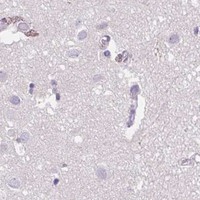


Comprehensive Human Protein Atlas IHC scoring per tissue (reliability: Approved — Low consistency between antibody staining and RNA expression data.). Rows are taken directly from the HPA tissue chart — click any row's HPA link to view the source.
| Tissue | Cell type | Level | Evidence | Source |
|---|---|---|---|---|
| Testis | Spermatogonia cells | High | Protein (IHC) | HPA → |
| Tissue | Cell type | Level | Evidence | Source |
|---|---|---|---|---|
| Adipose tissue | Adipocytes | Not detected | Protein (IHC) | HPA → |
| Appendix | Glandular cells | Not detected | Protein (IHC) | HPA → |
| Bone marrow | Hematopoietic cells | Not detected | Protein (IHC) | HPA → |
| Breast | Adipocytes | Not detected | Protein (IHC) | HPA → |
| Caudate | Glial cells | Not detected | Protein (IHC) | HPA → |
Troubleshoot SYNE3 staining by checking retrieval, nuclear envelope localisation, epitope access and cell-specific controls before interpreting chromogenic signal (UniProt Q6ZMZ3; HPA tissue IHC).
The IHC-validated anti-SYNE3 antibody has real paraffin-section images from human liver cancer, thyroid papillary carcinoma and spleen rupture tissue, and mouse liver (A13164-2 image captions).
A13164-2 is listed for IHC in human and mouse (catalog: applications and reactivity). Its IHC images show paraffin sections of human liver cancer, thyroid papillary carcinoma and spleen rupture tissue, and mouse liver (A13164-2 image captions).
Which to pick: Choose A13164-2 for paraffin-section IHC in human tissue or mouse liver; its captions document EDTA retrieval at pH 8.0 and primary antibody at 2 μg/ml (A13164-2 image captions). The fixative is unreported (A13164-2 image captions). No IF/ICC-validated SKU or IF image is supplied, so an IF/ICC choice would require separate validation; A13164-2 is the listed cross-species IHC option for human and mouse (catalog: applications, reactivity and IF images).