SYVN1 / E3 ubiquitin-protein ligase synoviolin · IHC design guide

Design Immunohistochemistry for SYVN1

Plan chromogenic FFPE IHC for SYVN1 using cytoplasmic staining and high signal in duodenal glandular cells as reference patterns (HPA tissue IHC). Start with the IHC-validated M02670 antibody at 1:50–1:200 (datasheet), then score staining by cell type and intensity.

Evidence assembled Oct 2026 · For research use; verify linked source records and product datasheet before use
Immunohistochemistry protocol sheet for SYVN1 (IHC for SYVN1): expected localisation Cytoplasmic tissue staining (HPA tissue IHC); ER membrane (UniProt), antibody M02670, validated IHC image, and IHC protocol steps
Printable SYVN1 IHC protocol sheet — expected localisation Cytoplasmic tissue staining (HPA tissue IHC); ER membrane (UniProt), antibody M02670, controls and protocol steps. Open the full SYVN1 IHC guide →

SYVN1 Immunohistochemistry Experimental Design Guide

Expected localisation, validated protocols, controls and antibodies — the at-a-glance facts below, then the full design guide.

Must know before staining
Expected localisation Cytoplasmic tissue staining (HPA tissue IHC); ER membrane (UniProt)
Staining pattern Cytoplasmic in most tissues; high in some immune cells (HPA tissue IHC)
Antigen retrieval EDTA pH 8.0 HIER, heat-mediated (datasheet M02670)
Positive control ⓘ Cerebellum+4 more · see all
Negative control ⓘ Esophagus+1 more · see all
Important caveats
Reasons your staining may differ from the expected pattern.
Fixation Matched tissue-IHC evidence does not establish the fixation claim. Validate the specimen-specific method before use. (selected-SKU IHC image M02670)
Caveat Immune-rich areas may dominate the signal (HPA tissue IHC)
Regulation Upregulated in rheumatoid synovium (UniProt)
Isoform / epitope 3 isoforms; ER-lumenal vs cytoplasmic epitope matters (UniProt)
Section 1

Recommended SYVN1 IHC & IF Protocols

The catalog antibody uses heat-mediated EDTA retrieval (datasheet M02670). The published IHC protocols below cover tissue sections, mouse lung, and NSCLC specimens (PMC12603505; PMC10693155; PMC12394631).

Recommended immunohistochemistry (IHC-P) protocol parameters
SampleParaffin-embedded human pancreas cancer tissue; fixative not specified (datasheet M02670)
FixationImage fixative and duration unreported (datasheet M02670); verify before use.
Sectioning4–5 µm sections on charged slides (standard)
DeparaffinisationXylene, graded ethanol series to water (standard)
Antigen retrievalHeat retrieval: EDTA pH 8.0 (datasheet M02670); 20 min, 95–100 °C (standard)
Peroxidase block3% H2O2, 10 min, room temperature (standard)
Blocking10% goat serum (datasheet M02670)
Primary antibodyRabbit monoclonal (clone 32S38) anti-SYVN1, 1:50-1:200 (datasheet M02670)
Primary incubationOvernight at 4 °C (datasheet M02670)
DetectionHRP-conjugated secondary, DAB chromogen (datasheet M02670)
CounterstainHematoxylin, blue, dehydrate and mount (standard)
Expected resultSYVN1-positive staining in cells in molecular layer of cerebellum (HPA tissue IHC: High). HPA tissue profile: Cytoplasmic expression in most tissues. Highly abundant in a subset of immune cells. No signal in the no-primary control.
💡Decision noteStart with EDTA pH 8.0 heat retrieval for the catalog antibody (datasheet M02670); the NSCLC protocol reports Tris-EDTA pressure-cooker retrieval (PMC12394631 methods).
Section 2

What Is the Expected SYVN1 Staining Pattern?

SYVN1 is an endoplasmic reticulum membrane protein with 6 transmembrane segments and a long cytoplasmic region (UniProt Q86TM6 topology). In paraffin section IHC, expect predominantly cytoplasmic staining in many tissues, including strongly stained glandular, neural and selected immune cells (HPA: tissue IHC). HPA rates the tissue pattern Enhanced, with medium consistency between antibody staining and RNA expression (HPA: tissue IHC reliability).

What am I looking at on my slide?
Cytoplasmic staining in duodenal glandular cells or lung alveolar type II cells, with discernible cell boundaries.This fits the reported High staining in those cell populations (HPA: duodenum and lung tissue IHC). An intracellular pattern also fits SYVN1 at the endoplasmic reticulum membrane (UniProt Q86TM6 subcellular location). Use cell identity and compartment together when judging a positive section.
Strong nuclear-only staining dominates a paraffin section.Review it as a possible compartment artifact because the tissue IHC profile is predominantly cytoplasmic (HPA: tissue IHC). Do not dismiss every nuclear observation outright: separate ICC-IF data report nucleoplasm as the main location (HPA: subcellular ICC-IF). Confirm the IHC pattern against a known-positive section and controls.
Strong staining appears chiefly in esophageal or vaginal squamous epithelial cells.HPA reports SYVN1 as Not detected in those specific cell populations (HPA: esophagus and vagina tissue IHC). Check cell identification, antibody specificity and endogenous chromogen-generating activity (standard IHC practice). This discrepancy alone does not establish cross-reactivity or prove the whole tissue is negative.
Diffuse color covers cells and surrounding tissue without a readable intracellular pattern.The section cannot reliably establish SYVN1 localization; HPA describes a cytoplasmic tissue pattern (HPA: tissue IHC). Consider nonspecific antibody binding, residual endogenous detection activity or excessive chromogen development as general IHC possibilities (standard IHC practice). Compare a no-primary control and a known-positive section.
No detectable staining in a section expected to contain duodenal glandular cells.HPA reports High staining in duodenal glandular cells (HPA: duodenum tissue IHC), so a blank result warrants a run-level check. Verify that the expected cells are present, then check antibody application, retrieval, detection and controls (standard IHC practice). One negative run does not establish absent SYVN1.
💡Expected SYVN1 appearanceCall a section positive when the expected cells show readable, predominantly cytoplasmic staining, potentially strong in duodenal glandular or lung alveolar type II cells (HPA: tissue IHC); isolated strong staining in HPA-undetected squamous cell populations is a finding to investigate, not an automatic positive (HPA: esophagus and vagina tissue IHC).
How each factor affects the staining
Membrane topology and compartmentSYVN1 spans the endoplasmic reticulum membrane 6 times, with residues 246–617 on the cytoplasmic side (UniProt Q86TM6 topology). This supports an intracellular interpretation; the record does not identify the catalog antibody's epitope, so epitope accessibility cannot be predicted.
Cell population and tissue choiceSYVN1 is broadly expressed, with highest protein levels in liver and kidney according to UniProt (UniProt Q86TM6 tissue specificity). For a cell-resolved IHC comparison, HPA specifically reports High duodenal glandular and lung alveolar type II staining, versus undetected esophageal squamous staining (HPA: tissue IHC).
Strength of tissue-pattern evidenceHPA labels the tissue IHC profile Enhanced but describes only medium staining–RNA consistency (HPA: tissue IHC reliability). Its listed cell-level intensities are observed patterns, not guaranteed results for every specimen or antibody (HPA: tissue IHC; standard IHC practice). Interpret an unexpected result with controls.
Isoforms and antibody coverageUniProt lists 3 SYVN1 isoforms (UniProt Q86TM6 isoforms). Their inclusion in an IHC signal depends on the antibody's recognized epitope, which the supplied record does not specify. Avoid assigning a weak or absent stain to a particular isoform from these data alone.
IF/ICC Q&A: where may fluorescence appear?HPA reports mainly nucleoplasmic fluorescence, plus endoplasmic reticulum and plasma membrane localization (HPA: subcellular ICC-IF). UniProt places SYVN1 at the endoplasmic reticulum membrane (UniProt Q86TM6 subcellular location). These sources differ in emphasis; the IF/ICC pattern does not redefine the reported tissue IHC pattern.
Why is my staining missing, weak or wrong?
SituationLikely causeNext action
The known-positive section is blank.The staining run may have failed, or the expected cell population may be absent from the section (standard IHC practice).Confirm the target cells on the counterstained section and review antibody application, retrieval and detection steps (standard IHC practice). Use a tissue and cell population reported High by HPA, such as duodenal glandular cells (HPA: tissue IHC).
The test section is blank but the positive control stains.The sampled cell population may have low or undetected staining; HPA reports variable levels by cell type (HPA: tissue IHC).Identify and score the relevant cells before interpreting the section. Compare their result with the corresponding HPA cell-level observation, while allowing for specimen variation (HPA: tissue IHC; standard IHC practice).
Color appears in the no-primary control.An antibody-independent detection signal, including endogenous enzyme activity when applicable, is possible (standard chromogenic IHC practice).Resolve the control signal before scoring SYVN1; check the detection chemistry, endogenous activity blocking and wash steps for the chosen method (standard IHC practice).
Most structures show uniform color without clear cellular detail.Nonspecific background or excessive detection development can obscure a compartment pattern (standard IHC practice).Compare control slides, review blocking and washes, and adjust antibody or development conditions within the validated IHC workflow (standard IHC practice). Reassess whether the remaining signal is predominantly cytoplasmic (HPA: tissue IHC).
Staining is nuclear-only in the IHC section.This differs from the predominantly cytoplasmic tissue IHC profile (HPA: tissue IHC), although nucleoplasmic localization is reported separately by ICC-IF (HPA: subcellular ICC-IF).Check the known-positive section, no-primary control and antibody validation before calling it SYVN1-specific IHC staining (standard IHC practice). Record the nuclear finding separately from the expected tissue pattern.
A nominally low or undetected cell population stains strongly.Misidentified cells, cross-reactivity or endogenous detection activity are possible (standard IHC practice). HPA reports undetected esophageal and vaginal squamous cells (HPA: tissue IHC).Confirm cell identity, inspect the no-primary control and compare staining with a High cell population in the same run (standard IHC practice; HPA: tissue IHC). Treat the discrepancy as unresolved until those checks support interpretation.

Sample controls for SYVN1 IHC & IF

🧪Run duodenum first: glandular cells should stain (HPA: High in duodenal glandular cells). Use esophagus squamous epithelial cells as the negative tissue (HPA: Not detected); on the positive slide, cells used as internal background comparators should show counterstain without specific DAB signal, but the supplied HPA row does not identify a verified negative cell type within duodenum (HPA: duodenum row).
Positive control tissue: Cerebellum (Cells in molecular layer, HPA High)
Negative control tissue: Esophagus (HPA Not detected)
ICC-IF cell lines (HPA subcellular resource): HPA ICC-IF images show SYVN1 in U-251MG, U2OS, A-431, NIH 3T3, with annotated localisation: Nucleoplasm (supported) (HPA subcellular).
Technical controls: Include a no-primary secondary-only control, a control immunoglobulin matched to the primary’s host species, class, and monoclonal or polyclonal format, and SYVN1-knockout material as a biological negative (standard IHC control practice). For chromogenic duodenum sections, quench endogenous peroxidase and check the no-primary slide for residual tissue background (standard IHC practice).
⚠️Feasibility: A target-specific fixation window or fixation effect is unreported, and the selected M02670 tissue-IHC caption does not state a fixative (selected-SKU tissue-IHC caption). That paraffin-section example uses heat retrieval in EDTA at pH 8.0 before 1:50 primary incubation, so retrieval is part of the reported example, but its necessity across specimens is unreported (selected-SKU tissue-IHC caption). The supplied evidence does not establish whether frozen sections or IF are easier; interpret IF localization cautiously because HPA reports nucleoplasmic, ER, and plasma-membrane signal while UniProt places SYVN1 in the ER membrane (HPA: subcellular; UniProt Q86TM6: subcellular location).

HPA tissue IHC evidence for SYVN1

Comprehensive Human Protein Atlas IHC scoring per tissue (reliability: Enhanced — Medium consistency between antibody staining and RNA expression data.). Rows are taken directly from the HPA tissue chart — click any row's HPA link to view the source.

Positive expression · recommended positive controls

TissueCell typeLevelEvidenceSource
Cerebellum Cells in molecular layer High Protein (IHC) HPA →
Cerebral cortex Glial cells High Protein (IHC) HPA →
Cervix Glandular cells High Protein (IHC) HPA →
Duodenum Glandular cells High Protein (IHC) HPA →
Fallopian tube Ciliated cells (cell body) High Protein (IHC) HPA →

Undetected expression · recommended negative controls

TissueCell typeLevelEvidenceSource
Esophagus Squamous epithelial cells Not detected Protein (IHC) HPA →
Vagina Squamous epithelial cells Not detected Protein (IHC) HPA →
Section 3

Advanced SYVN1 IHC Tips

Troubleshoot SYVN1 staining in paraffin sections by checking retrieval, cellular pattern, controls and scoring before interpreting chromogenic signal.

Which retrieval conditions should I try first for weak SYVN1 staining?
Start with heat-mediated antigen retrieval in EDTA at pH 8.0 for SYVN1 paraffin-section IHC (datasheet M02670). The selected image used that retrieval, followed by 1:50 primary antibody overnight at 4°C; keep those documented conditions together when establishing a reference section (datasheet M02670). If staining remains weak, optimise heating time and cooling consistently across adjacent sections, while checking that tissue morphology remains intact (standard IHC practice). Include a no-primary control and compare signal in expected cell populations, because HPA reports cytoplasmic staining in most tissues but only medium consistency between antibody staining and RNA data (HPA tissue IHC).
How should I troubleshoot fixation-related loss of SYVN1 signal?
The selected SYVN1 image documents a paraffin-embedded section but does not state its fixative, so target-specific fixation sensitivity is unknown (datasheet M02670). Record the actual fixative and fixation duration for each specimen, then compare sections processed under matched conditions before attributing weak signal to fixation (standard IHC practice). Apply the documented EDTA pH 8.0 retrieval and 1:50 overnight primary incubation consistently during that comparison (datasheet M02670). Check whether weak staining affects several antigens or tissue morphology as well as SYVN1, and use a no-primary control to distinguish lost signal from nonspecific detection (standard IHC practice).
Where should SYVN1 staining appear, and how should nuclear signal be assessed?
For chromogenic tissue IHC, begin by assessing cytoplasmic signal: HPA describes cytoplasmic expression in most tissues, while UniProt places SYVN1 at the endoplasmic reticulum membrane (HPA tissue IHC; UniProt Q86TM6 subcellular location). A perinuclear or reticular cytoplasmic pattern can fit that location, but chromogenic resolution alone cannot prove endoplasmic reticulum localisation (UniProt Q86TM6 subcellular location; standard IHC practice). HPA separately reports nucleoplasm as its main subcellular ICC/IF location, with additional endoplasmic reticulum and plasma-membrane locations (HPA subcellular). Score nuclear and cytoplasmic compartments separately, and investigate dominant nuclear staining against controls and the tissue-IHC pattern before assigning it to SYVN1 (HPA tissue IHC; HPA subcellular; standard IHC practice).
Could epitope position or isoforms explain variable SYVN1 staining?
SYVN1 has 3 annotated isoforms, so an antibody's epitope must be checked before interpreting absent staining as absence of every isoform (UniProt Q86TM6 isoforms). Its 6 transmembrane segments span residues 5–245, whereas residues 246–617 face the cytoplasm; epitope accessibility may therefore depend on the recognised sequence and preparation (UniProt Q86TM6 topology; standard IHC practice). The supplied tissue caption gives the antibody dilution but no epitope map, so it cannot establish isoform coverage or which side of the membrane the antibody recognises (datasheet M02670). Compare sections with matched retrieval and consult an experimentally mapped epitope before explaining a compartment difference as isoform-specific staining (standard IHC practice).
How can IF help verify an ambiguous SYVN1 IHC pattern?
Use IF as a complementary localisation check when chromogenic IHC cannot resolve a suspected endoplasmic reticulum pattern (UniProt Q86TM6 subcellular location; standard IF practice). Multiplex SYVN1 with a validated marker for the cell population being scored, and select fluorophores after inspecting unstained tissue for autofluorescence in each channel (standard IF practice). Permeabilisation must allow access to the antibody's epitope: residues 246–617 are cytoplasmic, while several short loops are lumenal, but the supplied caption does not map this antibody's epitope (UniProt Q86TM6 topology; datasheet M02670). Keep no-primary and single-stain controls, and interpret IF alongside the IHC result because HPA reports nucleoplasmic, endoplasmic reticulum and plasma-membrane locations in its subcellular data (standard IF practice; HPA subcellular).
How do I reduce diffuse brown background without losing SYVN1 signal?
First inspect a no-primary section to separate detection-system background from staining associated with the primary antibody (standard IHC practice). The selected image used 10% goat serum blocking, a 1:50 primary dilution, a peroxidase-conjugated secondary and DAB development; those are documented starting conditions for this antibody's paraffin-section example (datasheet M02670). Include a peroxidase block before HRP/DAB detection, then optimise washing and primary-antibody concentration on matched sections while preserving the documented retrieval as the initial condition (standard IHC practice; datasheet M02670). Compare background with tissue morphology and expected cell staining, since HPA reports cytoplasmic expression across most tissues rather than an entirely blank tissue baseline (HPA tissue IHC).
What is a defensible way to score SYVN1 across tissue sections? ⚠ ANSWER MARKED FOR VERIFICATION
Define the cell population and compartment before scoring, because HPA describes cytoplasmic staining in most tissues and highly abundant expression in a subset of immune cells (HPA tissue IHC). For comparable regions, record the percentage of positive cells and staining intensity on a 0–3 scale; an H-score then spans 0–300 (standard IHC scoring practice). If counting positive cells by area, report density per mm² of viable tissue and identify the counted cell type, rather than pooling unlike compartments (standard IHC scoring practice). Normalise comparisons to matched tissue area, cell population, section processing and image thresholds, while retaining separate nuclear and cytoplasmic scores where both appear (standard IHC scoring practice; HPA subcellular).
When is SYVN1 staining convincing, and when might it be artefactual?
A convincing IHC result shows reproducible staining in intact cells, with cytoplasmic signal compatible with HPA tissue staining and SYVN1's endoplasmic reticulum membrane assignment (HPA tissue IHC; UniProt Q86TM6 subcellular location; standard IHC practice). Check the identity of stained cells: HPA reports high signal in several named cell populations and highly abundant expression in a subset of immune cells, so mixed tissue can confound a whole-section impression (HPA tissue IHC). Treat isolated edge staining, necrotic regions and signal reproduced in a no-primary control as possible artefacts, including endogenous enzyme activity during HRP/DAB detection (standard IHC practice). Investigate predominantly nuclear staining separately because HPA subcellular data support nucleoplasmic localisation, whereas its tissue-IHC profile describes mainly cytoplasmic expression (HPA subcellular; HPA tissue IHC).
Boster reagents

Best SYVN1 / E3 ubiquitin-protein ligase synoviolin IHC Antibodies

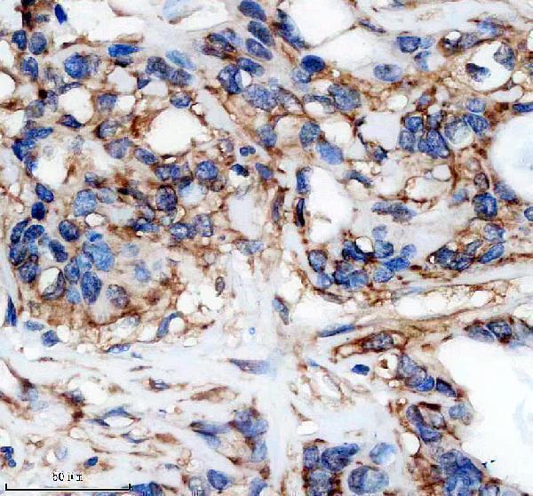
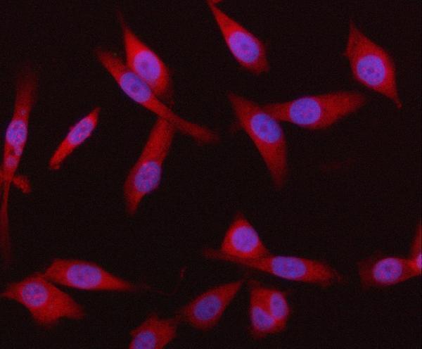
Two anti-SYVN1 antibodies have image evidence: paraffin-section IHC in human pancreas cancer tissue (M02670 image caption) and IF/ICC in PC-3 cells (A02670-3 image caption). Both list human, mouse and rat reactivity (catalog).

Real IHC data IHC analysis of HRD1/SYVN1 using anti-HRD1/SYVN1 antibody (M02670). HRD1/SYVN1 was detected in a paraffin-embedded section of human pancreas cancer tissue. Heat mediated antigen retrieval was performed in EDTA buffer (pH 8.0, epitope retrieval solution). The tissue section was blocked with 10% goat serum. The tissue section was then incubated with 1: 50 rabbit anti-HRD1/SYVN1 Antibody (M02670) overnight at 4°C. Peroxidase Conjugated Goat Anti-rabbit IgG was used as secondary antibody and incubated for 30 minutes at 37°C. The tissue section was developed using HRP Conjugated Rabbit IgG Super Vision Assay Kit (Catalog # SV0002) with DAB as the chromogen.
Anti-SYVN1 / HRD1 Rabbit Monoclonal Antibody
Cat # M02670
Real IF data IF analysis of HRD1/SYVN1 using anti-HRD1/SYVN1 antibody (A02670-3). HRD1/SYVN1 was detected in an immunocytochemical section of PC-3 cells. Enzyme antigen retrieval was performed using IHC enzyme antigen retrieval reagent (AR0022) for 15 mins. The cells were blocked with 10% goat serum. And then incubated with 5 μg/mL rabbit anti-HRD1/SYVN1 Antibody (A02670-3) overnight at 4°C. DyLight550 Conjugated Goat Anti-Rabbit IgG (BA1135) was used as secondary antibody at 1:500 dilution and incubated for 30 minutes at 37°C. The section was counterstained with DAPI. Visualize using a fluorescence microscope and filter sets appropriate for the label used.
Anti-HRD1/SYVN1 Antibody ®
Cat # A02670-3

M02670 will render with IHC data from a paraffin-embedded section of human pancreas cancer tissue (M02670 image caption). A02670-3 will render with IF data from PC-3 cells (A02670-3 image caption).

Which to pick: For tissue IHC, choose M02670: it is a rabbit monoclonal with IHC listed and a paraffin-section IHC image (M02670 catalog and image caption); the fixative is unreported (M02670 image caption). For IF/ICC, choose A02670-3 for its PC-3 cell IF image and listed IF/ICC applications (A02670-3 catalog and image caption). Both list human, mouse and rat reactivity (catalog), but the supplied images show only the stated tissue and cell samples (image captions).

Each figure is that product's own IHC / IF validation image from its datasheet.

References

  1. UniProt Consortium. UniProt entry Q86TM6 (SYVN1_HUMAN, E3 ubiquitin-protein ligase synoviolin).
  2. Human Protein Atlas. SYVN1 tissue IHC expression (reliability: Enhanced).
  3. Human Protein Atlas. SYVN1 subcellular location (ICC-IF): Mainly localized to the nucleoplasm. In addition localized to the plasma membrane and endoplasmic reticulum..
  4. Human Protein Atlas. SYVN1 antibody validation summary (3 antibodies).
  5. SYVN1 aggravates esophageal squamous cell carcinoma development by activating NF-κB pathway to facilitate macrophage M2 polarization. Journal of thoracic disease 2025 — PMC12603505.
  6. Ubiquitin ligase SYVN1/HRD1 facilitates degradation of the SERPINA1 Z variant/α-1-antitrypsin Z variant via SQSTM1/p62-dependent selective autophagy. Autophagy 2017 — PMC5388218.
  7. Role of SYVN1 in the control of airway remodeling in asthma protection by promoting SIRT2 ubiquitination and degradation. Biological research 2023 — PMC10693155.
  8. Targeting the SYVN1-EGFR axis: a breakthrough strategy for TKI-resistant NSCLC. Cell death & disease 2025 — PMC12394631.
  9. PubMed PMID:12459480 — UniProt-cited evidence.
  10. PubMed PMID:12646171 — UniProt-cited evidence.
  11. PubMed PMID:12975321 — UniProt-cited evidence.