This website uses cookies to ensure you get the best experience on our website.


- Table of Contents
Plan TBX21 chromogenic IHC in paraffin sections around nuclear staining in a subset of immune cells (HPA tissue IHC). Use spleen as a positive tissue reference (HPA tissue IHC) and score nuclear staining within the relevant cell population.

Expected localisation, validated protocols, controls and antibodies — the at-a-glance facts below, then the full design guide.
| Expected localisation | Nuclear staining in a subset of immune cells (HPA tissue IHC) | |
| Staining pattern | Selective staining of immune-cell nuclei (HPA tissue IHC) | |
| Antigen retrieval | Tris-EDTA pH 9.0 HIER, 95–98 °C, 20 min (rule: nuclear antigen) | |
| Positive control | Bone marrow+4 more · see all | |
| Negative control | Adipose tissue+4 more · see all |
| Fixation | Matched tissue-IHC evidence does not establish the fixation claim. Validate the specimen-specific method before use. (selected-SKU IHC image M00404) | |
| Caveat | Kidney proximal tubules also show medium staining (HPA tissue IHC) | |
| Regulation | T-cell-specific expression (UniProt) | |
| Isoform / epitope | No annotated isoforms; one 1–535 chain (UniProt) |
The catalog antibody’s IHC-P protocol is supplemented by one published TBX21 IHC workflow (PMC10948251).
| Sample | Paraffin-embedded human spleen tissue; fixative not specified (datasheet M00404) |
| Fixation | Image fixative and duration unreported (datasheet M00404); verify before use. |
| Sectioning | 4–5 µm sections on charged slides (standard) |
| Deparaffinisation | Xylene, graded ethanol series to water (standard) |
| Antigen retrieval | Heat-induced epitope retrieval in Tris-EDTA buffer, pH 9.0, 20 min at 95–98 °C (standard rule: nuclear antigen) |
| Peroxidase block | 3% H2O2, 10 min, room temperature (standard) |
| Blocking | 10% normal serum of the secondary host, 30 min, room temperature (standard) |
| Primary antibody | Rabbit monoclonal (clone AFHO-20) anti-TBX21, 1:50-1:200 (datasheet M00404) |
| Primary incubation | Overnight at 4 °C (standard) |
| Detection | HRP-polymer secondary, DAB chromogen 5–10 min (standard) |
| Counterstain | Hematoxylin, blue, dehydrate and mount (standard) |
| Expected result | TBX21-positive staining in hematopoietic cells of bone marrow (HPA tissue IHC: Medium). HPA tissue profile: Selective nuclear expression in a subset of immune cells. No signal in the no-primary control. |
TBX21 is nuclear and lacks a transmembrane segment (UniProt Q9UL17 topology). Expect selective nuclear staining in a subset of immune cells (HPA tissue IHC: Enhanced reliability), consistent with T-cell specificity (UniProt Q9UL17). HPA also reports staining in kidney proximal tubules (HPA tissue IHC).
| Nuclear stain in lymph-node non-germinal center cells (HPA tissue IHC). | Expected selective pattern; reported level is medium (HPA tissue IHC). |
| Predominantly cytoplasmic stain. | Suspect nonspecific signal; TBX21 is nuclear (UniProt Q9UL17; HPA tissue IHC). |
| Adipocytes or bronchial respiratory epithelium stain. | Suspect cross-reactivity or endogenous activity; both are not detected (HPA tissue IHC). |
| Diffuse stain across the section. | Suspect background rather than selective nuclear staining (HPA tissue IHC). |
| No stain in lymph-node non-germinal center cells. | Check assay performance; medium staining is reported there (HPA tissue IHC). |
| Cell distribution (HPA tissue IHC) | Immune-cell staining is selective; kidney proximal tubules also show medium stain (HPA tissue IHC). |
| Antibody validation (HPA antibodies) | CAB009524 and CAB030043 are IHC Enhanced (HPA antibodies). |
| Processing (UniProt Q9UL17) | One chain spans residues 1–535; no signal peptide or propeptide (UniProt Q9UL17). |
| Situation | Likely cause | Next action |
|---|---|---|
| No nuclear signal in lymph node. | Expected cells show medium stain (HPA tissue IHC). | Check a positive control, retrieval, primary incubation and detection (standard IHC practice). |
| Strong cytoplasmic stain. | Expected location is nuclear (UniProt Q9UL17; HPA tissue IHC). | Check the counterstain and a primary-omission control (standard IHC practice). |
| Diffuse background. | Blocking or detection conditions may contribute (standard IHC practice). | Optimize blocking and primary concentration (standard IHC practice). |
| Adipocytes or bronchial epithelium stain. | These cells are not detected by HPA tissue IHC; suspect nonspecific or endogenous signal. | Use primary-omission controls; check endogenous peroxidase (standard IHC practice). |
| Kidney proximal tubules stain. | HPA reports medium staining there (HPA tissue IHC). | Score this compartment against the HPA tissue IHC pattern. |
| What IF/ICC pattern is established? | No ICC-IF images or main location are available (HPA subcellular). | Use the separate IF/ICC guide; do not infer an IF pattern from IHC. |
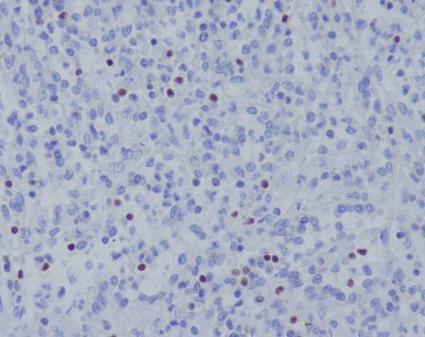
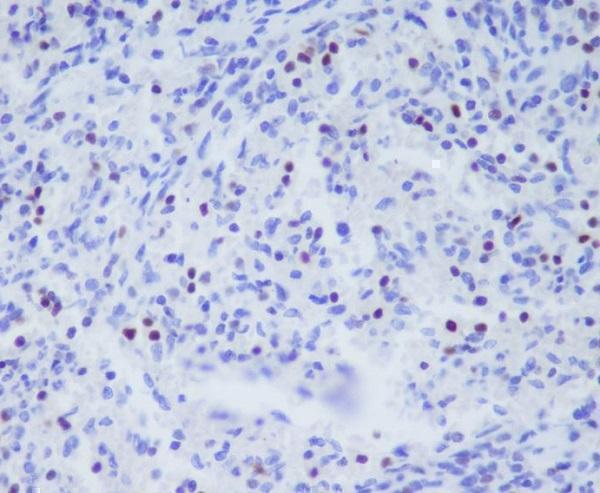


Comprehensive Human Protein Atlas IHC scoring per tissue (reliability: Enhanced — High consistency between antibody staining and RNA expression data.). Rows are taken directly from the HPA tissue chart — click any row's HPA link to view the source.
| Tissue | Cell type | Level | Evidence | Source |
|---|---|---|---|---|
| Bone marrow | Hematopoietic cells | Medium | Protein (IHC) | HPA → |
| Kidney | Proximal tubules (cell body) | Medium | Protein (IHC) | HPA → |
| Lymph node | Non-germinal center cells | Medium | Protein (IHC) | HPA → |
| Spleen | Cells in red pulp | Medium | Protein (IHC) | HPA → |
| Tonsil | Non-germinal center cells | Medium | Protein (IHC) | HPA → |
| Tissue | Cell type | Level | Evidence | Source |
|---|---|---|---|---|
| Adipose tissue | Adipocytes | Not detected | Protein (IHC) | HPA → |
| Adrenal gland | Glandular cells | Not detected | Protein (IHC) | HPA → |
| Appendix | Glandular cells | Not detected | Protein (IHC) | HPA → |
| Breast | Adipocytes | Not detected | Protein (IHC) | HPA → |
| Bronchus | Respiratory epithelial cells | Not detected | Protein (IHC) | HPA → |
Troubleshoot TBX21 chromogenic IHC in paraffin sections by checking nuclear staining, tissue controls, processing, and detection before interpreting cell counts.
Two human-reactive anti-TBX21 antibodies have IHC images from paraffin-embedded human spleen (catalog: M00404 and M00404-1 image captions).
M00404 is listed for human IHC and Flow Cytometry, with an IHC image from paraffin-embedded human spleen (catalog: applications, reactivity, M00404 image caption). M00404-1 is listed for human IHC, with its own image from paraffin-embedded human spleen (catalog: applications, reactivity, M00404-1 image caption).
Which to pick: For human paraffin-section IHC, both rabbit monoclonals have supporting images; M00404 lists 1:50–1:200 (catalog: clone AFHO-20, dilution; M00404 image caption), while M00404-1 lists 1:50 (catalog: clone 20T77, dilution; M00404-1 image caption). Neither caption reports the fixative (catalog: M00404 and M00404-1 image captions). Neither SKU lists IF/ICC or species beyond human, so the payload supports no IF/ICC or cross-species pick (catalog: applications, reactivity).