THEM4 / Acyl-coenzyme A thioesterase THEM4 · IHC design guide

Design Immunohistochemistry for THEM4

Plan chromogenic IHC on paraffin sections with the IHC-validated antibody A07814, starting at 2.5 μg/mL (datasheet). Use pancreatic exocrine cells as a positive tissue control (HPA tissue IHC), and assess subcellular staining cautiously because a tissue IHC compartment pattern is unreported (HPA tissue IHC).

Evidence assembled Oct 2026 · For research use; verify linked source records and product datasheet before use
Immunohistochemistry protocol sheet for THEM4 (IHC for THEM4): expected localisation Cytoplasm and mitochondria; tissue IHC pattern unestablished (UniProt), antibody A07814, validated IHC image, and IHC protocol steps
Printable THEM4 IHC protocol sheet — expected localisation Cytoplasm and mitochondria; tissue IHC pattern unestablished (UniProt), antibody A07814, controls and protocol steps. Open the full THEM4 IHC guide →

THEM4 Immunohistochemistry Experimental Design Guide

Expected localisation, validated protocols, controls and antibodies — the at-a-glance facts below, then the full design guide.

Must know before staining
Expected localisation Cytoplasm and mitochondria; tissue IHC pattern unestablished (UniProt)
Staining pattern High in pancreatic exocrine cells; compartment unreported (HPA tissue IHC)
Antigen retrieval Citrate pH 6.0 HIER, 95–98 °C, 20 min (rule: cytoplasmic / membrane antigen)
Positive control ⓘ Pancreas+4 more · see all
Negative control ⓘ Adipose tissue+4 more · see all
Important caveats
Reasons your staining may differ from the expected pattern.
Fixation Keep fixation consistent across sections (standard IHC practice; not target-specific). Selected-image fixative and duration unreported (datasheet A07814); verify before use.
Caveat Staining may disagree with RNA expression (HPA tissue IHC)
Regulation Glioma: methylation lowers expression (UniProt)
Isoform / epitope No isoforms; check epitope against mature chain 37–240 (UniProt)
Section 1

Recommended THEM4 IHC & IF Protocols

The catalog antibody’s IHC-P protocol is accompanied by one published THEM4 tumor-tissue IHC protocol (PMC8267908).

Recommended immunohistochemistry (IHC-P) protocol parameters
SampleTissue sections; selected-image fixative not specified (standard IHC workflow)
FixationImage fixative and duration unreported (datasheet A07814); verify before use.
Sectioning4–5 µm sections on charged slides (standard)
DeparaffinisationXylene, graded ethanol series to water (standard)
Antigen retrievalHeat-induced epitope retrieval in citrate buffer, pH 6.0, 20 min at 95–98 °C (standard rule: cytoplasmic / membrane antigen)
Peroxidase block3% H2O2, 10 min, room temperature (standard)
Blocking10% normal serum of the secondary host, 30 min, room temperature (standard)
Primary antibodyRabbit anti-THEM4, 2.5 μg/mL (datasheet A07814)
Primary incubationOvernight at 4 °C (standard)
DetectionHRP-polymer secondary, DAB chromogen 5–10 min (standard)
CounterstainHematoxylin, blue, dehydrate and mount (standard)
Expected resultTHEM4-positive staining in exocrine glandular cells of pancreas (HPA tissue IHC: High). HPA tissue profile: Expressed in several different tissues, including kidney and testis. No signal in the no-primary control.
💡Decision noteStart with citrate pH 6.0 retrieval at 95–98 °C for 20 min (page antigen-retrieval rule).
Section 2

What Is the Expected THEM4 Staining Pattern?

THEM4 is annotated in mitochondria, cytoplasm and at cell or ruffle membranes; it has no transmembrane segment (UniProt Q5T1C6 subcellular location and topology). In paraffin sections, look for staining in pancreatic exocrine, placental decidual, and small intestinal or stomach glandular cells, all scored High (HPA tissue IHC). Treat the pattern as a guide: the IHC antibody is Approved, with low consistency between staining and RNA expression (HPA tissue IHC).

What am I looking at on my slide?
Strong signal in pancreatic exocrine cells or placental decidual cells, chiefly within the cell body.This fits cells scored High by HPA (HPA tissue IHC) and is compatible with mitochondrial and cytoplasmic THEM4 (UniProt Q5T1C6 subcellular location). Chromogenic IHC cannot assign signal to a mitochondrial membrane or space from appearance alone; compare stained and unstained cells on the same section.
Predominantly nuclear staining, with little signal in expected positive cell bodies.A predominantly nuclear pattern lacks support in the supplied location record (UniProt Q5T1C6 subcellular location). Check morphology, counterstain, staining controls and antibody conditions before calling it THEM4; an apparent compartment mismatch alone does not identify the cause.
Strong staining confined to adipocytes or to cells in the cerebellar granular layer.Those cells are listed as Not detected (HPA tissue IHC). Recheck cell identity and compare a High tissue in the same run. If the unexpected signal persists, investigate cross-reactivity or endogenous detection activity; a single discordant section cannot distinguish them.
Uniform color across cells, stroma and section edges, obscuring cell boundaries.This distribution is difficult to score as cellular THEM4. As general IHC practice, examine a no-primary control for detection-system background and review blocking and washing. Do not interpret broad background as proof of expression in a tissue.
No cellular signal in pancreatic exocrine cells or stomach glandular cells.Both are scored High (HPA tissue IHC), so first verify that the section contains the specified cells. If it does, review antibody dilution, retrieval and detection controls as general IHC troubleshooting. HPA reports low staining–RNA consistency, so one negative section does not establish absence (HPA tissue IHC).
💡Expected THEM4 appearanceA convincing positive is cell-associated staining in a High-scoring population, such as pancreatic exocrine or stomach glandular cells (HPA tissue IHC), with a cytoplasmic pattern compatible with THEM4 location (UniProt Q5T1C6); broad stromal color or dominant nuclear staining calls for control checks before scoring.
How each factor affects the staining
Compartment and topologyMitochondrial, cytoplasmic, cell-membrane and ruffle-membrane locations are annotated, with no transmembrane segment (UniProt Q5T1C6). Expect possible within-cell variation, but do not assign a precise membrane compartment from chromogenic IHC alone.
Tissue and cell choicePancreatic exocrine, placental decidual, small intestinal glandular and stomach glandular cells are High; adipocytes are Not detected (HPA tissue IHC). Score the named cell population, since a tissue-wide average could hide the relevant pattern.
Antibody evidenceThe listed rabbit pAb, HPA028161, has Approved IHC status; the supplied record does not report Enhanced IHC validation (HPA antibodies; HPA tissue IHC). Low staining–RNA consistency also limits confidence in a single unexpected positive or negative (HPA tissue IHC).
Processing and modificationThe annotated chain begins at residue 37, and modified residues include phosphoserines at 37 and 38 (UniProt Q5T1C6 processing and modified residues). The supplied record gives no antibody epitope, so it cannot predict whether processing or modification changes this antibody's staining.
Fixation and retrieval evidenceTarget-specific fixation sensitivity and an effective retrieval condition are unreported in the supplied UniProt and HPA records. Retrieval optimization is general paraffin-IHC practice; do not infer a THEM4-specific fixation effect from topology or HPA tissue scores.
Why is my staining missing, weak or wrong?
SituationLikely causeNext action
A High-scoring tissue has no visible staining (HPA tissue IHC).The named positive cells may be absent, or a staining step may have failed; the supplied sources do not establish a THEM4-specific fixation cause.Locate the scored cell type, then check the IHC-validated antibody's dilution and the run's detection control. If needed, optimize retrieval as general IHC practice; compare sections processed together.
All cells and empty areas acquire similar chromogen color.Background or endogenous detection activity can produce color unrelated to cell-specific THEM4; this is a general chromogenic-IHC concern.Inspect a no-primary control. Review blocking, washes and detection reagents, then judge the test slide only where cellular signal clearly exceeds background.
A Not detected comparator stains strongly, such as adipocytes (HPA tissue IHC).Cell misidentification, cross-reactivity or detection background are possible; HPA's low staining–RNA consistency limits a definitive call (HPA tissue IHC).Confirm morphology and compare a High population on the same run (HPA tissue IHC). Repeat with appropriate controls before classifying the unexpected cell population as positive.
The dominant signal appears nuclear.Nuclear localisation is not among the supplied THEM4 locations (UniProt Q5T1C6 subcellular location). Counterstain overlap or nonspecific staining may mislead visual scoring.Inspect counterstain and no-primary control, then reassess cell boundaries at suitable magnification. Record a persistent nuclear-only pattern as discordant rather than forcing a mitochondrial interpretation.
Pancreatic exocrine cells stain, but adjacent compartments differ (HPA: High in exocrine glandular cells).HPA's High score applies to a named cell population, and THEM4 has several annotated cellular locations (HPA tissue IHC; UniProt Q5T1C6 subcellular location).Score the identified exocrine cells separately. Describe cytoplasmic or peripheral distribution at the resolution the slide permits; avoid claiming inner-membrane or intermembrane-space localisation from chromogenic IHC.
Can the same appearance be required in IF/ICC?HPA provides no main ICC-IF location or cell-line images, and the listed antibody has no ICC validation entry (HPA subcellular; HPA antibodies).Use the separate IF/ICC guide for that application. Here, treat any IF compartment match as exploratory and assess it with appropriate imaging controls; do not transfer the IHC tissue score into an IF validation claim.

Sample controls for THEM4 IHC & IF

🧪Run pancreas first: exocrine glandular cells should stain (HPA: High in pancreatic exocrine glandular cells). Use adipose tissue as the negative tissue, focusing on adipocytes (HPA: Not detected in adipocytes); on the pancreas slide, score non-exocrine cells as internal background comparators without assuming they are THEM4-negative.
Positive control tissue: Pancreas (Exocrine glandular cells, HPA High)
Negative control tissue: Adipose tissue (HPA Not detected)
ICC-IF cell lines (HPA subcellular resource): HPA carries no ICC-IF cell line for THEM4; derive a cell-line control from the positive tissue's cell type (Exocrine glandular cells) and confirm it by RNA or western blot first.
Technical controls: Include no-primary (secondary-only) and host-species- and isotype-matched controls, plus a THEM4 knockout sample or a peptide-block control if the antibody’s immunizing peptide is available (standard IHC practice). Quench endogenous peroxidase in the pancreas section and check the no-primary slide for residual chromogen (standard chromogenic IHC practice).
⚠️Feasibility: The supplied target and application evidence reports no target-specific fixation window or fixation effect, and the A07814 liver IHC caption leaves the fixative unreported (A07814 caption). Retrieval dependence is unreported; optimize antigen retrieval using the pancreatic positive and adipose negative tissues (HPA: High in pancreatic exocrine glandular cells; Not detected in adipocytes). The evidence does not establish whether frozen sections or IF/ICC are easier for THEM4; check pancreatic exocrine cells for background chromogen before scoring (standard IHC practice).

HPA tissue IHC evidence for THEM4

Comprehensive Human Protein Atlas IHC scoring per tissue (reliability: Approved — Low consistency between antibody staining and RNA expression data.). Rows are taken directly from the HPA tissue chart — click any row's HPA link to view the source.

Positive expression · recommended positive controls

TissueCell typeLevelEvidenceSource
Pancreas Exocrine glandular cells High Protein (IHC) HPA →
Placenta Decidual cells High Protein (IHC) HPA →
Small intestine Glandular cells High Protein (IHC) HPA →
Stomach Glandular cells High Protein (IHC) HPA →
Adrenal gland Glandular cells Medium Protein (IHC) HPA →

Undetected expression · recommended negative controls

TissueCell typeLevelEvidenceSource
Adipose tissue Adipocytes Not detected Protein (IHC) HPA →
Caudate Glial cells Not detected Protein (IHC) HPA →
Cerebellum Cells in granular layer Not detected Protein (IHC) HPA →
Cerebral cortex Endothelial cells Not detected Protein (IHC) HPA →
Fallopian tube Glandular cells Not detected Protein (IHC) HPA →
Section 3

Advanced THEM4 IHC Tips

Troubleshoot THEM4 staining in paraffin section IHC by checking retrieval, compartment, controls and cell specific scoring; use IF as a separate validation workflow.

What retrieval conditions should I try first if THEM4 staining is weak?
Start with citrate buffer at pH 6.0, heated to 95–98 °C for 20 min (page retrieval rule: cytoplasmic / membrane antigen). Let sections cool in the retrieval solution, then compare a retrieved section with an otherwise matched section processed without heat (standard IHC practice). Evaluate signal in the relevant cells alongside morphology: exocrine glandular cells in pancreas show high staining in the tissue profile (HPA: High in pancreatic exocrine glandular cells). If signal remains weak, test a more alkaline retrieval buffer as a documented fallback, changing one condition at a time and watching for tissue damage (standard IHC practice).
How should I troubleshoot weak staining across differently fixed IHC samples?
THEM4 specific fixation sensitivity is unknown from the supplied evidence; the liver IHC caption reports an antibody concentration but does not state a fixative (A07814 tissue IHC caption). Record fixative, fixation duration, tissue thickness and processing history for each block before comparing staining (standard IHC practice). Process matched control sections together, because differences in fixation and processing can change epitope accessibility and tissue morphology in paraffin IHC (standard IHC practice). If a batch stains weakly, compare it with a previously successful block under the same pH 6.0, 95–98 °C, 20 min retrieval conditions before changing antibody concentration (page retrieval rule; standard IHC practice).
Which staining compartments are plausible for THEM4 in tissue sections?
Assess cytoplasmic, mitochondrial region and membrane associated staining, because THEM4 is annotated in the cytoplasm, mitochondrion, cell membrane and ruffle membrane (UniProt Q5T1C6 subcellular location). Mitochondrial staining may appear granular in chromogenic IHC, but that appearance alone cannot establish an organelle identity at light microscope resolution (standard IHC interpretation). Compare the pattern within the relevant cell population: pancreatic exocrine glandular cells have high staining in the tissue profile (HPA: High in pancreatic exocrine glandular cells). A predominantly nuclear signal warrants control review because the supplied subcellular annotation does not list a nuclear location (UniProt Q5T1C6 subcellular location; standard IHC interpretation).
Could processing or modifications explain inconsistent staining with this antibody?
The supplied record annotates 0 isoforms and a processed THEM4 chain spanning residues 37–240 (UniProt Q5T1C6 isoforms and processing). It also lists phosphorylation at residues 37 and 38, succinylation at 55 and 66, and acetylation at 74 (UniProt Q5T1C6 modified residues). The antibody epitope is unspecified in the supplied evidence, so these annotations cannot establish whether processing or a modification affects binding (supplied antibody evidence; UniProt Q5T1C6). If staining varies, compare matched sections with the same retrieval and detection settings, then seek epitope mapping or an independent antibody before assigning a molecular cause (standard IHC validation practice).
How can I check a THEM4 pattern by multiplex IF?
Plan IF as a separate validation experiment: the supplied HPA subcellular record lists no ICC/IF image bearing cell lines (HPA subcellular record). Pair THEM4 with a marker identifying pancreatic exocrine glandular cells if using that tissue, because those cells show high IHC staining in the tissue profile (HPA: High in pancreatic exocrine glandular cells). Choose spectrally separated fluorophores, favor a far red channel when tissue autofluorescence interferes, and include single stain and unstained controls (standard IF practice). For an intracellular epitope, assess permeabilisation; for a membrane facing epitope, first establish which side is exposed, since the epitope is unspecified and THEM4 has no annotated transmembrane segment (supplied antibody evidence; UniProt Q5T1C6 topology; standard IF practice).
How do I separate diffuse THEM4 signal from chromogenic background?
Run a no primary control and inspect unstained tissue for pigment before attributing diffuse brown signal to THEM4 (standard IHC practice). Include a peroxidase blocking step before enzyme based chromogenic detection, and check whether DAB develops in the no primary control (standard chromogenic IHC practice). Titrate the primary antibody around the reported 2.5 µg/mL liver image concentration while holding retrieval and detection constant; that concentration documents an image, not an optimized range (A07814 tissue IHC caption; standard IHC practice). Treat low signal in HPA listed negative cell populations as a useful comparison, not proof of absolute absence, particularly given the reported low staining–RNA consistency (HPA tissue IHC profile and reliability description).
How should I quantify THEM4 staining across tissue regions? ⚠ ANSWER MARKED FOR VERIFICATION
Define the scored cell population and compartment before scoring, using matched section regions and the same detection settings throughout (standard IHC quantification practice). For cell staining, report percentage positive and intensity on a 0–3 scale, then calculate an H-score from 0–300 across those cells (standard IHC scoring practice). For sparse positive cells, report positive cell density per mm² of viable tissue and specify whether stromal and glandular cells were counted separately (standard IHC quantification practice). Normalize comparisons to viable tissue area or the number of eligible cells, and include a consistent control section because staining–RNA consistency is low in the supplied profile (standard IHC practice; HPA reliability description).
What evidence distinguishes true THEM4 staining from an artefact?
A credible pattern should occur in the expected cell population and plausible cytoplasmic or membrane associated compartments, with controls remaining appropriately clear (UniProt Q5T1C6 subcellular location; standard IHC interpretation). High staining in pancreatic exocrine glandular cells offers a reference pattern, whereas several listed cell populations are undetected in the HPA tissue profile (HPA tissue IHC profile). Review sharp section edge staining, necrotic areas and DAB deposition in a no primary control before calling a positive result (standard IHC practice). Interpret discordance cautiously because HPA rates staining–RNA consistency as low, and the supplied AKT1 reports describe opposing effects on its activity (HPA reliability description; UniProt Q5T1C6 function).
Boster reagents

Best THEM4 / Acyl-coenzyme A thioesterase THEM4 IHC Antibodies

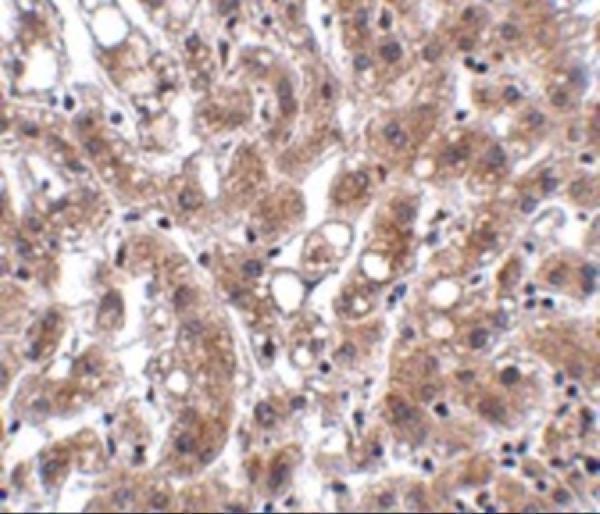
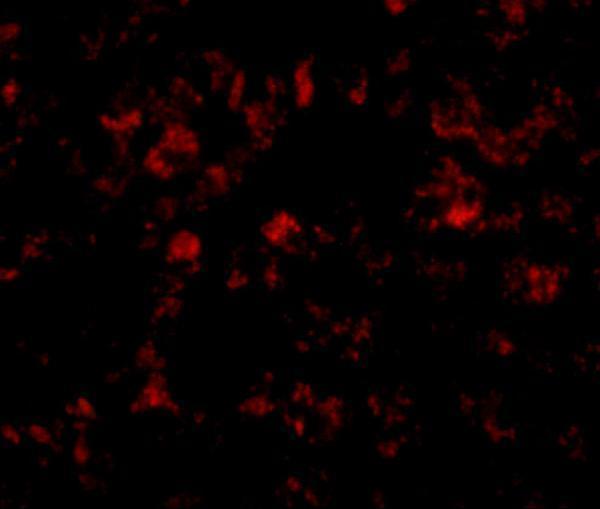
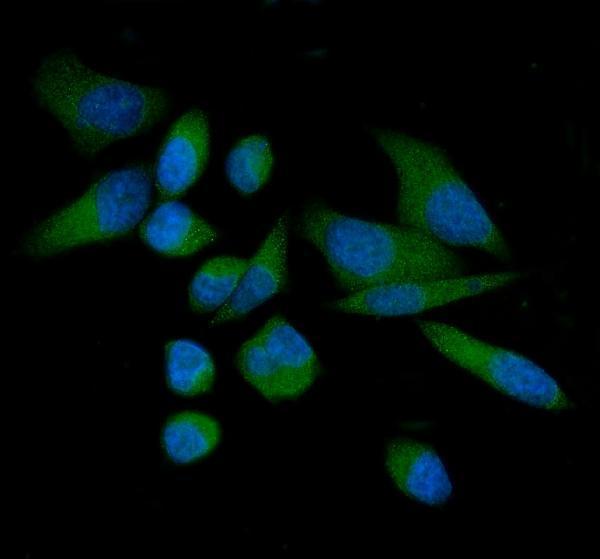
Anti-THEM4 antibodies have IHC images of human liver tissue and an IF/ICC image of PC-3 cells; A07814 also lists mouse and rat reactivity (catalog image captions; catalog reactivity).

Real IHC data Immunohistochemistry of THEM4 in human liver tissue with THEM4 antibody at 2.5 μg/mL.
Anti-THEM4 Antibody
Cat # A07814
Real IF data IF analysis of THEM4 using anti-THEM4 antibody (A07814-1). THEM4 was detected in an immunocytochemical section of PC-3 cells. Enzyme antigen retrieval was performed using IHC enzyme antigen retrieval reagent (AR0022) for 15 mins. The cells were blocked with 10% goat serum. And then incubated with 5 μg/mL rabbit anti-THEM4 Antibody (A07814-1) overnight at 4°C. DyLight®488 Conjugated Goat Anti-Rabbit IgG (BA1127) was used as secondary antibody at 1:100 dilution and incubated for 30 minutes at 37°C. The section was counterstained with DAPI. Visualize using a fluorescence microscope and filter sets appropriate for the label used.
Anti-THEM4 Antibody ®
Cat # A07814-1

A07814 lists IHC-P and IF for human, mouse and rat, with IHC and IF images of human liver tissue (catalog applications/reactivity; A07814 image captions). A07814-1 lists human IF/ICC, with an IF image of PC-3 cells (catalog applications/reactivity; A07814-1 IF caption).

Which to pick: For paraffin-section IHC, choose A07814: it lists IHC-P, and its own IHC image shows human liver tissue at 2.5 μg/mL; the caption does not report the fixative (catalog applications; A07814 IHC caption). For human-cell IF/ICC, choose A07814-1: it lists both applications, and its own IF image shows PC-3 cells at 5 μg/mL (catalog applications; A07814-1 IF caption). For mouse or rat work, A07814 lists reactivity with both species; clonality is unreported for either antibody (catalog reactivity; catalog clone fields).

Each figure is that product's own IHC / IF validation image from its datasheet.

References

  1. UniProt Consortium. UniProt entry Q5T1C6 (THEM4_HUMAN, Acyl-coenzyme A thioesterase THEM4).
  2. Human Protein Atlas. THEM4 tissue IHC expression (reliability: Approved).
  3. Human Protein Atlas. THEM4 subcellular location (ICC-IF): Highest expression in SK-MEL-30: 34.7 nTPM.
  4. Human Protein Atlas. THEM4 antibody validation summary (1 antibodies).
  5. A mitochondria-related gene-based signature predicts pancreatic ductal adenocarcinoma clinical outcome and revealed CAMK2A/THEM4 regulates progression phenotypes and mitophagy in vivo and in vitro. Journal of translational medicine 2025 — PMC12798084.
  6. Exosomal miR-183-5p Shuttled by M2 Polarized Tumor-Associated Macrophage Promotes the Development of Colon Cancer via Targeting THEM4 Mediated PI3K/AKT and NF-κB Pathways. Frontiers in oncology 2021 — PMC8267908.
  7. PubMed PMID:11598301 — UniProt-cited evidence.
  8. PubMed PMID:14702039 — UniProt-cited evidence.
  9. PubMed PMID:16710414 — UniProt-cited evidence.