THRB / Thyroid hormone receptor beta · IHC design guide

Design Immunohistochemistry for THRB

Plan THRB staining in paraffin sections around its nuclear tissue pattern (HPA tissue IHC). Use late spermatids in testis as an observed positive population, and interpret cytoplasmic staining cautiously (HPA tissue IHC).

Evidence assembled Oct 2026 · For research use; verify linked source records and product datasheet before use
Immunohistochemistry protocol sheet for THRB (IHC for THRB): expected localisation Nuclear in a few tissues, including CNS (HPA tissue IHC), antibody A01181-1, validated IHC image, and IHC protocol steps
Printable THRB IHC protocol sheet — expected localisation Nuclear in a few tissues, including CNS (HPA tissue IHC), antibody A01181-1, controls and protocol steps. Open the full THRB IHC guide →

THRB Immunohistochemistry Experimental Design Guide

Expected localisation, validated protocols, controls and antibodies — the at-a-glance facts below, then the full design guide.

Must know before staining
Expected localisation Nuclear in a few tissues, including CNS (HPA tissue IHC)
Staining pattern Nuclear staining in few tissues; late spermatids stain in testis (HPA tissue IHC)
Antigen retrieval Tris-EDTA pH 9.0 HIER, 95–98 °C, 20 min (rule: nuclear antigen)
Positive control ⓘ Testis
Negative control ⓘ Bone marrow+4 more · see all
Important caveats
Reasons your staining may differ from the expected pattern.
Fixation Keep fixation consistent across sections (standard IHC practice; not target-specific)
Caveat Cytoplasmic staining may be off target (HPA tissue IHC)
Regulation T3 regulates activity; intensity unknown (UniProt)
Isoform / epitope Two isoforms, β1 and β2; epitope effects unknown (UniProt)
Section 1

Recommended THRB IHC & IF Protocols

Start with the catalog antibody’s IHC-P protocol (datasheet), then compare the three published THRB staining workflows below (PMC10881509; PMC8703850; PMC11765354).

Recommended immunohistochemistry (IHC-P) protocol parameters
SampleParaffin-embedded human lung carcinoma tissue; fixative not specified (datasheet A01181-1)
FixationImage fixative and duration unreported (datasheet A01181-1); verify before use.
Sectioning4–5 µm sections on charged slides (standard)
DeparaffinisationXylene, graded ethanol series to water (standard)
Antigen retrievalHeat-induced epitope retrieval in Tris-EDTA buffer, pH 9.0, 20 min at 95–98 °C (standard rule: nuclear antigen)
Peroxidase block3% H2O2, 10 min, room temperature (standard)
Blocking10% normal serum of the secondary host, 30 min, room temperature (standard)
Primary antibodyRabbit anti-THRB, 1:50-1:200 (datasheet A01181-1)
Primary incubationOvernight at 4 °C (standard)
DetectionHRP-polymer secondary, DAB chromogen 5–10 min (standard)
CounterstainHematoxylin, blue, dehydrate and mount (standard)
Expected resultTHRB-positive staining in elongated or late spermatids of testis (HPA tissue IHC: Medium). HPA tissue profile: Nuclear expression in few tissues, including CNS. No signal in the no-primary control.
💡Decision noteTry Tris-EDTA pH 9.0 retrieval at 95–98 °C for 20 min first (page retrieval; UniProt P10828: nucleus).
Section 2

What Is the Expected THRB Staining Pattern?

THRB is a nuclear receptor with no transmembrane segment (UniProt P10828). In paraffin sections, expect cell-specific nuclear staining: HPA reports medium staining in elongated or late spermatids in testis and low staining in several other cell populations (HPA tissue IHC). HPA rates its tissue IHC data Approved, while reporting low agreement with RNA data and describing observed cytoplasmic staining as off target (HPA tissue IHC).

What am I looking at on my slide?
Nuclear signal in elongated or late spermatids, with moderate chromogenic intensity.This matches the clearest listed positive population: HPA reports medium staining in these testis cells (HPA tissue IHC). Assess the signal within the identified cells; staining elsewhere in the section does not establish the same pattern.
Predominantly cytoplasmic staining, with little or no convincing nuclear signal.This conflicts with THRB's nuclear location (UniProt P10828). HPA specifically viewed cytoplasmic tissue staining as off target (HPA tissue IHC). Reassess antibody specificity and detection background before counting these cells as positive.
Strong, widespread staining in a population reported as negative, such as bone-marrow hematopoietic cells.That distribution conflicts with the reported lack of staining in that population (HPA tissue IHC). Consider cross-reactivity or endogenous detection activity. The HPA result is a comparison point, not proof that every individual cell must be negative.
Diffuse color across tissue, including areas without a clear cellular or nuclear pattern.A diffuse deposit cannot be assigned confidently to THRB-positive nuclei. Review reagent background, blocking, washes and detection controls as general IHC practice. HPA's reported nuclear pattern and off-target cytoplasmic staining warrant a conservative interpretation (HPA tissue IHC).
No nuclear signal in a section containing identifiable elongated or late spermatids.The expected medium signal is absent from HPA's listed positive population (HPA tissue IHC). First check that the relevant cells are present and preserved; then review section processing, antigen retrieval, antibody incubation and detection as general IHC practice.
💡Expected THRB appearanceCall a result positive when elongated or late spermatids show medium nuclear staining (HPA tissue IHC); predominant cytoplasmic staining is a false-positive warning (HPA tissue IHC).
How each factor affects the staining
Cell and compartment selectionScore identified nuclei in the relevant cell population: HPA reports medium staining in elongated or late spermatids, low staining in selected other populations, and nuclear expression in few tissues including CNS (HPA tissue IHC).
Tissue IHC evidence qualityThe tissue profile is Approved, yet HPA reports low consistency between staining and RNA expression and calls cytoplasmic staining off target (HPA tissue IHC). Treat unexpected positives cautiously rather than broadening the expected pattern.
Antibody-specific validationCAB002008 and CAB002009 have Approved IHC status; HPA061035 has Supported ICC status with no listed IHC status (HPA antibodies). A result from one assay or antibody does not automatically validate another.
Isoforms and receptor locationUniProt lists 2 isoforms, beta-1 and beta-2, and places THRB in the nucleus without a transmembrane segment (UniProt P10828). The supplied sources do not map an antibody epitope or establish isoform-specific staining.
IF localisationHPA reports a supported nuclear-body location by ICC-IF (HPA subcellular). This informs an IF localisation check; it does not establish an IHC-P protocol, a chromogenic puncta requirement, or agreement between IF and tissue IHC.
Why is my staining missing, weak or wrong?
SituationLikely causeNext action
Testis positive population shows no detectable nuclear signal.The relevant cells may be absent from the examined area, or a general IHC processing or detection step may have failed.Locate elongated or late spermatids first (HPA tissue IHC). Then review section processing, retrieval, primary-antibody incubation and detection using suitable controls (general IHC practice); no THRB-specific retrieval condition is supplied.
Chromogen is mainly cytoplasmic.HPA viewed cytoplasmic tissue staining as off target (HPA tissue IHC); reagent background is another general IHC possibility.Do not score cytoplasm as the expected THRB pattern (UniProt P10828; HPA tissue IHC). Compare nuclear signal with an appropriate negative detection control and reassess antibody specificity (general IHC practice).
A reported negative population stains strongly throughout.Cross-reactivity or endogenous detection activity may contribute (general IHC practice). HPA reports bone-marrow hematopoietic cells as not detected (HPA tissue IHC).Confirm the cell identity and compartment, then inspect negative detection controls and endogenous-activity blocking appropriate to the detection chemistry (general IHC practice). Interpret the HPA comparison at the stated cell-population level.
Weak nuclear staining appears outside the main testis positive population.Low staining is reported in several populations, including thyroid glandular and hippocampal glial cells (HPA tissue IHC). A weak signal alone cannot establish specificity.Compare the exact cell type and nuclear location with the HPA profile, and assess controls and repeatability (general IHC practice). Avoid classifying every weakly stained tissue as either confirmed positive or failed staining.
Diffuse color obscures nuclear scoring.Nonspecific reagent binding, incomplete washing or detection background can produce diffuse deposits (general IHC practice).Review blocking, wash steps, detection controls and counterstain so individual nuclei can be assessed (general IHC practice). Require a cell-specific nuclear pattern before comparing the slide with HPA tissue IHC.
What should IF/ICC show?HPA reports supported localisation to nuclear bodies in ICC-IF (HPA subcellular), while its tissue IHC summary describes nuclear expression and flags cytoplasmic staining as off target (HPA tissue IHC).On the separate IF/ICC guide page, assess nuclear-body localisation against an appropriate nuclear counterstain and controls (HPA subcellular; general IF practice). Do not use that IF observation as an IHC-P staining protocol.

Sample controls for THRB IHC & IF

🧪Run testis first: elongated or late spermatids should stain (HPA: Medium in elongated or late spermatids). Use bone marrow hematopoietic cells as the negative tissue (HPA: Not detected); other cells on the testis slide should remain near background, although HPA does not identify a specific internal negative cell type.
Positive control tissue: Testis (Elongated or late spermatids, HPA Medium)
Negative control tissue: Bone marrow (HPA Not detected)
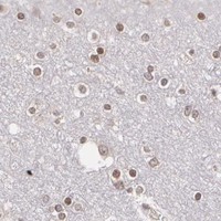
ICC-IF cell lines (HPA subcellular resource): HPA ICC-IF images show THRB in RT-4, SiHa, U2OS, with annotated localisation: Nuclear bodies (supported) (HPA subcellular).
Technical controls: Include a no-primary (secondary-only) control, nonimmune IgG matched to the primary antibody’s host species and class (and isotype for a monoclonal), and THRB-knockout material or an immunizing-peptide block if available (standard IHC controls). Quench endogenous peroxidase and assess residual background in the testis section with the no-primary control (standard chromogenic IHC practice).
⚠️Feasibility: A target-specific fixation window or fixation effect is unreported, and the fixative is unreported in the selected A01181-1 paraffin-section caption (caption: paraffin-embedded human lung carcinoma at 1:100). Retrieval dependence is unreported; compare retrieval conditions while checking the expected nuclear signal (UniProt P10828: Nucleus). The supplied evidence does not establish that frozen sections or IF are easier; crowded late spermatids may complicate nuclear scoring (HPA: Medium in elongated or late spermatids; UniProt P10828: Nucleus).

HPA tissue IHC evidence for THRB

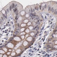
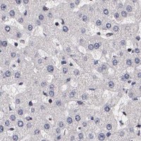
Comprehensive Human Protein Atlas IHC scoring per tissue (reliability: Approved — Low consistency between antibody staining and RNA expression data. Cytoplasmic staining was observed and viewed as off target.). Rows are taken directly from the HPA tissue chart — click any row's HPA link to view the source.

Positive expression · recommended positive controls

TissueCell typeLevelEvidenceSource
Testis Elongated or late spermatids Medium Protein (IHC) HPA →

Undetected expression · recommended negative controls

TissueCell typeLevelEvidenceSource
Bone marrow Hematopoietic cells Not detected Protein (IHC) HPA →
Cerebellum Cells in granular layer Not detected Protein (IHC) HPA →
Cervix Glandular cells Not detected Protein (IHC) HPA →
Endometrium Cells in endometrial stroma Not detected Protein (IHC) HPA →
Epididymis Glandular cells Not detected Protein (IHC) HPA →
Section 3

Advanced THRB IHC Tips

For THRB chromogenic IHC, assess nuclear staining against the expected cell pattern and treat cytoplasmic signal cautiously (UniProt P10828; HPA tissue IHC).

What retrieval conditions should I use if nuclear THRB staining is weak?
Start with Tris-EDTA pH 9.0 heat-induced epitope retrieval at 95–98 °C for 20 min (page retrieval rule: nuclear antigen). Cool sections consistently before antibody incubation, then compare a positive control and a no-primary control in the same run (standard IHC practice). If staining remains weak, optimize antibody concentration and detection before trying a different retrieval buffer or pH (standard IHC practice). Score nuclear signal separately from cytoplasmic color because THRB is nuclear and cytoplasmic HPA staining was viewed as off target (UniProt P10828 subcellular location; HPA tissue IHC). Record the final retrieval conditions with each scored slide (standard IHC practice).
How should I troubleshoot THRB staining when fixation history varies?
THRB-specific sensitivity to fixation is unknown from the supplied evidence; the catalog image identifies paraffin-embedded human lung carcinoma tissue but does not state its fixative (selected IHC caption, A01181-1). Document the fixative and processing history for each specimen, and compare sections processed together when assessing a staining difference (standard IHC practice). Keep section thickness, retrieval, antibody incubation, and chromogen development consistent during that comparison (standard IHC practice). Include a known staining control and a no-primary control in each run to distinguish assay variation from specimen variation (standard IHC practice). Do not assign a fixation cause from cytoplasmic color alone, which HPA viewed as off target (HPA tissue IHC).
Should diffuse cytoplasmic or punctate nuclear color count as THRB?
Prioritize signal within identifiable nuclei: UniProt places THRB in the nucleus, while HPA subcellular imaging supports localization to nuclear bodies (UniProt P10828 subcellular location; HPA subcellular). Punctate nuclear staining is therefore plausible, but chromogenic IHC alone may not resolve individual nuclear bodies reliably (HPA subcellular; standard IHC practice). Exclude diffuse cytoplasmic color from the primary THRB score because HPA observed cytoplasmic staining and viewed it as off target (HPA tissue IHC). Check the hematoxylin counterstain and focus through the section before assigning color to a nucleus (standard IHC practice). Compare the compartment pattern with no-primary and tissue controls processed in the same run (standard IHC practice).
Can this IHC stain distinguish THRB beta-1 from beta-2?
Do not call an isoform from this stain without an epitope map and isoform-specific validation for the antibody (UniProt P10828 isoforms; standard IHC validation practice). The record lists 2 isoforms, beta-1 and beta-2, but supplies no antibody epitope or differential recognition data (UniProt P10828 isoforms; supplied antibody evidence). THRB has no transmembrane segment, signal peptide, or annotated glycosylation sites in this record; those features do not establish which isoform an antibody detects (UniProt P10828 topology and processing). Its annotated nuclear receptor ligand-binding domain spans residues 217–461, but staining cannot identify an epitope within that domain (UniProt P10828 domains; standard IHC interpretation). Report the result as THRB immunoreactivity unless specificity evidence supports a narrower label (standard IHC validation practice).
How should I check THRB localisation in a multiplex IF follow-up?
For a separate IF experiment, pair THRB with a marker that identifies the expected cell population, such as elongated or late spermatids when examining testis (HPA tissue IHC; standard IF practice). Choose spectrally separated fluorophores and favor a far-red channel for a weak target when tissue autofluorescence is prominent; include single-channel and no-primary controls (standard IF practice). THRB is nuclear and has no transmembrane segment, so use controlled permeabilisation that permits antibody access to nuclear epitopes, then check that nuclear morphology remains intact (UniProt P10828 topology and subcellular location; standard IF practice). Compare any nuclear-body pattern with HPA's supported IF localization while checking for channel bleed-through and cytoplasmic background (HPA subcellular; standard IF practice).
What explains widespread brown staining outside THRB-positive nuclei?
First separate nuclear staining from cytoplasmic or stromal color, because THRB is nuclear and HPA viewed cytoplasmic staining as off target (UniProt P10828 subcellular location; HPA tissue IHC). Run no-primary and detection-only controls to identify background from the detection system, and inspect whether color appears around tissue folds or section edges (standard IHC practice). Confirm that endogenous peroxidase was blocked before chromogenic detection, then shorten development if DAB obscures nuclear boundaries (standard chromogenic IHC practice). Titrate the primary antibody against an appropriate control; the catalog's 1:100 lung carcinoma image documents that image, not a universal optimum (selected IHC caption, A01181-1; standard IHC practice).
How should I quantify THRB across sections with different cell compositions? ⚠ ANSWER MARKED FOR VERIFICATION
Define the eligible cell population before scoring and count only staining assigned to nuclei within that population (UniProt P10828 subcellular location; standard IHC scoring practice). Report the percentage of positive eligible nuclei and, when intensity is reproducible, an H-score from 0–300 using intensity grades 0–3 (standard IHC scoring practice). Normalize counts to the number of eligible nuclei, or report positive nuclei per mm² of evaluated tissue when area is the planned denominator (standard IHC scoring practice). Record excluded necrosis, folds, and edge regions, and apply the same threshold across sections stained together (standard IHC scoring practice). Keep cytoplasmic color out of the THRB nuclear score (HPA tissue IHC; UniProt P10828 subcellular location).
When is a THRB-positive result credible rather than artefactual?
A credible result has reproducible nuclear signal in identifiable cells, with expected controls behaving appropriately; THRB is nuclear and HPA reports nuclear expression in a few tissues (UniProt P10828 subcellular location; HPA tissue IHC; standard IHC practice). HPA reports medium staining in elongated or late spermatids in testis, while several listed cell populations are not detected, so interpret staining in its specified cell context (HPA tissue IHC). Review cytoplasmic color skeptically because HPA viewed it as off target (HPA tissue IHC). Exclude staining confined to section edges, necrosis, or areas showing endogenous enzyme signal in controls (standard chromogenic IHC practice). The catalog's paraffin-section lung carcinoma image at 1:100 supports that image condition alone (selected IHC caption, A01181-1).
Boster reagents

Best THRB / Thyroid hormone receptor beta IHC Antibodies

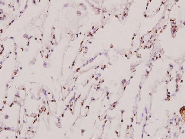
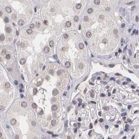
A01181-1 has real IHC data from paraffin-embedded human lung carcinoma tissue (IHC image caption). Its listed reactivity covers human, mouse, and rat (catalog: reactivity).

Real IHC data Immunohistochemistry (IHC) analyzes of THRB (L37) pAb in paraffin-embedded human lung carcinoma tissue at 1:100.
Anti-THRB (L37) Antibody
Cat # A01181-1

A01181-1 is listed for IHC and WB in human, mouse, and rat (catalog: applications and reactivity). Its IHC figure shows paraffin-embedded human lung carcinoma tissue at 1:100 (IHC image caption).

Which to pick: For tissue IHC, choose A01181-1: it is a rabbit polyclonal antibody listed for IHC, with an image from a human paraffin section (catalog: host and applications; IHC image caption: pAb, paraffin-embedded human lung carcinoma). For IF/ICC, there is no documented option here (catalog: A01181-1 applications and IF image alts). For mouse or rat IHC, A01181-1 lists reactivity with both species, but its IHC image documents only human tissue; the fixative is unreported (catalog: reactivity; IHC image caption).

Each figure is that product's own IHC / IF validation image from its datasheet.

References

  1. UniProt Consortium. UniProt entry P10828 (THB_HUMAN, Thyroid hormone receptor beta).
  2. Human Protein Atlas. THRB tissue IHC expression (reliability: Approved).
  3. Human Protein Atlas. THRB subcellular location (ICC-IF): Localized to the nuclear bodies..
  4. Human Protein Atlas. THRB antibody validation summary (3 antibodies).
  5. Development of a necroptosis-related prognostic model for uterine corpus endometrial carcinoma. Scientific reports 2024 — PMC10881509.
  6. Prognostic Relevance of Thyroid-Hormone-Associated Proteins in Adenoid Cystic Carcinoma of the Head and Neck. Journal of personalized medicine 2021 — PMC8703850.
  7. Canagliflozin alleviates progestin resistance by suppressing RARβ/CRABP2 signaling in THRB knockout endometrial cancer cells. Frontiers in pharmacology 2025 — PMC12075957.
  8. The Prognostic Significance of TRs in Hepatocellular Carcinoma: Insights from TCGA and GEO Databases. Biomarker insights 2025 — PMC11765354.
  9. PubMed PMID:3034496 — UniProt-cited evidence.
  10. PubMed PMID:2879243 — UniProt-cited evidence.
  11. PubMed PMID:1973914 — UniProt-cited evidence.