This website uses cookies to ensure you get the best experience on our website.


- Table of Contents
Plan paraffin-section TMEM175 IHC using caudate glia or testis Sertoli cells as high-staining references (HPA tissue IHC). Assess the reported nuclear and nuclear-rim pattern in light of TMEM175’s annotated endosomal and lysosomal membrane location (HPA tissue IHC; UniProt).

Expected localisation, validated protocols, controls and antibodies — the at-a-glance facts below, then the full design guide.
| Expected localisation | Nuclear/rim in tissue (HPA tissue IHC); endolysosomal membrane (UniProt) | |
| Staining pattern | General nuclear and nuclear rim staining (HPA tissue IHC) | |
| Antigen retrieval | EDTA pH 8.0 HIER, heat-mediated (datasheet A11712-1) | |
| Positive control | Caudate+4 more · see all | |
| Negative control | Adipose tissue+4 more · see all |
| Fixation | Keep fixation consistent across sections. (standard IHC practice; not target-specific) | |
| Caveat | Nuclear tissue staining differs from the annotated membrane location (HPA tissue IHC; UniProt) | |
| Regulation | Staining-linked expression regulation is unreported (UniProt) | |
| Isoform / epitope | 2 isoforms; map the epitope to lumenal or cytoplasmic regions (UniProt) |
The catalog antibody protocol is paired with one published chromogenic IHC method for TMEM175 (PMC13018187).
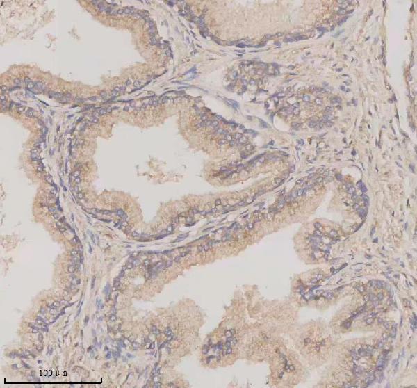
| Sample | Paraffin-embedded human prostate cancer tissue; fixative not specified (datasheet A11712-1) |
| Fixation | Image fixative and duration unreported (datasheet A11712-1); verify before use. |
| Sectioning | 4–5 µm sections on charged slides (standard) |
| Deparaffinisation | Xylene, graded ethanol series to water (standard) |
| Antigen retrieval | Heat retrieval: EDTA pH 8.0 (datasheet A11712-1); 20 min, 95–100 °C (standard) |
| Peroxidase block | 3% H2O2, 10 min, room temperature (standard) |
| Blocking | 10% goat serum (datasheet A11712-1) |
| Primary antibody | Rabbit anti-TMEM175, 2-5 μg/ml (datasheet A11712-1) |
| Primary incubation | Overnight at 4 °C (datasheet A11712-1) |
| Detection | HRP-conjugated secondary, DAB chromogen (datasheet A11712-1) |
| Counterstain | Hematoxylin, blue, dehydrate and mount (standard) |
| Expected result | TMEM175-positive staining in glial cells of caudate (HPA tissue IHC: High). HPA tissue profile: General nuclear and nuclear membranous expression. No signal in the no-primary control. |
TMEM175 is a 12-pass endosome/lysosome membrane protein (UniProt Q9BSA9 topology and subcellular location). In paraffin IHC, HPA reports general nuclear and nuclear-membranous staining, with high signal in caudate glial cells and testicular Sertoli cells (HPA tissue IHC). HPA rates the tissue IHC pattern Approved, with medium consistency against RNA data and external verification pending (HPA tissue IHC). Interpret the observed nuclear pattern in light of its difference from the UniProt location.
| Prominent nuclear and nuclear-rim staining in caudate glial cells or testicular Sertoli cells (HPA tissue IHC). | This matches HPA's reported compartment and High staining in those cells (HPA tissue IHC). Record the cell type, intensity and compartment together. The pattern is an IHC observation; UniProt places TMEM175 on endosome and lysosome membranes (UniProt Q9BSA9). |
| Only coarse cytoplasmic deposits, diffuse cytosolic fill or extracellular chromogen in a reference-positive region. | These appearances do not match HPA's reported nuclear and nuclear-membranous IHC profile (HPA tissue IHC). Endolysosomal puncta remain biologically plausible from the UniProt location, so an alternate compartment alone does not prove artefact (UniProt Q9BSA9). Review morphology and detection controls before scoring (standard IHC practice). |
| Strong staining in adipocytes or colon glandular cells. | HPA reports TMEM175 as Not detected in those specific cell populations (HPA tissue IHC). Consider cross-reactivity or endogenous detection activity, especially if the product also appears in a no-primary control (standard IHC practice). Avoid treating every cell in the tissue as an established negative. |
| Widespread, similarly intense staining across cells and tissue structures, with little compartment detail. | A uniform haze is difficult to reconcile with HPA's cell-specific staining levels and nuclear profile (HPA tissue IHC). It may reflect nonspecific antibody binding or detection background; compare the no-primary control and assess whether the stain follows intact cell boundaries (standard IHC practice). |
| No signal in caudate glial cells or testicular Sertoli cells. | Those are HPA's High reference populations (HPA tissue IHC). A blank result there does not establish biological absence. Check the tissue section, detection controls and assay conditions before interpreting a negative result (standard IHC practice); HPA's Approved rating still awaits external verification (HPA tissue IHC). |
| Reference pattern and validation | HPA reports a nuclear and nuclear-membranous tissue IHC pattern and rates antibody HPA057160 Approved for IHC; its ICC rating is Uncertain (HPA tissue IHC; HPA antibodies). Approved here carries medium RNA–staining consistency and pending external verification, so the pattern is a reference for comparison rather than independent proof of molecular location (HPA tissue IHC). |
| Membrane topology and unknown epitope | TMEM175 has 12 transmembrane segments and alternating cytoplasmic and lumenal regions (UniProt Q9BSA9 topology). The supplied evidence does not map the antibody epitope, so topology cannot identify which side the antibody recognizes or predict a TMEM175-specific retrieval condition. |
| Expression breadth and cell-level controls | UniProt describes TMEM175 as widely expressed, and HPA assigns low tissue RNA specificity (UniProt Q9BSA9; HPA tissue IHC). Those broad descriptions do not override HPA's observed differences between named cell populations: select comparison regions by the reported cell type, not tissue name alone (HPA tissue IHC). |
| Antigen retrieval and assay conditions | Antigen retrieval can alter visibility in paraffin IHC, so document and optimize it with appropriate controls (standard IHC practice). Neither HPA tissue staining nor UniProt topology establishes a TMEM175-specific retrieval method or fixation sensitivity; do not infer either from a positive or negative tissue result. |
| Situation | Likely cause | Next action |
|---|---|---|
| The reference-positive caudate or testis section is blank. | HPA reports High staining in caudate glial cells and testicular Sertoli cells (HPA tissue IHC); a blank assay may reflect tissue selection or a failed detection step (standard IHC practice). | Confirm that the relevant cells are present, then check control reactions and review retrieval, primary-antibody dilution and detection conditions as general IHC variables (standard IHC practice). |
| Chromogen forms a diffuse haze over most of the section. | Uniform background obscures the cell and compartment pattern reported by HPA (HPA tissue IHC). Nonspecific binding or detection background are general IHC possibilities (standard IHC practice). | Compare a no-primary control, check blocking and washing, and adjust primary-antibody or detection conditions if controls indicate background (standard IHC practice). |
| Adipocytes or colon glandular cells stain strongly. | HPA reports those cell populations as Not detected (HPA tissue IHC). Cross-reactivity or endogenous detection activity may account for an unexpected chromogenic result (standard IHC practice). | Check the no-primary control and inspect cell identity and compartment; repeat comparison against an HPA-positive cell population before assigning the signal to TMEM175 (HPA tissue IHC; standard IHC practice). |
| Staining appears only as cytoplasmic puncta, with no HPA-like nuclear pattern. | Puncta differ from HPA's tissue IHC profile but could fit the endosome/lysosome location in UniProt (HPA tissue IHC; UniProt Q9BSA9). The supplied evidence cannot resolve that discrepancy. | Document both observations, inspect assay controls and avoid calling puncta either confirmed TMEM175 or definite artefact from location alone (standard IHC practice). |
| Adjacent sections show inconsistent staining intensity. | HPA already notes medium consistency between tissue staining and RNA data (HPA tissue IHC). Section composition or variable assay conditions can also change apparent intensity (standard IHC practice). | Compare the same named cell population across sections and review section quality, retrieval, incubation and detection records before making a biological interpretation (standard IHC practice). |
| Q: Does a nuclear-rim IF/ICC image validate the paraffin IHC result? | HPA lists nucleoplasm and nuclear membrane as approved ICC-IF locations, but rates antibody HPA057160's ICC validation Uncertain (HPA subcellular; HPA antibodies). | A: Treat the IF/ICC image as a separate localisation observation. Compare the paraffin result with HPA's tissue IHC cell types and its Approved IHC pattern; neither readout resolves the conflict with UniProt's endosome/lysosome annotation (HPA tissue IHC; UniProt Q9BSA9). |


Comprehensive Human Protein Atlas IHC scoring per tissue (reliability: Approved — Medium consistency between antibody staining and RNA expression data. Pending external verification.). Rows are taken directly from the HPA tissue chart — click any row's HPA link to view the source.
| Tissue | Cell type | Level | Evidence | Source |
|---|---|---|---|---|
| Caudate | Glial cells | High | Protein (IHC) | HPA → |
| Testis | Sertoli cells | High | Protein (IHC) | HPA → |
| Adrenal gland | Glandular cells | Medium | Protein (IHC) | HPA → |
| Breast | Glandular cells | Medium | Protein (IHC) | HPA → |
| Bronchus | Respiratory epithelial cells | Medium | Protein (IHC) | HPA → |
| Tissue | Cell type | Level | Evidence | Source |
|---|---|---|---|---|
| Adipose tissue | Adipocytes | Not detected | Protein (IHC) | HPA → |
| Appendix | Glandular cells | Not detected | Protein (IHC) | HPA → |
| Colon | Glandular cells | Not detected | Protein (IHC) | HPA → |
| Duodenum | Glandular cells | Not detected | Protein (IHC) | HPA → |
| Oral mucosa | Squamous epithelial cells | Not detected | Protein (IHC) | HPA → |
Troubleshoot TMEM175 staining in paraffin sections using the catalog IHC conditions, cellular controls, and the reported compartment patterns.
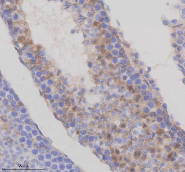
The IHC-validated anti-TMEM175 antibody A11712-1 has paraffin-section images from human prostate cancer tissue and mouse and rat testis (catalog IHC captions).
A11712-1 is the only SKU that will render and lists IHC reactivity for human, mouse, and rat (catalog applications/reactivity). Its IHC captions document staining in paraffin sections of human prostate cancer tissue, mouse testis, and rat testis (catalog IHC captions).
Which to pick: Choose A11712-1 for paraffin-section IHC; its captions document EDTA pH 8.0 retrieval, 2 μg/ml primary antibody, and DAB detection, but do not report the fixative (catalog IHC captions). The same SKU covers human, mouse, and rat IHC (catalog applications/reactivity). No listed SKU has IF/ICC validation or an IF image, so this catalog does not support an IF/ICC recommendation (catalog applications and IF image records).