This website uses cookies to ensure you get the best experience on our website.


- Table of Contents
Plan TMEM87A staining in paraffin sections using the granular cytoplasmic tissue pattern reported by HPA (HPA tissue IHC). This guide covers cell selection, antibody use and interpretation alongside its annotated Golgi membrane location (UniProt).

Expected localisation, validated protocols, controls and antibodies — the at-a-glance facts below, then the full design guide.
| Expected localisation | Granular cytoplasm (HPA tissue IHC); Golgi membrane (UniProt) | |
| Staining pattern | Granular cytoplasm in glandular, neuronal and glial cells (HPA tissue IHC) | |
| Antigen retrieval | EDTA pH 8.0 HIER, heat-mediated (datasheet A14716-1) | |
| Positive control | Adrenal gland+4 more · see all | |
| Negative control | Adipose tissue+1 more · see all |
| Fixation | Keep fixation consistent across sections. (standard IHC practice; not target-specific) | |
| Caveat | Brain signal depends on the cell population sampled (HPA tissue IHC) | |
| Regulation | Expression regulation unreported (UniProt) | |
| Isoform / epitope | 3 isoforms; check lumenal versus cytoplasmic epitope coverage (UniProt) |
The catalog antibody’s IHC-P protocol (datasheet: A14716-1) is accompanied by a published TMEM87A IHC method for human lung carcinoma tissue microarrays (PMC13221295).
| Sample | Paraffin-embedded human colon adenocarcinoma tissue; fixative not specified (datasheet A14716-1) |
| Fixation | Image fixative and duration unreported (datasheet A14716-1); verify before use. |
| Sectioning | 4–5 µm sections on charged slides (standard) |
| Deparaffinisation | Xylene, graded ethanol series to water (standard) |
| Antigen retrieval | Heat retrieval: EDTA pH 8.0 (datasheet A14716-1); 20 min, 95–100 °C (standard) |
| Peroxidase block | 3% H2O2, 10 min, room temperature (standard) |
| Blocking | 10% goat serum (datasheet A14716-1) |
| Primary antibody | Rabbit anti-TMEM87A, 1-2 μg/ml (datasheet A14716-1) |
| Primary incubation | Overnight at 4 °C (datasheet A14716-1) |
| Detection | HRP-conjugated secondary, DAB chromogen (datasheet A14716-1) |
| Counterstain | Hematoxylin, blue, dehydrate and mount (standard) |
| Expected result | TMEM87A-positive staining in glandular cells of adrenal gland (HPA tissue IHC: High). HPA tissue profile: General cytoplasmic expression with a granular pattern. No signal in the no-primary control. |
TMEM87A should appear mainly as granular cytoplasmic staining in positive cells on paraffin sections (HPA: tissue IHC profile). This fits its supported Golgi localization in ICC-IF and its annotation as a seven-transmembrane protein of the Golgi and cell membranes (HPA: subcellular; UniProt Q8NBN3 topology and location). HPA rates the tissue IHC profile Enhanced, citing agreement with RNA expression and similar staining by paired antibodies (HPA: tissue IHC reliability).
| Granular cytoplasmic staining in adrenal, appendix, colon or duodenal glandular cells (HPA: High in these cells). | This matches the reported tissue pattern and offers several positive-reference choices (HPA: tissue IHC). Golgi localization supports a cytoplasmic distribution, but a chromogenic section alone does not identify each granule as Golgi (HPA: subcellular; general IHC interpretation). |
| Predominantly nuclear staining, or a uniform membrane outline with little granular cytoplasm. | This differs from the reported general granular cytoplasmic IHC profile (HPA: tissue IHC). UniProt also places TMEM87A at the cell membrane, so some membrane-associated signal is plausible; an isolated membrane or nuclear pattern needs antibody and control review before assignment to TMEM87A (UniProt Q8NBN3 location; general IHC interpretation). |
| Strong staining in adipocytes or chondrocytes despite weak staining in the selected positive-reference cells. | HPA reports TMEM87A as Not detected in adipocytes and chondrocytes (HPA: tissue IHC). Treat a strong signal there as discordant with that reference; check cross-reactivity and endogenous detection activity with appropriate controls (general IHC practice). |
| Diffuse color across tissue, including spaces or cell types without a convincing granular pattern. | This is difficult to assign to the reported TMEM87A pattern (HPA: tissue IHC). Background from the detection system or nonspecific binding can obscure cellular scoring; compare the section with reagent controls and the positive-reference tissue (general IHC practice). |
| No staining in a well-preserved section containing an HPA High cell population. | An absent signal in, for example, colon glandular cells warrants a technical check before calling an experimental sample negative (HPA: High in colon glandular cells; general IHC practice). Review antibody application, detection and counterstain, then compare with a run control; the supplied sources give no TMEM87A-specific fixation sensitivity. |
| Tissue and cell choice (HPA: tissue IHC) | HPA reports High staining in adrenal, appendix, colon, duodenal and epididymal glandular cells, cortical neurons, and caudate and hippocampal glia. It reports Low staining in several other listed cell populations and Not detected staining in adipocytes and chondrocytes; score the specified cell type rather than treating an entire tissue as uniformly positive or negative (HPA: tissue IHC). |
| Localization and topology (UniProt Q8NBN3; HPA: subcellular) | UniProt annotates seven transmembrane segments, a lumenal region at residues 22–225 and a cytoplasmic tail at 459–555; HPA supports mainly Golgi localization by ICC-IF. The recognized epitope is unspecified, so topology cannot predict this antibody's staining strength or whether a retrieval step is needed (UniProt Q8NBN3 topology; HPA: subcellular). |
| Isoforms and processing (UniProt Q8NBN3) | UniProt lists three isoforms, a signal peptide at residues 1–21 and a chain at 22–555. Without the antibody's epitope and isoform coverage, do not infer that every isoform will stain equally or that loss of the signal peptide explains a negative section (UniProt Q8NBN3 processing and isoforms). |
| Glycosylation (UniProt Q8NBN3) | Four glycosylation sites are annotated at residues 79, 127, 157 and 160 (UniProt Q8NBN3 glycosylation). Their existence does not establish an effect on this antibody's binding, retrieval response or tissue intensity; no such target-specific effect is supplied. |
| IF/ICC question: should fluorescence be Golgi-enriched? (HPA: subcellular) | Yes: HPA reports TMEM87A mainly at the Golgi apparatus in ICC-IF and lists A-431, U-251MG and U2OS among imaged cell lines (HPA: subcellular). That supports a Golgi-enriched expectation for IF/ICC, while the tissue IHC observations above remain the reference for scoring paraffin sections (HPA: tissue IHC). |
| Situation | Likely cause | Next action |
|---|---|---|
| The positive-reference glandular cells have no signal. | The section may have a detection or antibody-application problem; HPA reports High staining in these selected cells (HPA: tissue IHC; general IHC practice). | Inspect morphology and the run's positive control, then check reagent order, antibody application and detection development before interpreting experimental negatives (general IHC practice). |
| Only weak staining appears in lung alveolar type I cells or liver cholangiocytes. | These cells are reported as Low, so weak staining alone is not discordant (HPA: tissue IHC). | Compare the run with a listed High cell population and score intensity within the named cell type, using consistent viewing conditions (HPA: tissue IHC; general IHC practice). |
| Strong color appears in adipocytes or chondrocytes. | This conflicts with their Not detected HPA observations and may reflect nonspecific binding or detection activity (HPA: tissue IHC; general IHC practice). | Compare serial or matched sections using an appropriate reagent control; review whether color follows cells, tissue edges or deposits before assigning it to TMEM87A (general IHC practice). |
| Granular signal is hidden by diffuse background. | Nonspecific antibody binding or the detection system may reduce contrast (general IHC practice). | Review blocking, washes and chromogen development against the run controls; retain the reported granular cytoplasmic pattern as the interpretive reference (general IHC practice; HPA: tissue IHC). |
| Staining is predominantly nuclear or exclusively outlines the plasma membrane. | That pattern does not match HPA's general granular cytoplasmic IHC profile, although UniProt also annotates cell-membrane localization (HPA: tissue IHC; UniProt Q8NBN3 location). | Check the positive-reference tissue and reagent controls; record any membrane signal separately from the granular cytoplasmic pattern rather than assuming that compartment alone validates the section (general IHC practice). |
| Two antibodies give different tissue patterns. | HPA's Enhanced rating reflects agreement between paired antibodies and RNA expression for its assessed profile; it does not guarantee agreement in every new run (HPA: tissue IHC reliability). | Compare the same named cell populations and controls across matched sections, and investigate discordant compartments or background before choosing a pattern for interpretation (HPA: tissue IHC; general IHC practice). |


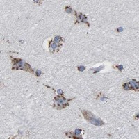


Comprehensive Human Protein Atlas IHC scoring per tissue (reliability: Enhanced — High consistency between antibody staining and RNA expression data. Paired antibodies with high similarity supports the protein expression profile.). Rows are taken directly from the HPA tissue chart — click any row's HPA link to view the source.
| Tissue | Cell type | Level | Evidence | Source |
|---|---|---|---|---|
| Adrenal gland | Glandular cells | High | Protein (IHC) | HPA → |
| Appendix | Glandular cells | High | Protein (IHC) | HPA → |
| Caudate | Glial cells | High | Protein (IHC) | HPA → |
| Cerebral cortex | Neuronal cells | High | Protein (IHC) | HPA → |
| Colon | Glandular cells | High | Protein (IHC) | HPA → |
Troubleshoot chromogenic TMEM87A IHC in paraffin sections using the catalog antibody’s tissue example, protein topology, and reported staining patterns.
A14716-1 has IHC images from paraffin sections of human colon adenocarcinoma, human liver cancer, mouse brain and rat brain, plus an IF/ICC image from U87 cells (catalog image captions).
A14716-1 has IHC images from paraffin sections of human colon adenocarcinoma and liver cancer, and mouse and rat brain (catalog IHC captions). A14716-1 also has an IF/ICC image from U87 cells (catalog IF caption).
Which to pick: Choose A14716-1 for tissue IHC because its images document paraffin sections with EDTA retrieval at pH 8.0 and 2 μg/ml primary antibody (catalog IHC captions); the fixative is unreported (catalog IHC captions). Choose A14716-1 for IF/ICC in U87 cells because its IF image documents staining at 5 μg/ml (catalog IF caption). For cross-species IHC, A14716-1 lists human, mouse and rat reactivity and has paraffin-section images for all three (catalog applications/reactivity; catalog IHC captions).