This website uses cookies to ensure you get the best experience on our website.


- Table of Contents
Plan TNIK immunohistochemistry in paraffin sections using tissue staining and catalog antibody evidence. The guide covers controls, detection and interpretation of the predominantly cytoplasmic tissue pattern (HPA tissue IHC).

Expected localisation, validated protocols, controls and antibodies — the at-a-glance facts below, then the full design guide.
| Expected localisation | Cytoplasmic staining in most tissues (HPA tissue IHC) | |
| Staining pattern | Strong cytoplasmic staining in colon glandular cells (HPA tissue IHC) | |
| Antigen retrieval | EDTA pH 8.0 HIER, heat-mediated (datasheet A03934-3) | |
| Positive control | Appendix+4 more · see all | |
| Negative control | Adipose tissue+4 more · see all |
| Fixation | Keep paraffin-section fixation consistent across specimens. (selected-SKU IHC image A03934-3) | |
| Caveat | Staining shows low consistency with RNA expression (HPA tissue IHC) | |
| Regulation | Intensity varies by tissue (HPA tissue IHC) | |
| Isoform / epitope | 8 isoforms; verify epitope coverage (UniProt) |
The catalog antibody uses EDTA pH 8.0 heat retrieval (datasheet A03934-3). Three published TNIK IHC protocols cover gastric, thyroid and colorectal specimens (PMC5424097; PMC10272969; PMC4619995).
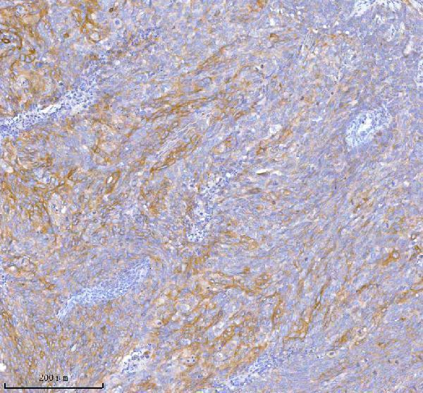
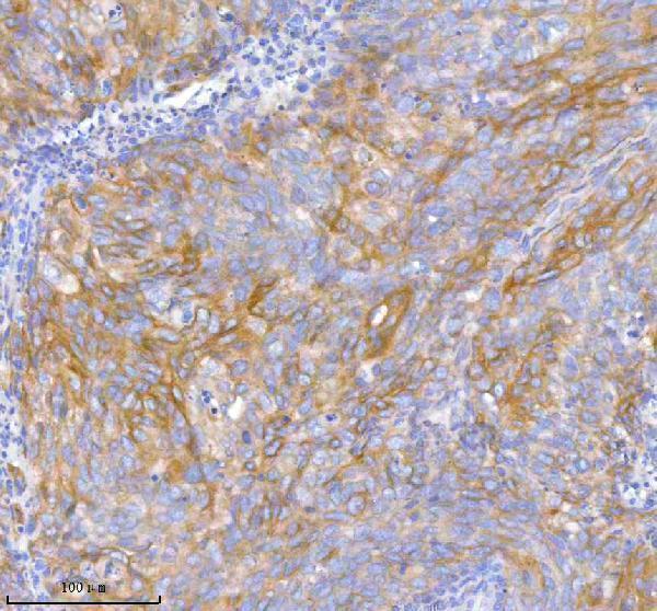
| Sample | Paraffin-embedded human cervix squamous cell carcinoma tissue; fixative not specified (datasheet A03934-3) |
| Fixation | Image fixative and duration unreported (datasheet A03934-3); verify before use. |
| Sectioning | 4–5 µm sections on charged slides (standard) |
| Deparaffinisation | Xylene, graded ethanol series to water (standard) |
| Antigen retrieval | Heat retrieval: EDTA pH 8.0 (datasheet A03934-3); 20 min, 95–100 °C (standard) |
| Peroxidase block | 3% H2O2, 10 min, room temperature (standard) |
| Blocking | 10% goat serum (datasheet A03934-3) |
| Primary antibody | Rabbit anti-TNIK, 2-5 μg/ml (datasheet A03934-3) |
| Primary incubation | Overnight at 4 °C (datasheet A03934-3) |
| Detection | HRP-conjugated secondary, DAB chromogen (datasheet A03934-3) |
| Counterstain | Hematoxylin, blue, dehydrate and mount (standard) |
| Expected result | TNIK-positive staining in glandular cells of appendix (HPA tissue IHC: High). HPA tissue profile: Cytoplasmic expression in most tissues. No signal in the no-primary control. |
TNIK is a non-transmembrane protein found in the cytoplasm and nucleus (UniProt Q9UKE5 topology; UniProt Q9UKE5 subcellular location). In paraffin-section IHC, expect mainly cytoplasmic staining in glandular cells, especially in appendix, colon, duodenum, gallbladder, rectum and small intestine (HPA: tissue IHC). HPA rates its IHC evidence Approved but reports low consistency between antibody staining and RNA expression; interpret unexpected patterns cautiously (HPA: tissue IHC reliability).
| Strong cytoplasmic staining in intestinal or gallbladder glandular cells. | This matches the reported high glandular-cell staining in colon, duodenum, rectum, small intestine and gallbladder (HPA: tissue IHC). Judge the result by cell type and compartment as well as intensity; a dark chromogen deposit alone does not establish TNIK localization (standard IHC interpretation). |
| Predominantly extracellular deposits or sharply outlined cell membranes, with little intracellular signal. | Treat this as a pattern mismatch: TNIK has no transmembrane segment, and HPA describes mainly cytoplasmic tissue staining (UniProt Q9UKE5 topology; HPA: tissue IHC). Check the matched negative control and staining distribution before attributing the deposits to TNIK (standard IHC practice). |
| Strong staining in a cell population reported as undetected, such as adipocytes or skin fibroblasts. | This is discordant with those HPA observations (HPA: adipocytes and skin fibroblasts not detected). Consider nonspecific antibody binding or endogenous detection activity, then inspect controls. HPA's low antibody-staining/RNA consistency means a single discordant specimen is insufficient to prove cross-reactivity (HPA: tissue IHC reliability). |
| Pale, widespread chromogen obscures cell boundaries and appears in several compartments. | Diffuse background makes compartment and cell-type scoring unreliable (standard IHC interpretation). Compare a section processed without primary antibody, review blocking and washing, and optimize antibody concentration and detection conditions as general IHC workflow checks (standard IHC practice). |
| No convincing signal in appendix or colon glandular cells. | HPA reports high staining in these glandular cells, so this is a useful sensitivity check (HPA: tissue IHC). Confirm that the expected cells are present and that detection and counterstaining worked; then review the IHC-validated antibody's instructions. Tissue-level expectations do not guarantee every specimen will stain (HPA: low antibody-staining/RNA consistency). |
| Tissue and cell selection | HPA reports high glandular-cell staining in appendix, colon, duodenum, gallbladder, rectum and small intestine, but no detected staining in adipocytes or skin fibroblasts (HPA: tissue IHC). Select and score the named cell population within each section; an unlabeled whole-tissue average can hide a cell-specific pattern (standard IHC practice). |
| Subcellular interpretation | HPA describes mainly cytoplasmic staining across tissues, while UniProt places TNIK in the nucleus, cytoplasm and recycling endosome; its endosome and cytoskeletal association is reported upon RAP2A overexpression (HPA: tissue IHC; UniProt Q9UKE5 subcellular location). Nuclear staining can be biologically plausible, but nuclear-only tissue IHC needs control review before acceptance. |
| Topology and processing | UniProt lists no transmembrane segment, signal peptide or propeptide and records a chain spanning residues 1–1360 (UniProt Q9UKE5 topology and processing). This supports an intracellular interpretation; it does not identify the catalog antibody's epitope or establish how paraffin processing affects that epitope. |
| Isoforms and antibody evidence | UniProt lists eight TNIK isoforms (UniProt Q9UKE5 isoforms). Their presence alone cannot predict which forms the catalog antibody detects because its epitope is unspecified. HPA lists HPA012128 and HPA012297 as IHC Approved; that status should be read alongside HPA's stated low antibody-staining/RNA consistency (HPA: antibodies; HPA: tissue IHC reliability). |
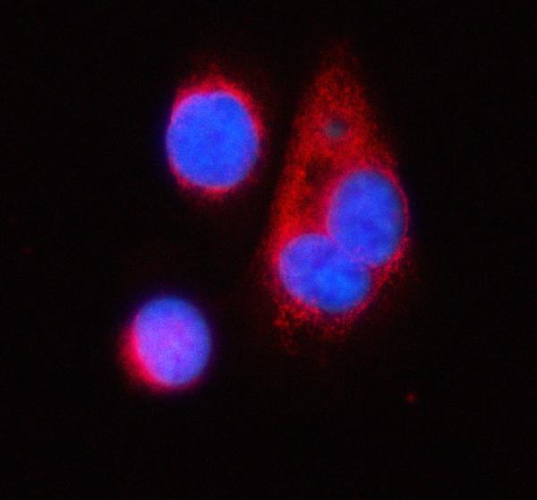
| IF/ICC Q&A: where should signal appear? | For the separate IF/ICC application, HPA reports supported nucleoplasmic localization with additional supported cytosolic localization (HPA: subcellular ICC-IF). The IF/ICC distribution can therefore differ from HPA's mainly cytoplasmic tissue IHC profile; use the application-specific guide to interpret IF/ICC images (HPA: tissue IHC; HPA: subcellular ICC-IF). |
| Situation | Likely cause | Next action |
|---|---|---|
| Expected glandular cells are unstained. | The section may lack the intended cells, or an IHC workflow step may have failed; HPA reports high staining in several glandular-cell populations (HPA: tissue IHC; standard IHC practice). | Verify glandular morphology, counterstain and a positive-control section; check the IHC-validated antibody's instructions and detection reagents before interpreting the sample as negative (standard IHC practice). |
| Only nuclei stain in tissue IHC. | UniProt permits nuclear TNIK, and ICC-IF supports nucleoplasmic localization, but HPA describes mainly cytoplasmic tissue staining (UniProt Q9UKE5 subcellular location; HPA: subcellular ICC-IF; HPA: tissue IHC). | Record the nuclear pattern as discordant with the usual tissue profile; inspect a matched control and repeat on an HPA high-staining glandular tissue before assigning specificity (standard IHC practice). |
| Signal outlines membranes or pools outside cells. | That distribution conflicts with TNIK's lack of a transmembrane segment and HPA's mainly cytoplasmic IHC profile (UniProt Q9UKE5 topology; HPA: tissue IHC). | Check the primary-antibody omission control, tissue morphology and chromogen deposits; optimize washing or detection if the control also develops color (standard IHC practice). |
| Diffuse staining prevents compartment scoring. | Broad background can arise from nonspecific binding or excess detection signal (standard IHC practice). | Compare controls, then adjust blocking, washing, antibody concentration or detection exposure one variable at a time; score only when cell boundaries and compartment can be distinguished (standard IHC practice). |
| A reported undetected cell population stains strongly. | HPA reports no detection in adipocytes and skin fibroblasts, but also warns of low consistency between antibody staining and RNA expression (HPA: tissue IHC). Endogenous activity or nonspecific binding remains possible (standard IHC practice). | Confirm cell identity, inspect the omission control and compare an HPA high-staining glandular section processed alongside it; describe unresolved staining as discordant rather than specific TNIK signal (HPA: tissue IHC; standard IHC practice). |
| Staining varies across specimens or regions. | HPA reports cytoplasmic expression in most tissues with cell-dependent intensity, and its IHC reliability note limits confidence in strict RNA concordance (HPA: tissue IHC). | Score the same cell population and compartment across sections, document intensity and distribution, and include a shared reference section in the run to separate sample variation from workflow variation (standard IHC practice). |


Comprehensive Human Protein Atlas IHC scoring per tissue (reliability: Approved — Low consistency between antibody staining and RNA expression data.). Rows are taken directly from the HPA tissue chart — click any row's HPA link to view the source.
| Tissue | Cell type | Level | Evidence | Source |
|---|---|---|---|---|
| Appendix | Glandular cells | High | Protein (IHC) | HPA → |
| Colon | Glandular cells | High | Protein (IHC) | HPA → |
| Duodenum | Glandular cells | High | Protein (IHC) | HPA → |
| Gallbladder | Glandular cells | High | Protein (IHC) | HPA → |
| Rectum | Glandular cells | High | Protein (IHC) | HPA → |
| Tissue | Cell type | Level | Evidence | Source |
|---|---|---|---|---|
| Adipose tissue | Adipocytes | Not detected | Protein (IHC) | HPA → |
| Cervix | Glandular cells | Not detected | Protein (IHC) | HPA → |
| Esophagus | Squamous epithelial cells | Not detected | Protein (IHC) | HPA → |
| Ovary | Ovarian stroma cells | Not detected | Protein (IHC) | HPA → |
| Skin | Fibroblasts | Not detected | Protein (IHC) | HPA → |
Troubleshooting TNIK staining in paraffin sections requires attention to retrieval, compartment, cell identity, and the limits of antibody validation.
A03934-3 has IHC data from human paraffin sections of cervix squamous cell carcinoma and placenta, plus IF data from PC-3 cells (catalog image captions). Human and mouse reactivity are listed (catalog: reactivity).
A03934-3 will render with its IHC figure from a human paraffin-embedded cervix squamous cell carcinoma section (A03934-3 IHC caption). The same SKU lists IHC and ICC/IF applications, with additional IHC data from human placenta and IF data from PC-3 cells (catalog: applications; A03934-3 image captions).
Which to pick: For tissue IHC, choose A03934-3: its IHC caption documents a paraffin section, EDTA retrieval at pH 8.0, and primary antibody at 2 μg/ml; the fixative is unreported (A03934-3 IHC caption). For IF/ICC, choose A03934-3 because its IF caption documents staining in PC-3 cells at 5 μg/ml (A03934-3 IF caption). For cross-species planning, A03934-3 lists human and mouse reactivity, although the supplied IHC tissue examples are human; its host is rabbit and clonality is unreported (catalog: reactivity and host; A03934-3 IHC captions; catalog: clone).