This website uses cookies to ensure you get the best experience on our website.


- Table of Contents
Plan chromogenic IHC on paraffin sections using heart muscle as a positive tissue control: cardiomyocytes show high cytoplasmic staining (HPA tissue IHC). Testis pachytene spermatocytes also show medium staining, so interpret staining outside the heart in context (HPA tissue IHC).

Expected localisation, validated protocols, controls and antibodies — the at-a-glance facts below, then the full design guide.
| Expected localisation | Cytoplasm of cardiac myocytes (HPA tissue IHC) | |
| Staining pattern | Cytoplasmic staining in cardiac myocytes (HPA tissue IHC) | |
| Antigen retrieval | Citrate pH 6.0 HIER, 95–98 °C, 20 min (rule: cytoplasmic / membrane antigen) | |
| Positive control | Heart muscle+1 more · see all | |
| Negative control | Adipose tissue+4 more · see all |
| Fixation | Keep paraffin-section fixation consistent across samples. (standard IHC practice; not target-specific) | |
| Caveat | Pachytene spermatocytes also show medium staining (HPA tissue IHC) | |
| Regulation | Heart muscle enriched at RNA level (HPA tissue RNA) | |
| Isoform / epitope | 0 isoforms annotated; mature chain spans aa 2–210 (UniProt) |
The catalog antibody’s IHC-P protocol is paired with published protocols for mouse heart sections (PMC3376223) and mouse lung sections (PMC5592927).
| Sample | Tissue sections; selected-image fixative not specified (standard IHC workflow) |
| Fixation | Image fixative and duration unreported (datasheet M01720-3); verify before use. |
| Sectioning | 4–5 µm sections on charged slides (standard) |
| Deparaffinisation | Xylene, graded ethanol series to water (standard) |
| Antigen retrieval | Heat-induced epitope retrieval in citrate buffer, pH 6.0, 20 min at 95–98 °C (standard rule: cytoplasmic / membrane antigen) |
| Peroxidase block | 3% H2O2, 10 min, room temperature (standard) |
| Blocking | 10% normal serum of the secondary host, 30 min, room temperature (standard) |
| Primary antibody | Rabbit anti-TNNI3, 1:200-1:1000 (datasheet M01720-3) |
| Primary incubation | Overnight at 4 °C (standard) |
| Detection | HRP-polymer secondary, DAB chromogen 5–10 min (standard) |
| Counterstain | Hematoxylin, blue, dehydrate and mount (standard) |
| Expected result | TNNI3-positive staining in cardiomyocytes of heart muscle (HPA tissue IHC: High). HPA tissue profile: Cytoplasmic expression in cardiac myocytes. No signal in the no-primary control. |
In paraffin-section IHC, expect TNNI3 staining in the cytoplasm of cardiomyocytes, with strong signal in heart muscle (HPA: cytoplasmic expression in cardiac myocytes; High in cardiomyocytes). HPA rates the tissue pattern Enhanced, citing high consistency between antibody staining and RNA expression (HPA: tissue IHC reliability). UniProt describes TNNI3 as a thin-filament regulatory protein without a transmembrane segment, but provides no subcellular localization annotation (UniProt P19429: function, topology, subcellular record).
| Strong cytoplasmic staining in cardiomyocytes, concentrated in the expected heart-muscle cells. | This matches the reported IHC pattern and is the clearest positive readout (HPA: cytoplasmic expression in cardiac myocytes; High in cardiomyocytes). Assess the stained cells and compartment together; color intensity alone does not establish that a result is specific (standard IHC interpretation). |
| Predominantly nuclear or cell-surface staining in cardiomyocytes, with little cytoplasmic signal. | Treat this as a compartment mismatch that warrants review for artefact or nonspecific detection (HPA: cytoplasmic cardiac-myocyte IHC; UniProt P19429: no transmembrane segment). UniProt does not supply a subcellular annotation, so its topology cannot independently define the microscopic pattern (UniProt P19429: subcellular record, topology). |
| Strong staining in cells expected to be negative, such as adipocytes in adipose tissue. | Consider antibody cross-reactivity or endogenous chromogenic detection activity (HPA: TNNI3 not detected in adipose-tissue adipocytes; standard IHC practice). A negative-tissue comparison and appropriate detection control can help distinguish these possibilities; an unexpected stain is not proof of either cause (standard IHC practice). |
| Diffuse color across tissue, extracellular spaces, or multiple unrelated cell types. | This obscures the cell-restricted pattern reported in heart muscle (HPA: High in cardiomyocytes). Uneven blocking, residual detection background, or overly concentrated reagents are general IHC possibilities; evaluate them with controls before assigning the color to TNNI3 (standard IHC practice). |
| No convincing cytoplasmic staining in a heart-muscle section that should be positive. | Target-specific fixation effects are not established by the supplied assay evidence. Verify with a matched IHC source before attributing a result to fixation. |
| Which tissue and cells anchor the IHC call? | Heart-muscle cardiomyocytes are the strongest supplied reference (HPA: High). Testis pachytene spermatocytes have Medium staining, so positivity outside heart muscle is not automatically false; judge each cell type against its own HPA observation (HPA: Medium in testis pachytene spermatocytes). Several other listed tissues are not detected, including adipose tissue and adrenal gland (HPA: tissue IHC). |
| How much confidence does the antibody evidence provide? | The reported tissue pattern has Enhanced reliability because antibody staining is highly consistent with RNA expression (HPA: tissue IHC reliability). HPA lists Enhanced IHC status for HPA046428 and CAB009349 (HPA: antibody validation). This supports the reference pattern, while an individual section still needs sound controls and cell-level review (standard IHC interpretation). |
| Does topology predict a membrane stain? | UniProt reports no transmembrane segment and identifies TNNI3 as the inhibitory subunit of the thin-filament troponin complex (UniProt P19429: topology, function). Use the observed cytoplasmic cardiomyocyte pattern for the IHC decision (HPA: tissue IHC). Neither statement identifies a target-specific antigen-retrieval condition or fixation response (UniProt P19429; HPA tissue IHC). |
| What should IF/ICC show? | For an IF/ICC image, HPA reports mainly cytosolic localization with additional vesicular localization (HPA: approved cytosol and vesicles in ICC-IF). That observation can inform compartment review, but it comes from ICC-IF rather than paraffin-section chromogenic IHC; use the cardiac-myocyte tissue result as the IHC reference (HPA: subcellular ICC-IF; tissue IHC). |
| Situation | Likely cause | Next action |
|---|---|---|
| Heart-muscle cardiomyocytes show no stain. | The result misses the reported High positive reference; a workflow failure is possible (HPA: High in cardiomyocytes; standard IHC practice). | Target-specific fixation effects are not established by the supplied assay evidence. Verify with a matched IHC source before attributing a result to fixation. |
| The heart section is stained, but nuclei dominate. | The compartment differs from the reported cytoplasmic IHC pattern (HPA: cytoplasmic cardiac-myocyte expression). | Compare cell boundaries and counterstain with the chromogen distribution; review background and antibody specificity using controls before scoring the nuclei as TNNI3-positive (standard IHC practice). |
| Adipocytes or other listed negative cells stain strongly. | This disagrees with the applicable HPA negative-cell observation and may reflect cross-reactivity or endogenous detection activity (HPA: adipocytes not detected; standard IHC practice). | Compare a negative tissue and a no-primary detection control; if background persists, review blocking and chromogenic detection conditions (standard IHC practice). Interpret testis separately because pachytene spermatocytes are Medium (HPA: testis). |
| Color is diffuse across the section. | Broad color makes the expected cardiac-myocyte cytoplasmic pattern difficult to resolve (HPA: tissue IHC); detection background is possible (standard IHC practice). | Inspect the no-primary control and reagent handling, then optimize general blocking, washes, or antibody concentration as indicated by those controls (standard IHC practice). |
| Only a few heart-muscle cells appear positive. | Patchiness may reflect section quality or uneven staining rather than the expected High cardiomyocyte pattern (HPA: High in heart-muscle cardiomyocytes; standard IHC practice). | Check morphology across the section and whether retrieval, antibody coverage, and chromogen development were uniform; compare with a well-stained positive control (standard IHC practice). |
| IF/ICC seems vesicular while the tissue IHC call is cytoplasmic. | HPA reports additional vesicular ICC-IF localization alongside its main cytosolic signal (HPA: subcellular ICC-IF). | Interpret each application against its own evidence: review the IF/ICC compartment against HPA subcellular data and score paraffin-section IHC against cytoplasmic cardiomyocytes (HPA: subcellular ICC-IF; tissue IHC). |
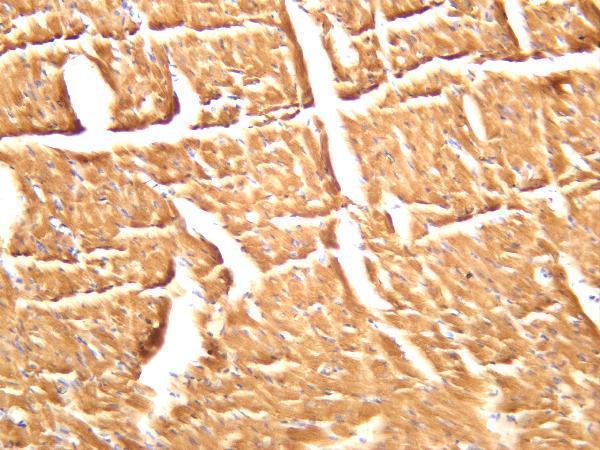
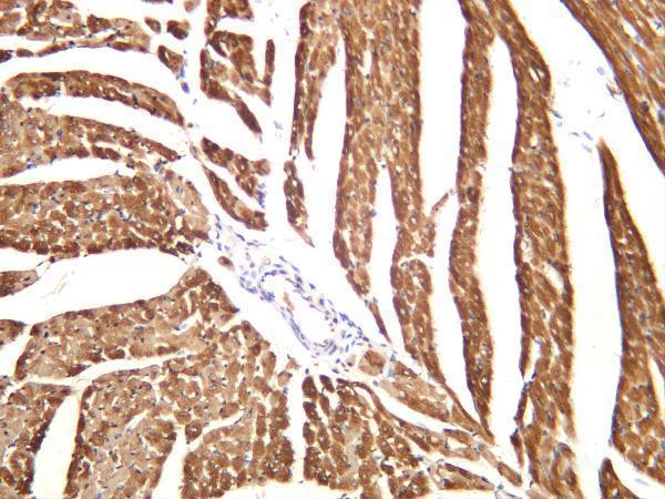
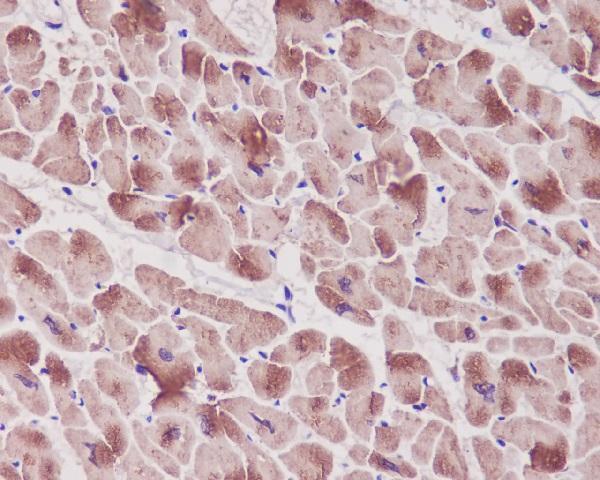


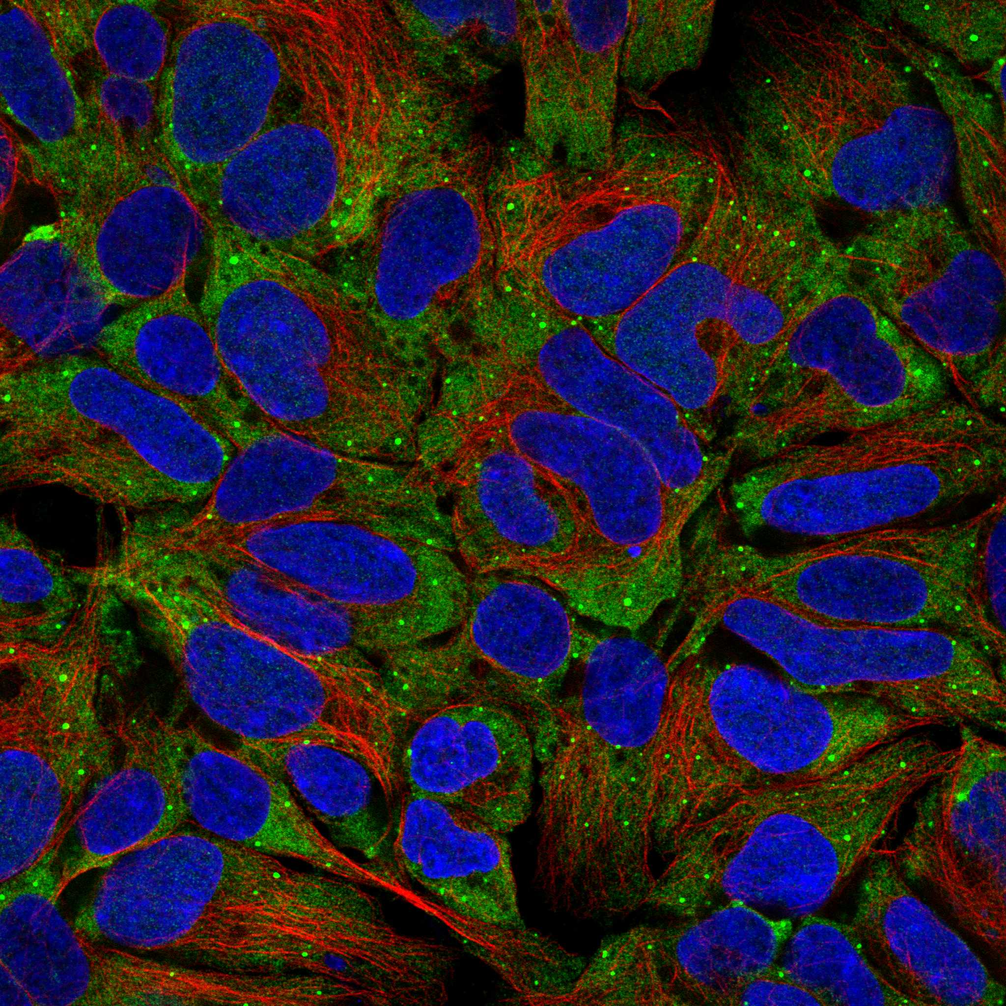
Comprehensive Human Protein Atlas IHC scoring per tissue (reliability: Enhanced — High consistency between antibody staining and RNA expression data.). Rows are taken directly from the HPA tissue chart — click any row's HPA link to view the source.
| Tissue | Cell type | Level | Evidence | Source |
|---|---|---|---|---|
| Heart muscle | Cardiomyocytes | High | Protein (IHC) | HPA → |
| Testis | Pachytene spermatocytes | Medium | Protein (IHC) | HPA → |
| Tissue | Cell type | Level | Evidence | Source |
|---|---|---|---|---|
| Adipose tissue | Adipocytes | Not detected | Protein (IHC) | HPA → |
| Adrenal gland | Glandular cells | Not detected | Protein (IHC) | HPA → |
| Appendix | Glandular cells | Not detected | Protein (IHC) | HPA → |
| Bone marrow | Hematopoietic cells | Not detected | Protein (IHC) | HPA → |
| Breast | Adipocytes | Not detected | Protein (IHC) | HPA → |
Use cardiac myocyte staining as the reference pattern while optimising paraffin-section IHC for TNNI3 (HPA tissue IHC).
Anti-TNNI3 antibodies have IHC images from human heart paraffin sections and mouse and rat cardiac muscle (catalog IHC captions). Both list IF, but neither supplies an IF image (catalog applications and image alts).
M01720 lists human IHC, IF and ICC, with an IHC image of paraffin-embedded human heart (catalog applications, reactivity and IHC caption). M01720-3 lists human, mouse and rat IHC and IF, with IHC images of mouse and rat cardiac muscle (catalog applications, reactivity and IHC captions).
Which to pick: For human paraffin-section IHC, choose monoclonal M01720: its own IHC image shows paraffin-embedded human heart; the fixative is unreported (catalog clone and IHC caption). For IF/ICC, M01720 lists both applications for human samples, while monoclonal M01720-3 lists IF for human, mouse and rat samples (catalog applications, reactivity and antibody descriptions). For cross-species tissue IHC, choose M01720-3: its own IHC images show mouse and rat cardiac muscle, and human reactivity is listed; section processing and fixative are unreported in those captions (catalog reactivity and IHC captions).