This website uses cookies to ensure you get the best experience on our website.
- Table of Contents
1 Citations 1 Q&As
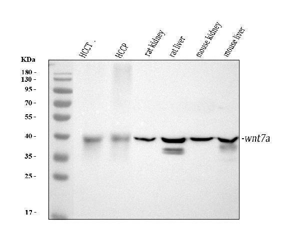
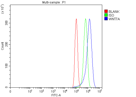
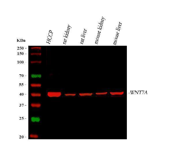
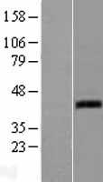
Facts about Protein Wnt-7a.
Required for central nervous system (CNS) angiogenesis and blood-brain barrier regulation (PubMed:30026314). Required for normal, sexually dimorphic development of the Mullerian ducts, and for normal fertility in both sexes (By similarity).
Human | |
---|---|
Gene Name: | WNT7A |
Uniprot: | O00755 |
Entrez: | 7476 |
Belongs to: |
---|
Wnt family |
protein Wnt-7a; proto-oncogene Wnt7a protein; wingless-type MMTV integration site family, member 7A; Wnt7a; Wnt-7a
Mass (kDA):
39.005 kDA
Human | |
---|---|
Location: | 3p25.1 |
Sequence: | 3; NC_000003.12 (13816258..13880071, complement) |
Expression is restricted to placenta, kidney, testis, uterus, fetal lung, and fetal and adult brain.
Secreted, extracellular space, extracellular matrix. Secreted.
Boster Bio Anti-Protein Antibody (WNT7A Antibody) allows you to run qRT-PCR analyses for miRNA, mRNA, circRNA. This article will show you how to use the WNT7A Marker from Boster Bio and the benefits. This method does not apply to scientists in the same country or lab. These methods can be used by all scientists, regardless of where they are located.
The Boster Bio Antitein Wnt-7aWnt7aAntibody monoclonal antibody detects WNT7a proteins. Wnt7a is a member of the Wnt group of secreted cysteinerich glycoproteins. These proteins are important for tumor growth and progression. The b.catenin gene product is an important component of the Wnt/b.catenin Signaling Pathway, which regulates embryonic and patterning development in vertebrates.
Boster Bio Anti-Protein Wnt-7a Antibody detects human Wnt-7a in direct ELISAs. It can be used in a PBS solution 0.2 um filter or lyophilized. The optimal dilutions for specific applications should be determined by the laboratory. The Technical Information section of our website has general protocols.
The Wnt gene family consists of structurally related genes. The proteins encode secreted signaling proteins. Wnt protein play an important role during development in the patterning and differentiation limbs. Fuhrmann syndrome is characterized by abnormal bone and cartilage development due to mutations in the Wnt genes. Wnt7a is responsible for sexually dimorphic development of mullerian ducts.
Wnt3a plays a crucial role in chondrogenesis. It also inhibits Wnt3a-signalling and mesenchymal precessor cell growth. It also inhibits micromass growth in limbbud micromass cultures. This antibody, Wnt3a, has many biological functions. It can be used in the laboratory to test Wnt7a-antibodies.
Tissue samples were fixed using 10% formaldehyde. Then, paraffin was embedded in the tissue. The sections were then cut in four-mm strips and dried in a incubator at 60°C. After drying, the membranes had to be dewaxed with xylene. Then, a graded series was applied of ethanol. Finally, samples of the second antibodies were incubated (1:5000; Boster Bio).
This study utilized has_circ_0068871 cell lines from the EJ (UMUC3) and UMUC3 celllines. RNase R was used at a concentration of 4 U/mg. The relative expression levels in each sample were compared to the standard curve using U6 and B-actin as internal control. For each sample, the qRTPCR was done three times.
Prostate cancer tumors expressed lower levels of cir-ITCH than normal tissue adjacent. These results indicate that circRNAs could be associated with PCa progression. Further studies are needed to examine the roles of circRNAs and their clinical applications. This new method has many potential applications, including identifying the source of prostate cancer and determining its treatment.
The data from qRTPCR for circRNA can be used to identify cancers in different tissues. They are a promising biomarker for tumours due to their long half life and specific expression in cancerous tissues. More research is needed to determine their role as early detection and treatment for cancer. If you are a physician, qRT-PCR for circRNA is an invaluable tool.
SYBR Green BioFACT 2x master mix was used to perform qRT-PCR of circRNA on the QIAGEN Rotor Gene Q real-timePCR system. To evaluate the expression levels for circRNA in the tissues, primers for circCPE (HIAT1) and FOXC1 were used. The Pfaffel method was used in order to calculate the fold changes for each gene. The real time qPCR products were evaluated with agarose gels containing 2.2% TBE buffer.
CircadianRNA, a non-coding class of RNA, is derived mainly from exons. Circadian RNA is unique and extremely diverse, unlike linear RNA. In recent years circRNA has been associated to many physiological functions. They regulate gene expression, splicing and transcription. Furthermore, some studies suggest that circRNA is associated with BCa.
Combining with PSA, circRNAs could be used as biomarkers for PCa. Further research is needed in order to determine their role as biomarkers for the disease. These two genes are thought to play different roles during the development of this disease. This is evident from the higher expression levels of circCDR1AS (in PCa) and circHIAT1 (in PCa). However, this work does not mean that qRT-PCR for circRNA is not a viable diagnostic tool.
The first step of RT-qPCR is to design primers. This can be accomplished for mRNA by designing an amplification prime that crosses the exon-exon boundary. This reduces the chance of false positives because contaminating DNA is not amplified by the amplification. It is important that the primers are specific for the desired gene/transcript.
Reverse transcriptase converts RNA to DNA. RNaseH is an option for some reverse transcriptase proteins, while others are not. Moloney mouse leukemiavirus (MLLV), and Avian myeloblastosisvirus virus (AMV), are two examples. Reverse transcriptase's important property is its high thermal stability. This property ensures the successful transcription of RNA having a secondary structural. A thermally stable enzyme has higher cDNA yields.
To use qRTPCR for the analysis of mRNA, RNA had to be isolated from a variety tissues and cell lines. After that, it was separated on an 12% polyacrylamide gel. Afterward, it was transferred to a Hybond-NX membrane (Amersham Biosciences) and probed with a gATP-labelled probe complementary to the miRNA. The membranes were exposed to a Kodak Phosphor Screen SD230, which were then scanned with a Molecular Imager FX reader.
qRT-PCR for mRNA enables researchers to analyze a range of genes. A single gene may be expressed in multiple tissues, including cancer and aging. qRTPCR can be used to detect the effect on microRNAs. It provides information about a wide range of biological processes, including genetic disorders. It can also identify new candidate miRNAs. If it is a circulating protein, it may affect the growth of cancer cells.
MiR140-5p, for instance, is a gene associated osteoarthritis. qRT-PCR uses RNA extracted from cartilage tissues for its highest quality. These miRNAs have been implicated in osteoarthritis and many other human diseases. Further, qRTPCR for mRNA can identify multiple candidate targets for the disease. For a further study, miR-140-5p and miR-142-5p were identified as candidates.
qRTPCR for miRNA is an effective tool to analyze miRNA expression levels in human body fluids. This method requires only a small amount of total RNA, usually a few picograms. This is a good choice when you need to collect small quantities of forensic samples. MiRNA expression in humans is highly stable, making them a useful biomarker for PMI estimation.
Although miRNA is usually found in cell membranes, some miRNA have been detected in bodily fluids. This has led to increased interest in miRNA as biomarkers. They are highly stable, and they are resistant to endogenous RNAse activity. They are carried in lipoprotein complexes and RNA-binding protein in extracellular vesicles. These circulating miRNAs can be detected by qRTPCR for miRNA, which allows researchers to determine their presence within human blood. Compared to conventional PCR methods, qRT-PCR for miRNA is highly sensitive and reproducible.
The two miRNAs studied in the study had excellent linearity over seven orders of magnitude. Two independent research staffs independently examined the panel containing diluted samples. Their results showed high agreement. The kappa statistic for concordance was 0.89. To normalize the expression of miRNA, qRTPCR using miRNA was done using a U6 standard.
A previous study showed that VEGF could regulate miR-17-5p expression. The three-hour and nine hour post-VEGF treatments showed a positive correlation. In addition, expression of miR-18a and hsa–miR–155 was increased in a time-dependent fashion. These miRNAs were also responsible for regulating the survival rate of tumor cells.
The study also looked at the molecular mechanisms of atrophic bone nonunion. Researchers discovered that osteogenic genes down-regulated and antagonists were associated to nonunion of bones. They also found a correlation between miRNA expression and predicted target genes in hBMSCs transfected by four types of synthetic double-strandedRNAs. These results indicate that up-regulated microRNAs play a key role in atrophic osteogenesis.
After stimulation by VEGF, VEGF regulated MIRNAs are expressed high in humans. Dicer inactivation decreases EC proliferation in vivo, which suggests that Dicer-dependent miRNAs are necessary for the appropriate angiogenic response to VEGF. However, it isn't known if Dicer inactivation alters the expression of these microRNAs.
PMID: 9161407 by Bui T.D., et al. Isolation of a full-length human WNT7A gene implicated in limb development and cell transformation, and mapping to chromosome 3p25.
PMID: 8893824 by Ikegawa S., et al. Isolation, characterization and chromosomal assignment of the human WNT7A gene.
*More publications can be found for each product on its corresponding product page