| SKU | A00318-1 |
|---|---|
| Application | Western Blot |
| Sample | Mouse brain tissue |
| Sample Processing Description | Mouse brain tissue was lysed in RIPA buffer supplemented with protease inhibitors at 4°C for 2 hours. The lysate was centrifuged to collect the supernatant, and protein concentration was determined. After adjusting the concentration, samples were mixed with 5× protein loading buffer and heated at 95–100°C for 10 minutes to denature. Then, 15 μL of each protein sample was loaded per lane for electrophoresis. |
| Other Reagents | Blocking buffer |
| Primary Antibody | PI 3 Kinase p85 alpha/PIK3R1 Antibody Picoband® |
| Primary Incubation | 1:2000, overnight at 4 ℃ |
| Secondary Antibody | HRP Goat Anti-Rabbit IgG |
| Secondary Incubation | 1:10000, 1 hour in room temperature |
| Detection | Substrate: ECL, Imaging system:ChemiDoc MP |
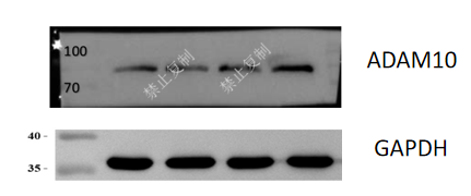
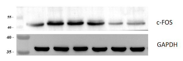
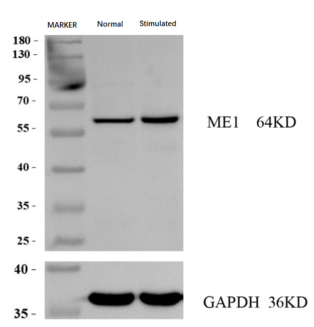
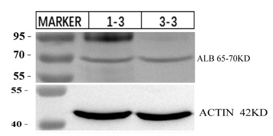
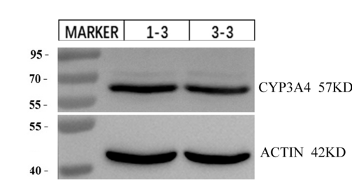
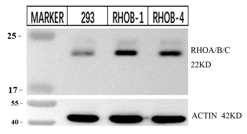
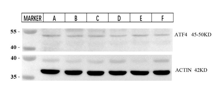
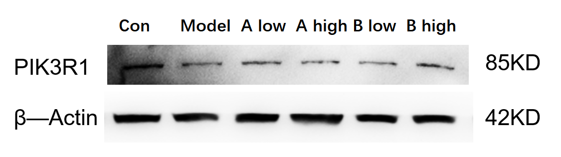
| Results Summary | The figure shows a schematic representation of Western blot results for PIK3R1 and the loading control β-actin in mouse brain tissues from normal mice, model group, and mice treated with high and low doses of drug AB. Although the expression differences between experimental groups are not pronounced, the Western blot results obtained with this antibody are still clear and well-defined. |