| SKU | A00318-1 |
|---|---|
| Application | Western Blot |
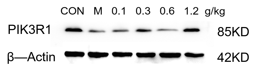
| Sample | Mouse hippocampus tissue |
| Sample Processing Description | The mouse hippocampus was lysed with RIPA lysis buffer containing protease inhibitors, followed by protein quantification. The samples were then mixed with 5× protein loading buffer and heated for 10 minutes for denaturation. Five microliters of protein sample were loaded into each lane for SDS-PAGE. |
| Primary Antibody | Anti-PI 3 Kinase p85 alpha/PIK3R1 Antibody Picoband® |
| Primary Incubation | overnight at 4 ℃ |
| Secondary Antibody | HRP-conjugated Anti-Rabbit IgG Secondary Antibody |
| Secondary Incubation | 1 hour in room temperature |
| Detection | Substrate: Ultra-sensitive ECL luminescent reagent , Imaging system:ChemiDoc MP |
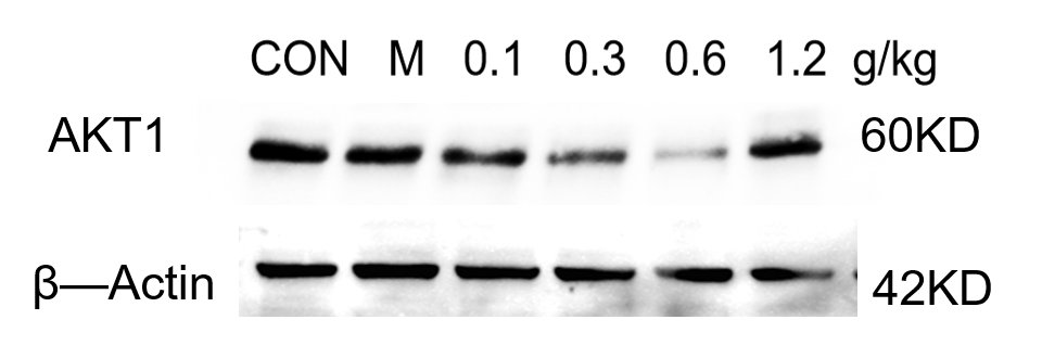
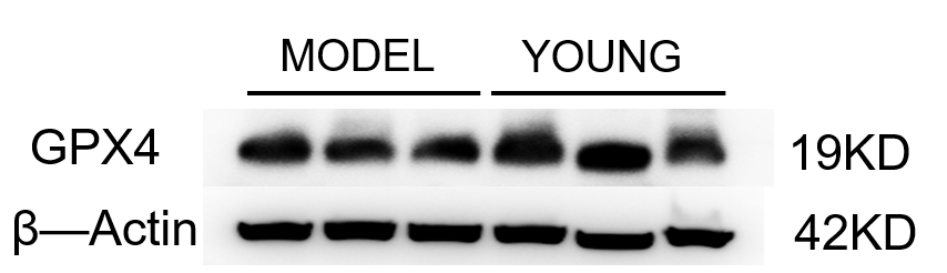
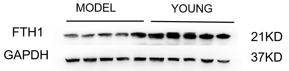
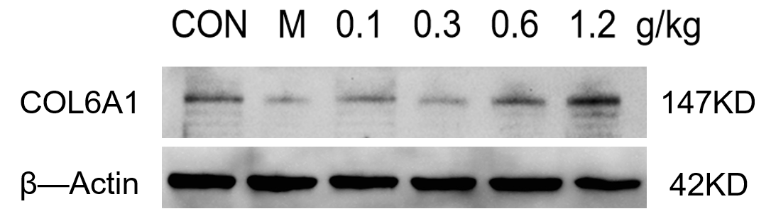
| Results Summary | The antibody shows clear bands with no non-specific signals in WB, and is suitable for detecting PIK3R1 protein in mouse hippocampus by Western blot. |