Customer Testimonials

Peer Reviews from Real Labs

Browse peer-submitted results linked to specific products—ratings, images, and practical notes included. Contribute your own result to strengthen the evidence base and claim rewards after verification.

Video Interviews

See real workflows and decision points—what researchers tried, what worked, and why. Share your story in a short interview and claim rewards!

Get Interview Rewards

Filter by category, reactivity, and host to find the most relevant peer results.

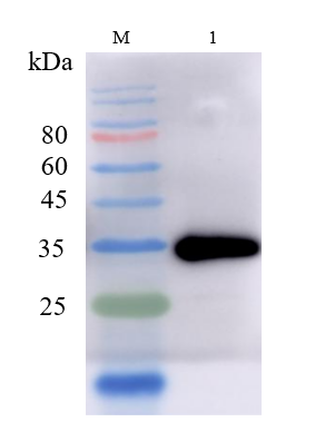
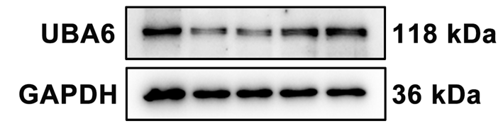
The antibody performs efficiently and specifically, with very few nonspecific bands.

Excellent, submitted by on
WB result image
SKU A03213-2
Application Western Blot
Sample MCF-7 cell
Sample Processing Description Cells were directly lysed in NP-40 buffer, mixed with loading buffer at the appropriate ratio, and denatured by heating at 98 °C. Then, 20 µL of protein sample was loaded per lane onto SDS-PAGE.
Primary Antibody UBA6 Antibody
Primary Incubation 1:1000, overnight at 4 °C
Blocking Agent 5% Non-fat milk
Secondary Antibody HRP-conjugated Goat Anti-Rabbit IgG
Secondary Incubation Incubate at room temperature for 1 hour
Detection Signal was developed using ECL substrate on a Tanon system.
Results Summary The antibody performs efficiently and specifically, with very few nonspecific bands.

This antibody shows excellent consistency and reproducibility across batches, ensuring reliable and stable experimental results. Its performance remains consistent across different experiments, providing strong technical support for our research.

Excellent, submitted by on
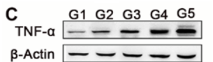
PA1079 wb
SKU PA1079
Application Western Blot
Sample Mouse lung tissue
Sample Processing Description Tissue samples were directly lysed in RIPA buffer, mixed with loading buffer at the appropriate ratio, and denatured by heating at 98 °C. Load 20 µL of protein sample per lane onto SDS-PAGE.
Primary Incubation The membrane was incubated with TNF-α primary antibodies (1:1000) overnight at 4 °C.
Secondary Antibody HRP-conjugated Goat Anti-Rabbit IgG Secondary Antibody
Secondary Incubation Incubate at room temperature for 1 hour
Other Reagents used 5% Non-fat milk
Detection Signal was developed using ECL substrate on an ChemiDoc MP system.
Results Summary This antibody shows excellent consistency and reproducibility across batches, ensuring reliable and stable experimental results. Its performance remains consistent across different experiments, providing strong technical support for our research.

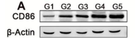
The antibody is highly efficient and specific, showing a clear target band with no nonspecific bands.

Excellent, submitted by on
SKU M00220-1
Application Western Blot
Sample Mouse lung cancer tissue
Sample Processing Description Tissue samples were directly lysed in RIPA buffer, mixed with loading buffer at the appropriate ratio, and denatured by heating at 98 °C. Load 20 µL of protein sample per lane onto SDS-PAGE.
Primary Incubation The membrane was incubated with the CD86 primary antibodies (1:2000) overnight at 4 °C.
Secondary Antibody HRP-conjugated Goat Anti-Rabbit IgG Secondary Antibody
Secondary Incubation Incubate at room temperature for 1 hour
Other Reagents used 5% non-fat milk
Detection Signal was developed using ECL substrate on an Tanon system.
Results Summary The antibody is highly efficient and specific, showing a clear target band with no nonspecific bands. I will definitely continue using BosterBio products and will recommend them to my classmates and colleagues.

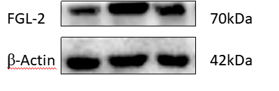
Works very well in Western blot for rat enteric glial cells and mouse FGL-2 protein, with only slight nonspecific bands.

Excellent, submitted by on
SKU A06303-3
Application Western Blot
Sample rat enteric glial cells and mouse tissue
Sample Processing Description Samples (5 µL per lane) were run on 10% resolving and stacking gels at 80 V for 20 min, then 150 V for 70 min, and transferred at 400 mA for 100 min.
Primary Incubation The membrane was incubated with the FGL-2 primary antibody (1:1000) overnight at 4 °C.
Secondary Antibody HRP-conjugated Goat Anti-Rabbit IgG Secondary Antibody
Secondary Incubation Incubate at room temperature for 2 hour
Other Reagents used 5% non-fat milk
Detection Signal was developed using ECL substrate on an ImageQuant LAS4000mini system.
Results Summary Works very well in Western blot for rat enteric glial cells and mouse FGL-2 protein, with only slight nonspecific bands.

The antibody shows high efficiency and specificity in Western blot detection of hemolymph cell proteins from Pacific oyster, with only slight nonspecific bands.

Excellent, submitted by on
SKU PB9148
Application Western Blot
Sample hemolymph of the Pacific oyster
Sample Processing Description Add 10 µL of hemolymph cells to each lane and directly lyse in 1× Laemmli sample buffer. After adding β-mercaptoethanol, load the samples onto SDS-PAGE.
Primary Incubation The membrane was incubated with the MyD88 primary antibody (1:1000) overnight at 4 °C.
Secondary Antibody HRP-conjugated Goat Anti-Rabbit IgG Secondary Antibody
Secondary Incubation Incubate at room temperature for 1 hour
Other Reagents used Milk
Detection Signal was developed using ECL substrate on a ChemiDoc MP system.
Results Summary The antibody shows high efficiency and specificity in Western blot detection of hemolymph cell proteins from Pacific oyster, with only slight nonspecific bands.

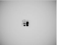
This Antibody From Boster Bio Demonstrates Proficiency in Recognizing and Expressing the Protein Precisely

Excellent, submitted by on

Source: Biocompare.com

SKU A05936-2
Application Western Blot
Sample Ripa buffer lysis transfected HEK293 cell
Primary Incubation +4°C overnight
Blocking Agent 5% milk in 1X TBST
Secondary Incubation 30 mins
Tertiary Incubation 1:1000
Detection Clarity Western ECL Substrate
Results Summary The antibody is excellent at recognizing and expressing the protein at the correct position. I have tried samples from different vendors, but only this vendor offers high-quality products.

Anti-C-Terminal CD44 Antibody for Western Blotting

Excellent, submitted by on

Source: Biocompare.com

SKU PA1021-2
Application Western Blot
Sample Tumor cell lysate
Primary Incubation Overnight at 8-10 degrees Celcius, with rocking, 1 ug/ml in 5% milk/BPS/Tw
Blocking Agent 5% milk in PBS/0.05% Tween-20 (5% milk/PBS/Tw)
Secondary Incubation Goat anti-Rabbit antibody conjugated with HRP at 1:3,000 in 5% milk/PBS/Tw
Tertiary Incubation HRP-bound secondary antibodies were detected by WestPico from ThermoScientific/Tw
Detection Chemiluminescence: West Pico from Thermo Scientific
Results Summary The antibody recognizes the expected ~80 kDa full-length CD44 and its low mol. wt. fragments containing C-terminal domain. The antibody is highly specific, produces "clean", definitive results; does not produce any non-specific bands. The antibody is sensitive and detects CD44 band when total protein per lane is loaded at 10-20 ug. The antibody is stable and could be re-used for blotting several times when stored in the original 5% milk/PBS/Tw solution at -20℃.

"The antibody was used to detect the full length and cleaved fragments of human transmembrane protein CD44. The rabbit antibody PA1021-2 is sensitive, i.e. detects CD44 protein bands under reducing conditions and also when tested material is loaded at low total protein per lane. The antibody is highly specific, i.e. does not recognize any bands of unknown nature on the membrane."

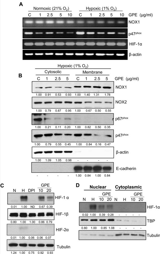
A Nice Antibody For Gp91phox Subunit Of NOX System

Excellent, submitted by on

Source: Biocompare.com

tr>
SKU PA1667
Application Western Blot
Sample 22RV1 cytosolic and membrane fractions
Primary Incubation 1:1000, overnight at 4 degree
Blocking Agent 5% Milk
Secondary Incubation 1:5000, 1 hour RT.
Detection ECL
Results Summary Please refer our paper (Scientific Reports 6, Article number: 23135 (2016)doi:10.1038/srep23135).

"We used this antibody to study the inhibitory effect of GPE on hypoxia induced translocation of gp91phox subunit from cytosol to membrane in our recently published paper (Scientific Reports 6, Article number: 23135 (2016); doi:10.1038/srep23135). This antibody is very much specific in our in vitro system."

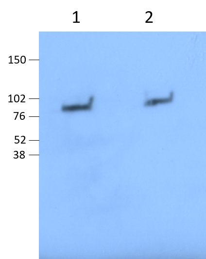
Very Clean Anti-Lactoferrin/LTF Antibody Picoband

Excellent, submitted by on

Source: Biocompare.com

SKU A00633-1
Application Western Blot
Sample Mouse Neutrophils
Primary Incubation 1:2000 1 hr
Blocking Agent 5% BSA
Secondary Incubation 1:3000
Detection HRP
Results Summary Nicely compared total LTF in neutrophils to degranulated LTF. In the image below, Lane 1: Total LTF from mouse neutrophil cell lysates. Lane 2: degranulated LTF. Ratio 1:20.

"Nicely detects degranulated LTF in supernatant mouse neutrophils stimulated with bacteria. Used at 1:2000 in 5% BSA."

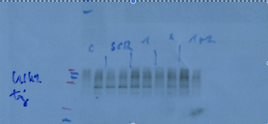
Anti-ULK2 Antibody Works Great for Western Blotting

Excellent, submitted by on

Source: Biocompare.com

SKU A05219
Application Western Blot
Sample HEK293T
Primary Incubation 1:5000 for 24hours at 4degrees celsius.
Blocking Agent Skim milk
Secondary Incubation 1 hour room temperature 1:3000
Detection ECL
Results Summary We silenced ULK1 and ULK2 genes in HEK293T cells, the antibody could differentiate the two proteins.

"Used for western blotting. The antibody worked good, used skim milk for blocking. It was used for investigating silenced HEK293T cells."