Customer Testimonials

Peer Reviews from Real Labs

Browse peer-submitted results linked to specific products—ratings, images, and practical notes included. Contribute your own result to strengthen the evidence base and claim rewards after verification.

Video Interviews

See real workflows and decision points—what researchers tried, what worked, and why. Share your story in a short interview and claim rewards!

Get Interview Rewards

Filter by category, reactivity, and host to find the most relevant peer results.

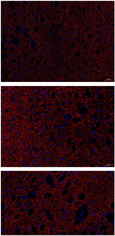
The Anti-TH antibody (M01917) demonstrated excellent specificity and clear fluorescence staining in IF analysis of paraffin-embedded mouse striatum sections, accurately labeling dopaminergic neurons with minimal background.

Excellent, submitted by on
SKU M01917
Application Immunofluorescence
Sample mouse brain
Sample Processing Description Sagittal sections of mouse striatum were fixed in formaldehyde for 48 hours and paraffin-embedded.
Other ReagentsGoat serum,DAPI,Anti-fade mounting medium
Primary Antibody Iba1 Rabbit Monoclonal Antibody
Primary Incubation 1:200, overnight at 4 ℃
Secondary Antibody 1:500, Goat Anti-Rabbit IgG (H+L) Secondary Antibody, Fluoro594 Conjugated
Secondary Incubation 45 min at 37℃
Detection Imaging system:Leica DM2500
Results Summary TH is a marker of catecholaminergic neurons and can specifically label and identify dopaminergic, noradrenergic, and adrenergic neurons. In this experiment, TH immunofluorescence was performed to observe the distribution of dopaminergic neurons in the striatum. The results show clear staining and precise localization.

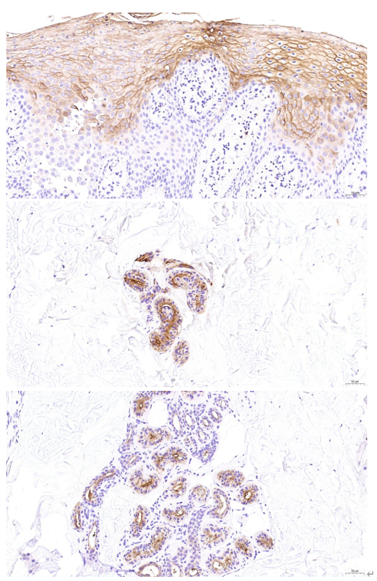
In IHC of human skin cancer sections, the CEACAM5 antibody (A00356) clearly distinguished melanoma from other skin cancers, showing specific staining with low background.

Excellent, submitted by on
SKU M12477-15
Application Immunohistochemistry
Sample normal and preterm human placentas
Sample Processing Description Clinically collected normal and preterm placentas were sectioned longitudinally (with amnion and chorion visible) and prepared as paraffin-embedded sections.
Other ReagentsTris-EDTA Antigen Retrieval Solution, DAB Chromogenic Solution
Primary Antibody Histone H3 (acetyl K27) Rabbit Monoclonal Antibody
Primary Incubation 1:200, overnight at 4 ℃
Secondary Antibody Polymer anti-rabbit IgG-HRP IHC detection kit
Secondary Incubation 45 min at 37℃
Detection Imaging system:Leica DM2500
Results Summary Histone H3 (acetyl K27) is the acetylated form of histone H3 at lysine 27 and serves as a sensitive indicator of epigenetic abnormalities in placental cells; IHC results showed higher expression in preterm placentas than in normal placentas.

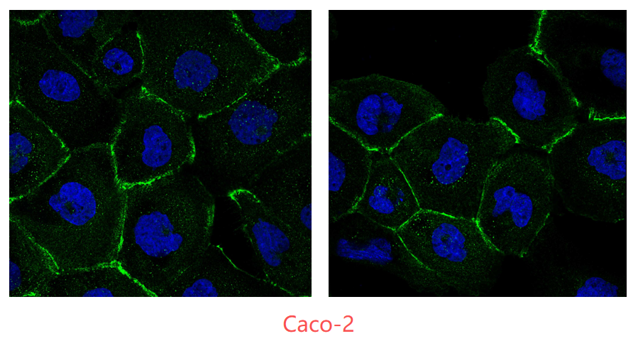
In an immunofluorescence experiment using normally cultured Caco-2 cells, the ZO-1 antibody (PB9234) showed clear and specific membrane staining with low background, demonstrating reliable performance.

Excellent, submitted by on
SKU PB9234
Application Immunofluorescence
Sample human Caco-2 cell line
Sample Processing Description Cells were normally cultured in 24-well plates using MEM supplemented with 20% fetal bovine serum. When the cell density reached approximately 60%, the culture was terminated. The medium was removed, cells were washed three times with PBS, fixed with 4% paraformaldehyde for 15 minutes, and then washed three times with PBS before further use.
Other ReagentsGoat serum, DAPI, and an anti-fade mounting medium.
Primary Antibody ZO1 tight junction protein/TJP1 Antibody Picoband®
Primary Incubation 1:200, overnight at 4 ℃
Secondary Antibody Goat Anti-Rabbit IgG (H+L) Secondary Antibody, Fluoro488 Conjugated
Secondary Incubation 45 min at 37℃
Detection Imaging system:Leica DM2500
Results Summary Caco-2 cells are a “gold standard” in vitro model for studying intestinal epithelial function, and ZO-1 is a key marker of barrier integrity. The images show immunofluorescence staining of ZO-1 in normally cultured Caco-2 cells to evaluate antibody performance. Clear membrane localization and accurate staining indicate that this antibody is suitable for downstream research applications.

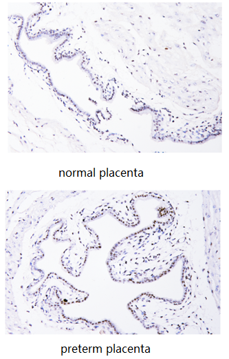
Using IHC on paraffin-embedded human placental tissues, the Anti-Histone H3 (acetyl K27) antibody (M12477-15) showed high specificity and clear staining, with higher H3K27 acetylation in preterm placentas than in normal controls.

Excellent, submitted by on
SKU M12477-15
Application Immunohistochemistry
Sample normal and preterm human placentas
Sample Processing Description Clinically collected normal and preterm placentas were sectioned longitudinally (with amnion and chorion visible) and prepared as paraffin-embedded sections.
Other ReagentsTris-EDTA Antigen Retrieval Solution, DAB Chromogenic Solution
Primary Antibody Histone H3 (acetyl K27) Rabbit Monoclonal Antibody
Primary Incubation 1:200, overnight at 4 ℃
Secondary Antibody Polymer anti-rabbit IgG-HRP IHC detection kit
Secondary Incubation 45 min at 37℃
Detection Imaging system:Leica DM2500
Results Summary Histone H3 (acetyl K27) is the acetylated form of histone H3 at lysine 27 and serves as a sensitive indicator of epigenetic abnormalities in placental cells; IHC results showed higher expression in preterm placentas than in normal placentas.

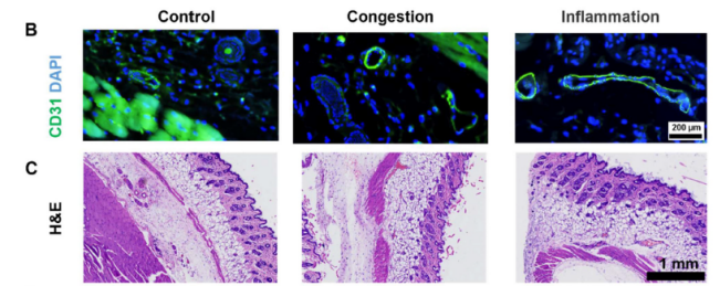
IF using Anti-CD31 antibody (A01513-2) showed specific, strong, and stable endothelial staining, revealing expanded and inflamed microvessels in treated mouse skin compared to controls.

Excellent, submitted by on
SKU A01513-2
Application Immunofluorescence
Sample Paraffin-embedded mouse skin sections
Sample Processing Description Mouse skin samples were divided into three groups: (i) normal control, (ii) congestion group, treated with ethanol, and (iii) inflammation group, stimulated with LPS.
Other Reagents Goat serum, DAPI, Anti-fade mounting medium
Primary Antibody CD31/Pecam1 Antibody Picoband®
Primary Incubation 1:200, overnight at 4 ℃
Secondary Antibody DyLight 488–conjugated Goat Anti-Rabbit IgG (H+L)) (Boster, BA1127)
Secondary Incubation 1:500, 45 min at 37℃
Detection Imaging system:Leica DM2500
Results Summary IF staining with Boster CD31 antibody produced high-quality vascular images. In the control group, vessels were small and well-defined. In the congestion group (ethanol-treated), CD31 staining revealed markedly dilated vascular lumens. In the inflammation group (LPS-treated), vessel dilation was accompanied by changes in tissue spacing. Combined with Prussian blue staining and magnetic signal measurements, we observed that despite extreme vessel dilation in the congestion group, magnetic signals were not significantly elevated; only the inflammation group, with extensive macrophage infiltration and uptake, showed high magnetic signals.

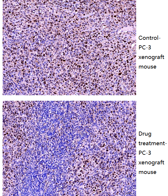
IHC using Anti-Ki67 antibody(A01505-3) showed clear nuclear staining with minimal background, revealing markedly reduced Ki67-positive tumor cells in docetaxel-treated PC-3 xenografts compared to control mouse.

Excellent, submitted by on
SKU A01505-3
Application Immunohistochemistry
Sample PC-3 xenograft mouse tissue
Sample Processing Description (1) PC-3 cells were implanted in nude mice to form tumors as the control group; (2) tumor-bearing mice were treated with the drug docetaxel.
Other Reagents Goat serum, DAPI, Anti-fade mounting medium
Primary Antibody Prostate Specific Antigen/KLK3 Antibody Picoband®
Primary Incubation 1:500, overnight at 4 ℃
Secondary Antibody Two-step IHC Detection Kit (Rabbit IgG)
Secondary Incubation 1:500, 45 min at 37℃
Detection Imaging system:Leica DM2500
Results Summary Ki67 is a nuclear protein directly associated with cell proliferation and is widely recognized as a marker of proliferating cells due to its importance in evaluating tumor growth rate and patient prognosis. Higher Ki67 indices indicate more actively proliferating tumor cells, usually correlating with faster growth and increased aggressiveness. After treatment with docetaxel, extensive tumor cell death occurred, and proliferative activity was partially suppressed, resulting in fewer Ki67-positive tumor cells. These results clearly reflect the inhibitory effect of the drug on tumor proliferation.

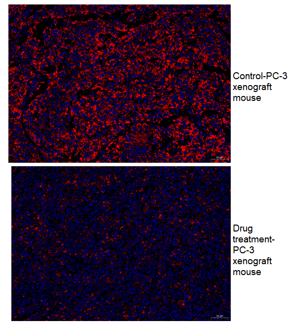
Immunofluorescence using KLK3 antibody (A01505-3) showed clear and specific staining in PC-3 xenograft tumors, with markedly reduced PSA expression in tumors treated with docetaxel compared to untreated controls, consistent with expected drug effects.

Excellent, submitted by on
SKU A01505-3
Application Immunofluorescence
Sample PC-3 xenograft mouse tissue
Sample Processing Description (1) PC-3 cells were implanted in nude mice to form tumors as the control group; (2) tumor-bearing mice were treated with the drug docetaxel.
Other Reagents Goat serum, DAPI, Anti-fade mounting medium
Primary Antibody Prostate Specific Antigen/KLK3 Antibody Picoband®
Primary Incubation 1:500, overnight at 4 ℃
Secondary Antibody DyLight 594–conjugated Goat Anti-Rabbit IgG (H+L))
Secondary Incubation 1:500, 45 min at 37℃
Detection Imaging system:Leica DM2500
Results Summary PSA, or prostate-specific antigen, is a multifaceted tumor marker that can promote the proliferation and migration of prostate cancer cells, driving tumor growth. After treatment with docetaxel, its secretion is expected to decrease, which is confirmed by the experimental results showing markedly reduced PSA expression in the xenograft tumor cells following treatment.

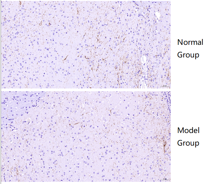
IHC using Tyrosine Hydroxylase/TH Antibody Picoband® (P00683) showed clear staining with no background, revealing markedly reduced TH expression in the medulla of Alzheimer’s mouse brain compared to normal mouse brain.

Excellent, submitted by on
SKU PB9449
Application Immunohistochemistry
Sample Normal mouse brain and Alzheimer’s model mouse brain tissue
Sample Processing Description Paraffin-embedded normal mouse brain and Alzheimer’s model mouse brain.
Other Reagents Goat serum, DAB
Primary Antibody Tyrosine Hydroxylase/TH Antibody Picoband®
Primary Incubation 1:500, overnight at 4 ℃
Secondary Antibody Two-step IHC kit
Secondary Incubation 37 minutes in 37 ℃
Detection Imaging system:Leica DM2500
Results Summary TH is a marker of catecholaminergic neurons, specifically labeling dopaminergic, noradrenergic, and adrenergic neurons. IHC results showed that TH expression in the medulla was markedly lower in Alzheimer’s mouse compared to normal mouse.

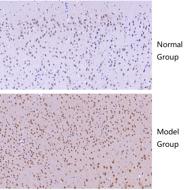
IHC with HDAC5 Antibody Picoband® (A01230-6) showed clear staining and low background, with significantly higher HDAC5 levels in the cortex of Alzheimer’s mice, suggesting potential as a biomarker.

Excellent, submitted by on
SKU A01230-6
Application Immunohistochemistry
Sample Normal mouse brain and Alzheimer’s model mouse brain
Sample Processing Description Paraffin-embedded normal mouse brain and Alzheimer’s model mouse brain.
Other Reagents Goat serum, DAB chromogen
Primary Antibody Anti-HDAC5 Antibody Picoband®
Primary Incubation 1:400, overnight at 4 ℃
Secondary Antibody Two-step IHC kit
Secondary Incubation 30 minutes in 37 ℃
Detection Imaging system: Leica DM2500
Results Summary The results showed that HDAC5 levels in the cortex were significantly higher in Alzheimer’s mice than in normal mice, suggesting its potential as a biomarker for further validation.

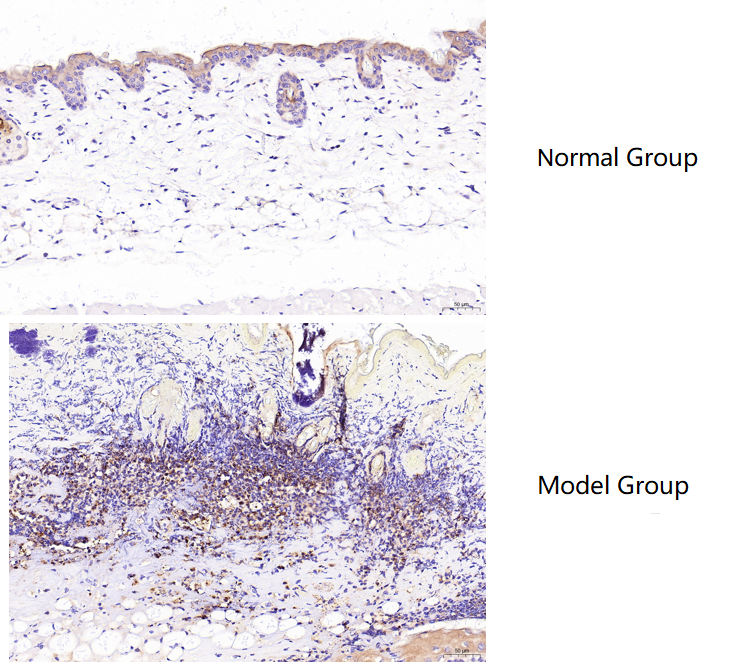
Using Myeloperoxidase/MPO Antibody Picoband® (PA1054) in IHC, MPO showed low expression in normal mouse skin and was markedly increased in burned skin, with clear staining and expected results.

Excellent, submitted by on
SKU PA1054
Application Immunohistochemistry
Sample mouse skin tissue
Sample Processing Description Male BALB/c mice aged 6–8 weeks were used. (1) Normal dorsal skin was collected from untreated mice. (2) Burn injury was induced on the dorsal skin using a burn device. After one week of housing, hair was removed with depilatory cream. Skin samples were collected from normal mice (dorsal skin) and burned mice (burned area), fixed in formalin for 72 hours, and embedded in paraffin for sectioning./td>
Other ReagentsTris-EDTA Antigen Retrieval Buffer (50×, pH 9.0), DAB Chromogen Kit
Primary Antibody Myeloperoxidase/MPO Antibody Picoband®
Primary Incubation 1:100, overnight at 4 ℃
Secondary Antibody Polymer Anti-Rabbit IgG–HRP Immunohistochemistry Kit
Detection Imaging system:Leica DM2500
Results Summary MPO (myeloperoxidase) is a lysosomal enzyme mainly present in neutrophils and monocytes/macrophages. Its core function is to generate reactive oxygen species, and therefore it plays a “double-edged sword” role in innate immune defense and inflammation-related diseases. MPO is present at very low levels in normal skin, but in burned skin, a large number of macrophages accumulate around the injured tissue, accompanied by a significant increase in MPO expression. Based on the immunohistochemical results, the staining is clear and the findings are consistent with expectations.