| SKU | A01513-2 |
|---|---|
| Application | Immunofluorescence |
| Sample | Paraffin-embedded mouse skin sections |
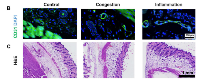
| Sample Processing Description | Mouse skin samples were divided into three groups: (i) normal control, (ii) congestion group, treated with ethanol, and (iii) inflammation group, stimulated with LPS. |
| Other Reagents | Goat serum, DAPI, Anti-fade mounting medium |
| Primary Antibody | CD31/Pecam1 Antibody Picoband® |
| Primary Incubation | 1:200, overnight at 4 ℃ |
| Secondary Antibody | DyLight 488–conjugated Goat Anti-Rabbit IgG (H+L)) (Boster, BA1127) |
| Secondary Incubation | 1:500, 45 min at 37℃ |
| Detection | Imaging system:Leica DM2500 |
| Results Summary | IF staining with Boster CD31 antibody produced high-quality vascular images. In the control group, vessels were small and well-defined. In the congestion group (ethanol-treated), CD31 staining revealed markedly dilated vascular lumens. In the inflammation group (LPS-treated), vessel dilation was accompanied by changes in tissue spacing. Combined with Prussian blue staining and magnetic signal measurements, we observed that despite extreme vessel dilation in the congestion group, magnetic signals were not significantly elevated; only the inflammation group, with extensive macrophage infiltration and uptake, showed high magnetic signals. |