| SKU | A01263 |
|---|---|
| Application | Western Blot |
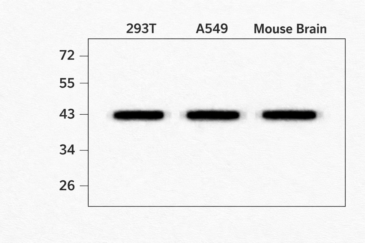
| Sample | Human 293T and A549 cell lysates, Mouse brain tissue |
| Sample Processing Description | Cells were lysed in RIPA buffer containing protease inhibitors. Lysates were clarified by centrifugation and quantified using a BCA assay. 30–35 µg of protein was loaded per lane on a 5–20% SDS-PAGE gel and transferred to nitrocellulose membranes. |
| Primary Antibody | Anti-beta Actin ACTB Antibody |
| Primary Incubation | 1:1000, incubated overnight at 4°C. |
| Secondary Antibody | Goat anti-rabbit IgG-HRP (Catalog # BA1054). |
| Secondary Incubation | 1:5000 dilution, 1 hour at room temperature. |
| Other Reagents used | 5% non-fat milk/TBST blocking buffer, ECL Plus substrate, Azure Biosystems c600 imaging system. |
| Detection | Chemiluminescent detection. Strong single band observed at ~42 kDa, corresponding to beta actin. |
| Results Summary | Produced clear, sharp bands with no background. Excellent specificity and consistency across replicates. Ideal as a loading control for WB. |