| SKU | MA1083 |
|---|---|
| Application | Immunohistochemistry |
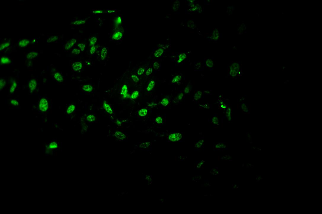
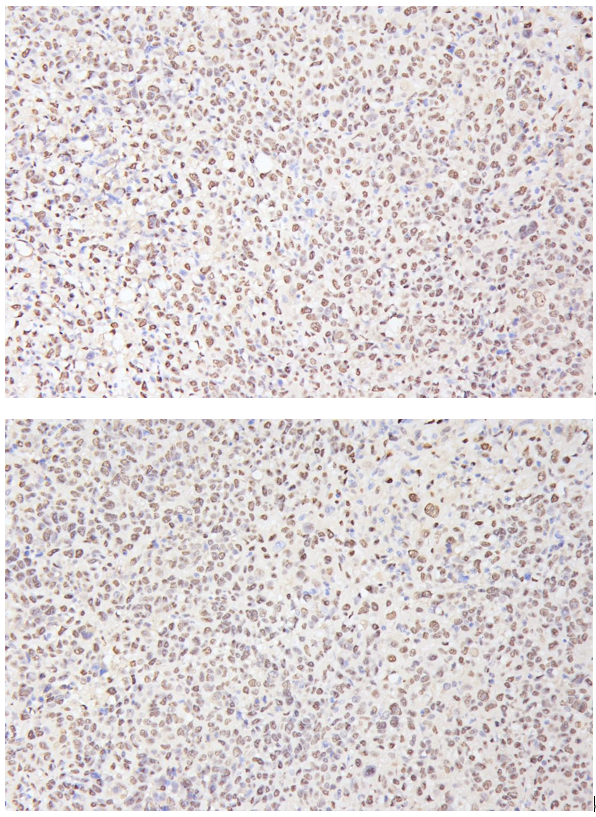
| Sample | HepG2 subcutaneous xenograft in nude mice |
| Sample Processing Description | HepG2 cells were expanded and then implanted subcutaneously into nude mice. After 2 weeks, tumors formed, which were excised, fixed in formalin for 48 hours, and processed for paraffin embedding and sectioning. |
| Other Reagents | Goat serum, DAB chromogen solution |
| Primary Antibody | PCNA Antibody (Monoclonal, PC 10) |
| Primary Incubation | 1:500, overnight at 4 ℃ |
| Secondary Antibody | Two-step IHC detection kit |
| Secondary Incubation | 30 min in 37℃ |
| Detection | Image system: Leica DM2500 |
| Results Summary | PCNA is a key marker for cell proliferation studies; however, besides being a proliferation marker, it is a core component of the DNA replication and repair complex. Its long half-life and involvement in DNA damage repair mean that PCNA positivity does not solely indicate cell proliferation but may also reflect cells responding to DNA damage. Combined with Ki67 staining, it can accurately indicate proliferative status. In our experiment, the staining was clear, with distinct nuclear positivity. |