| SKU | M00254-9 |
|---|---|
| Application | Immunohistochemistry |
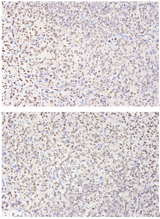
| Sample | HepG2 subcutaneous xenograft in nude mice |
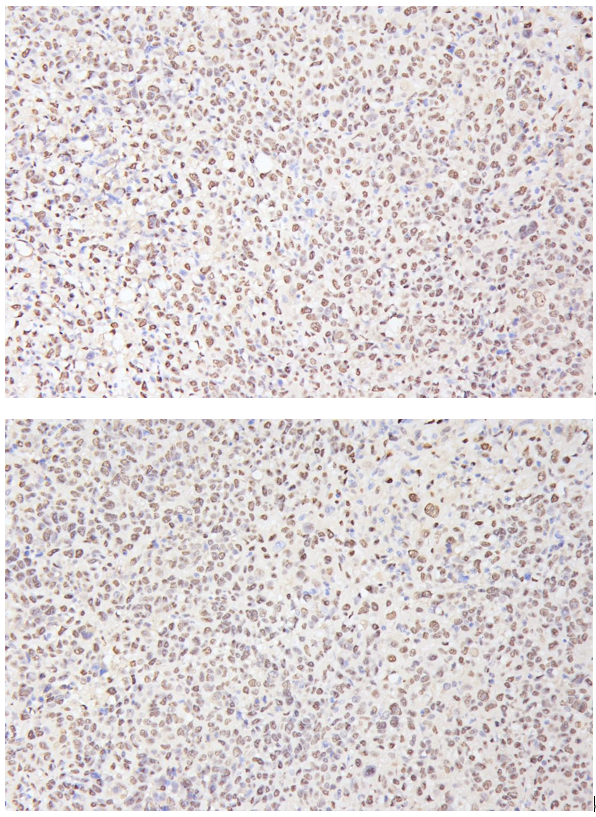
| Sample Processing Description | HepG2 cells were expanded in culture and subcutaneously implanted into nude mice. After 2 weeks of tumor formation, tumor tissues were harvested, fixed in 4% formaldehyde for 48 hours, and then processed for paraffin embedding and sectioning. |
| Other Reagents | Goat serum, DAB chromogen solution |
| Primary Antibody | Ki67 Antibody Picoband® (monoclonal, 5C7) |
| Primary Incubation | 1:200, overnight at 4 ℃ |
| Secondary Antibody | Two-step IHC detection kit |
| Secondary Incubation | 30 min in 37℃ |
| Detection | Image system: Leica DM2500 |
| Results Summary | Ki67 is a key marker of cell proliferation with high specificity, as it is expressed only in actively dividing cells and has a very short half-life, allowing accurate assessment of the proliferation index at the time of sampling. It is widely used in tumor prognosis evaluation (e.g., high Ki67 index in breast cancer is often associated with poor prognosis) and assessment of tissue regenerative activity. In this experiment, clear and distinct nuclear positivity was observed with well-defined staining. |