| SKU | PA2087 |
|---|---|
| Application | Western Blot |
| Sample | mouse brain tissue |
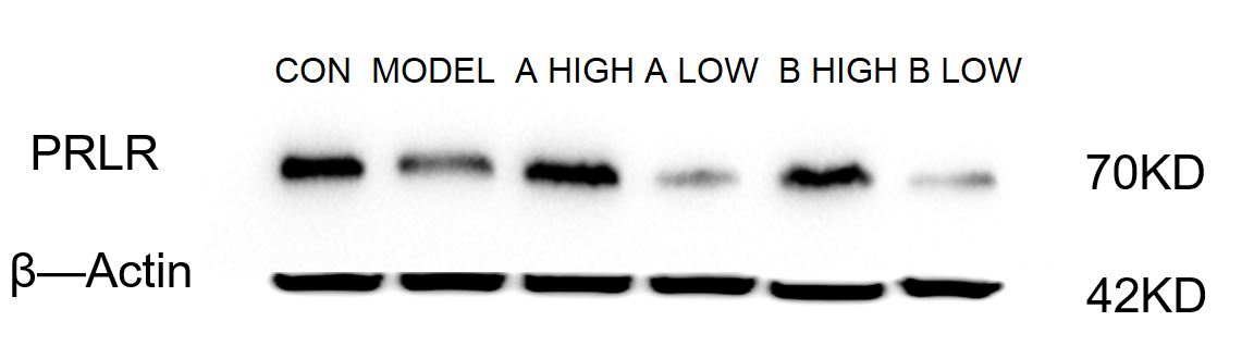
| Sample Processing Description | Mouse brain tissues were lysed in RIPA buffer containing a protease inhibitor cocktail at 4 °C for 2 hours. After centrifugation, the supernatant was collected for protein quantification. The protein concentration was adjusted accordingly, mixed with 5× protein loading buffer, and denatured by heating for 10 minutes. Then, 15 μl of protein sample was loaded per lane for electrophoresis. |
| Other Reagents | blocking buffer |
| Primary Antibody | Prolactin Receptor/PRLR Antibody Picoband® |
| Primary Incubation | 1:1000, overnight at 4 ℃ |
| Secondary Antibody | HRP Conjugated AffiniPure Goat Anti-Rabbit IgG (H+L) (BA1054) |
| Secondary Incubation | 1:2000, 1 h in RT |
| Detection | Substrate: ECL substrate, Image system:ChemiDoc MP |
| Results Summary | The figure shows representative Western blot results of PRLR and the internal control β-actin in brain tissues from normal mice, the model group, and mice treated with low and high doses of AB. The antibody produced clear bands, and distinct differences among the experimental groups were clearly observed. |