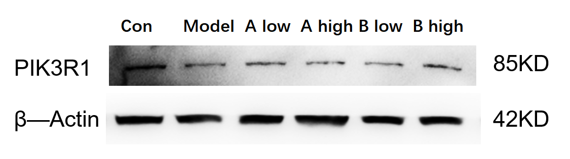
| SKU | M00297 |
|---|---|
| Application | Western Blot |
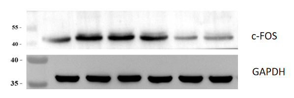
| Sample | Mouse hippocampal tissue |
| Sample Processing Description | An Alzheimer’s disease (AD) model was established by injecting streptozotocin (STZ) into the lateral ventricles of mice. Hippocampal tissues were collected after treatment with four different doses of QianCengTa compound, and total protein was extracted. |
| Other Reagents | RIPA lysis buffer,Protease inhibitor,Electrophoresis buffer,Transfer buffer,Blocking buffer |
| Primary Antibody | c-Fos Rabbit Monoclonal Antibody |
| Primary Incubation | 1:2000, overnight at 4 ℃ |
| Secondary Antibody | HRP Goat Anti-Rabbit IgG |
| Secondary Incubation | 1:10000, 1 hour in room temperature |
| Detection | Substrate: ECL, Imaging system:ChemiDoc MP |
| Results Summary | c-FOS is a marker of neuronal activation under pathological conditions. Its expression is low in normal brain tissue but elevated in the AD model, and decreases following drug treatment. Lane 1 represents normal hippocampus, lane 2 the AD model hippocampus, lanes 3, 4, and 5 correspond to increasing doses of QianCengTa compound, and lane 6 is the positive control drug. The Western blot results indicate that QianCengTa compound exhibits a therapeutic effect on AD. |