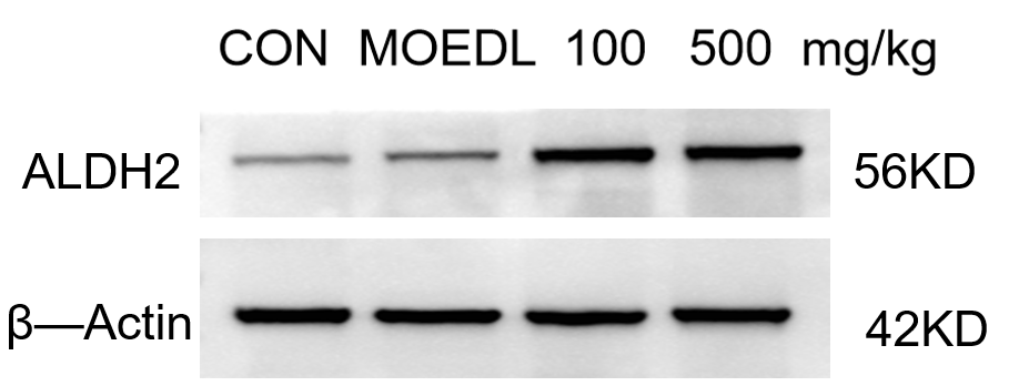
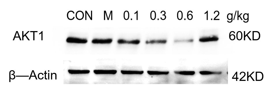
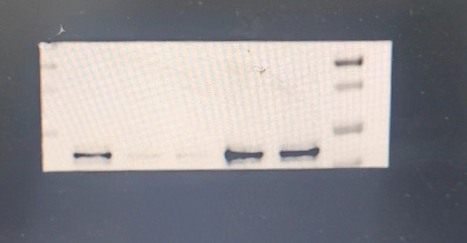
Customer Testimonials

Peer Reviews from Real Labs

Browse peer-submitted results linked to specific products—ratings, images, and practical notes included. Contribute your own result to strengthen the evidence base and claim rewards after verification.

Video Interviews

See real workflows and decision points—what researchers tried, what worked, and why. Share your story in a short interview and claim rewards!

Get Interview Rewards

Filter by category, reactivity, and host to find the most relevant peer results.

The SLC40A1 antibody was used to detect the expression of the target protein in human uterine tissue. Although two bands were observed, the differences in expression levels were still clearly discernible.

Excellent, submitted by on
SKU A01953-2
Application Western Blot
Sample human uterine tissue
Sample Processing Description The tissue was minced and further disrupted by sonication, then lysed on ice for 1 hour using RIPA buffer. After centrifugation, the supernatant was collected and quantified using the BCA method. The appropriate amount of loading buffer was added, and the samples were boiled in a water bath to denature the proteins. Finally, 15 μL of each protein sample was loaded into each lane of the SDS-PAGE gel.
Other Reagents 5% Non-fat milk
Primary Antibody Anti-SLC40A1 Antibody Picoband®
Primary Incubation overnight at 4 ℃
Secondary Antibody HRP-conjugated Anti-Rabbit IgG Secondary Antibody
Secondary Incubation 1 hour in room temperature
Detection Substrate: Ultra-sensitive ECL luminescent reagent (Cat# AR1191), Imaging system:Tanon
Results Summary The SLC40A1 antibody was used to detect the expression of the target protein in human uterine tissue. Although two bands were detected, the differences in expression levels were still clearly observable and did not affect the analysis of the experimental results.

This antibody is suitable for detecting PTEN protein in mouse hippocampus by Western blot, showing clear, distinct, and highly specific bands.

Excellent, submitted by on
SKU M00006-1
Application Western Blot
Sample Mouse hippocampus tissue
Sample Processing Description The mouse hippocampus was lysed with RIPA buffer containing a protease inhibitor cocktail. After protein quantification, samples were mixed with 5× protein loading buffer and heated for 10 minutes to denature. Load 5 μL of protein per lane and apply to SDS-PAGE.
Other Reagents 5% Non-fat milk
Primary Antibody Anti-PTEN Rabbit Monoclonal Antibody
Primary Incubation overnight at 4 ℃
Secondary Antibody HRP-conjugated Anti-Rabbit IgG Secondary Antibody
Secondary Incubation 1 hour in room temperature
Detection Substrate: Ultra-sensitive ECL luminescent reagent, Imaging system:ChemiDoc MP
Results Summary This antibody is suitable for detecting PTEN protein in mouse hippocampus by Western blot, showing clear, distinct, and highly specific bands.

The antibody shows clear bands with no non-specific signals in WB, and is suitable for detecting PIK3R1 protein in mouse hippocampus by Western blot.

Excellent, submitted by on
SKU A00318-1
Application Western Blot
Sample Mouse hippocampus tissue
Sample Processing Description The mouse hippocampus was lysed with RIPA lysis buffer containing protease inhibitors, followed by protein quantification. The samples were then mixed with 5× protein loading buffer and heated for 10 minutes for denaturation. Five microliters of protein sample were loaded into each lane for SDS-PAGE.
Primary Antibody Anti-PI 3 Kinase p85 alpha/PIK3R1 Antibody Picoband®
Primary Incubation overnight at 4 ℃
Secondary Antibody HRP-conjugated Anti-Rabbit IgG Secondary Antibody
Secondary Incubation 1 hour in room temperature
Detection Substrate: Ultra-sensitive ECL luminescent reagent , Imaging system:ChemiDoc MP
Results Summary The antibody shows clear bands with no non-specific signals in WB, and is suitable for detecting PIK3R1 protein in mouse hippocampus by Western blot.

The GPX4 antibody was used to detect the expression of the protein in mouse uterine tissue. The WB results showed clear bands, and the antibody could be reused after recovery with good performance, offering excellent cost-effectiveness.

Excellent, submitted by on
SKU M02059
Application Western Blot
Sample Mouse Uterus tissue
Sample Processing Description The GPX4 antibody was used to detect the expression of the protein in mouse uterine tissue. The Western blot results showed clear bands, and the antibody retained good performance after reuse, offering excellent cost-effectiveness.
Primary Antibody Anti-GPX4 Rabbit Monoclonal Antibody
Primary Incubation overnight at 4 ℃
Secondary Antibody HRP-conjugated Anti-Rabbit IgG Secondary Antibody
Secondary Incubation 1 hour in room temperature
Detection Substrate: Ultra-sensitive ECL luminescent reagent (Cat# AR1191), Imaging system:Tanon
The GPX4 antibody was used to detect the protein expression in mouse uterine tissue. The Western blot results showed clear bands, and the antibody maintained good performance after reuse, providing excellent cost-effectiveness.

The FTH1 antibody was used to detect the expression of the target protein in human uterine tissue. The WB bands were single and clear, and compared with other domestic and international brands, this antibody offers excellent cost-performance.

Excellent, submitted by on
SKU M02401
Application Western Blot
Sample Mouse hippocampus tissue
Sample Processing Description The tissue was minced and sonicated, then lysed on ice for 1 hour using RIPA buffer. After centrifugation to collect the supernatant and protein quantification by BCA, samples were mixed with loading buffer at the appropriate ratio and denatured by boiling in a water bath. Fifteen microliters of each protein sample were loaded per lane onto SDS-PAGE gel.
Primary Antibody Anti-Ferritin FTH1 Rabbit Monoclonal Antibody
Primary Incubation overnight at 4 ℃
Secondary Antibody HRP-conjugated Anti-Rabbit IgG Secondary Antibody
Secondary Incubation 1 hour in room temperature
Detection Substrate: Ultra-sensitive ECL luminescent reagent (Cat# AR1191), Imaging system:Tanon
Results Summary The FTH1 antibody was used to detect the expression of the target protein in human uterine tissue. The WB bands were single and clear, and compared with other domestic and international brands, this antibody offers excellent cost-performance.

Western blot analysis was performed using the COL6A1 antibody to detect COL6A1 protein expression in the mouse hippocampus.

Excellent, submitted by on
SKU M02226
Application Western Blot
Sample Mouse hippocampus tissue
Sample Processing Description The mouse hippocampus was lysed in RIPA buffer supplemented with a protease inhibitor cocktail. After protein quantification, samples were mixed with 5× protein loading buffer and denatured by heating at 100°C for 10 minutes. Five microliters of each protein sample were loaded per lane onto SDS-PAGE.
Primary Antibody Anti-Collagen VI COL6A1 Rabbit Monoclonal Antibody
Primary Incubation overnight at 4 ℃
Secondary Antibody HRP-conjugated Anti-Rabbit IgG Secondary Antibody
Secondary Incubation 1 hour in room temperature
Detection Substrate: Ultra-sensitive ECL luminescent reagent (Cat# AR1191), Imaging system:ChemiDoc MP
Results Summary Western blot analysis was performed using the COL6A1 antibody to detect COL6A1 protein expression in the mouse hippocampus. Although minor non-specific bands were observed, they did not affect the trend analysis, indicating that the antibody is suitable for detecting the target protein in this tissue.

The antibody was used to perform WB quantitative detection of ALDH2 protein in mouse hippocampus. The experimental results showed clear and distinct bands, indicating that it is suitable for WB detection of ALDH2 protein in mouse tissues.

Excellent, submitted by on
SKU PB9472
Application Western Blot
Sample Mouse hippocampus tissue
Sample Processing Description The mouse hippocampus tissue was lysed using RIPA lysis buffer supplemented with a protease inhibitor cocktail. Protein concentration was determined, and samples were mixed with 5× protein loading buffer and denatured by heating at 100°C for 10 minutes. Five microliters of each protein sample were loaded per lane onto SDS-PAGE.
Primary Antibody Anti-ALDH2 Antibody Picoband®
Primary Incubation overnight at 4 ℃
Secondary Antibody HRP-conjugated Anti-Rabbit IgG Secondary Antibody
Secondary Incubation 1 hour in room temperature
Detection Substrate: Ultra-sensitive ECL luminescent reagent (Cat# AR1191), Imaging system: ChemiDoc MP (Bio-Rad)
Results Summary The antibody was used for quantitative WB detection of ALDH2 protein in mouse hippocampus. The results showed clear and distinct bands, indicating its suitability for WB detection of ALDH2 protein in mouse tissues.

Western blot analysis of AKT1 protein in mouse hippocampus using the AKT1 antibody showed clear bands. The antibody is suitable for mouse hippocampal tissue samples.

Excellent, submitted by on
SKU M00024-1
Application Western Blot
Sample Mouse hippocampus tissue
Sample Processing Description The mouse hippocampus was lysed with RIPA buffer containing a protease inhibitor cocktail. After protein quantification, samples were mixed with 5× protein loading buffer and heated for 10 minutes to denature. Load 5 μL of protein per lane and apply to SDS-PAGE.
Primary Antibody Anti-AKT1 Monoclonal Antibody
Primary Incubation overnight at 4 ℃
Secondary Antibody HRP-conjugated Anti-Rabbit IgG Secondary Antibody
Secondary Incubation 1 hour in room temperature
Detection Substrate: Ultra-sensitive ECL luminescent reagent (Cat# AR1191), Imaging system: ChemiDoc MP (Bio-Rad)
Results Summary Western blot analysis of AKT1 protein in the mouse hippocampus using the AKT1 antibody showed clear bands. This antibody is suitable for mouse hippocampal tissue samples.

This antibody exhibits high specificity and clear bands, and the resulting experimental data are reliable, providing important support for elucidating the molecular mechanisms underlying the role of PTN in this study.

Excellent, submitted by on
SKU P30433
Application Western Blot
Sample Mouse Spinal
Sample Processing Description Tissue samples were directly lysed in RIPA lysis buffer, mixed with loading buffer at the appropriate ratio, and denatured by heating at 98°C. Then, 20 μl of each cell protein sample was loaded into each lane of the SDS-PAGE gel.
Primary Antibody Phospho GRF-1 (Tyr1105) Antibody
Primary Incubation 1:1000, overnight at 4 ℃
Blocking Agent 5% non-fat milk
Secondary Antibody HRP-conjugated anti-rabbit IgG
Secondary Incubation Incubate at room temperature for 1 hour
Detection Substrate: ECL substrate, Imaging system: Sangon Biotech
Results Summary I will purchase Boster products again and recommend them to my classmates and colleagues.

The staining is clear, highly specific, with almost no non-specific bands.

Excellent, submitted by on
SKU PA2290
Application Western Blot
Sample Protein extracts from LA795 and 16HBE cells.
Sample Processing Description After collecting the cells, lyse them with RIPA buffer containing protease inhibitors, quantify the protein using the BCA assay, and add loading buffer proportionally. Denature by heating at 98 °C. Load the samples onto SDS-PAGE, with 30 μg of total protein per lane.
Primary Antibody Anti-TMEM16A/ANO1 Antibody Picoband®
Primary Incubation 1:1000, overnight at 4 ℃
Blocking Agent 5% non-fat milk
Secondary Antibody HRP-conjugated goat anti-rabbit IgG
Secondary Incubation Incubate at room temperature for 1 hour
Detection Substrate: ECL substrate, Imaging system: Tanon
Results Summary The TMEM16A antibody we previously used was never ideal; despite long-term optimization, it was difficult to obtain clear bands. This time, by using BOSTER’s polyclonal antibody, we achieved much better experimental results. The bands were successfully exposed in essentially one attempt, with clear signals and no non-specific bands. We are very satisfied with Boster’s antibody.