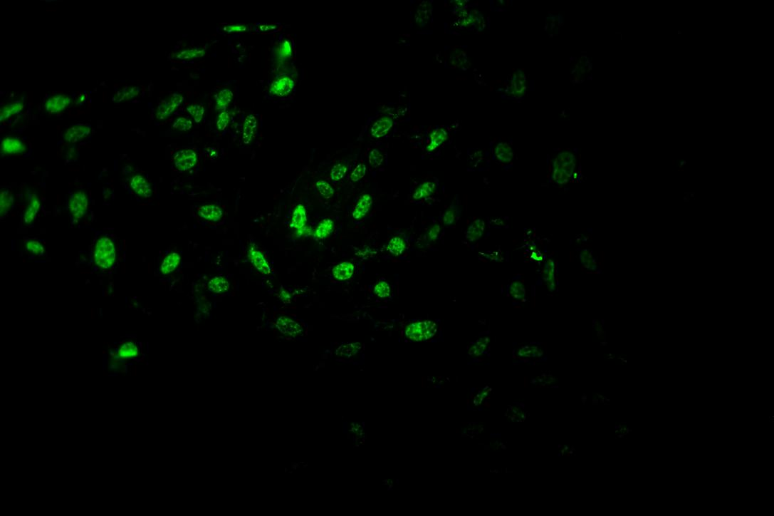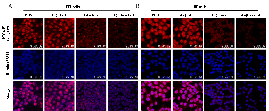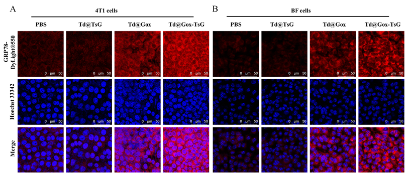
| SKU | PB9234 |
|---|---|
| Application | ICC/IF |
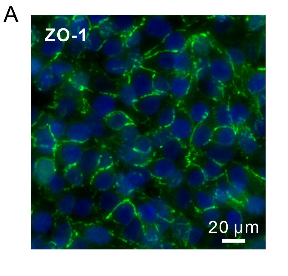
| Sample | Endothelial cells |
| Sample Processing Description | Endothelial cells were seeded in collagen gel-treated chips for 12 hours. |
| Primary Antibody | Anti-ZO1 tight junction protein/TJP1 Antibody Picoband® |
| Primary Incubation | 1:200, overnight at 4 ℃ |
| Blocking Agent | Ready-to-use Goat Serum |
| Secondary Antibody | Goat Anti-Rabbit IgG (H+L) Secondary Antibody, DyLight®488 Conjugated(BA1127) |
| Secondary Incubation | Incubate at room temperature for 1 hour |
| Other reagents | DAPI (AR1176), Anti-fade mounting medium |
| Detection | Fluorescence microscope |
| Results Summary | At a critical stage of our manuscript preparation, we needed to stain tight junction proteins in endothelial cells. After trying primary antibodies from many companies without achieving satisfactory results, it was ultimately Boster’s product that solved the problem (Figure 5A). I was so excited that I took the photo below — treating it like a true treasure. |