Customer Testimonials

Peer Reviews from Real Labs

Browse peer-submitted results linked to specific products—ratings, images, and practical notes included. Contribute your own result to strengthen the evidence base and claim rewards after verification.

Video Interviews

See real workflows and decision points—what researchers tried, what worked, and why. Share your story in a short interview and claim rewards!

Get Interview Rewards

Filter by category, reactivity, and host to find the most relevant peer results.

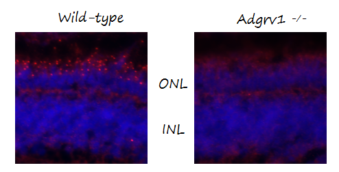
Both are confirmed specific in zebrafish using one of our KO lines.

Excellent, submitted by on
SKU DZ41032
Application Immunofluorescence
Sample wildtype and KO mutant zebrafish retina
Primary Incubation 1:100
Blocking Agent 10% normal goat serum and 2% bovine serum albumin in PBS
Secondary Antibody Alexa Fluor 568 goat anti-rabbit was used in a 1:800 dilution.

Anti-C-Terminal CD44 Antibody for Western Blotting

Excellent, submitted by on

Source: Biocompare.com

SKU PA1021-2
Application Western Blot
Sample Tumor cell lysate
Primary Incubation Overnight at 8-10 degrees Celcius, with rocking, 1 ug/ml in 5% milk/BPS/Tw
Blocking Agent 5% milk in PBS/0.05% Tween-20 (5% milk/PBS/Tw)
Secondary Incubation Goat anti-Rabbit antibody conjugated with HRP at 1:3,000 in 5% milk/PBS/Tw
Tertiary Incubation HRP-bound secondary antibodies were detected by WestPico from ThermoScientific/Tw
Detection Chemiluminescence: West Pico from Thermo Scientific
Results Summary The antibody recognizes the expected ~80 kDa full-length CD44 and its low mol. wt. fragments containing C-terminal domain. The antibody is highly specific, produces "clean", definitive results; does not produce any non-specific bands. The antibody is sensitive and detects CD44 band when total protein per lane is loaded at 10-20 ug. The antibody is stable and could be re-used for blotting several times when stored in the original 5% milk/PBS/Tw solution at -20℃.

"The antibody was used to detect the full length and cleaved fragments of human transmembrane protein CD44. The rabbit antibody PA1021-2 is sensitive, i.e. detects CD44 protein bands under reducing conditions and also when tested material is loaded at low total protein per lane. The antibody is highly specific, i.e. does not recognize any bands of unknown nature on the membrane."

A Nice Antibody For Gp91phox Subunit Of NOX System

Excellent, submitted by on

Source: Biocompare.com

tr>
SKU PA1667
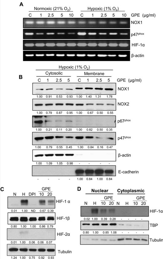
Application Western Blot
Sample 22RV1 cytosolic and membrane fractions
Primary Incubation 1:1000, overnight at 4 degree
Blocking Agent 5% Milk
Secondary Incubation 1:5000, 1 hour RT.
Detection ECL
Results Summary Please refer our paper (Scientific Reports 6, Article number: 23135 (2016)doi:10.1038/srep23135).

"We used this antibody to study the inhibitory effect of GPE on hypoxia induced translocation of gp91phox subunit from cytosol to membrane in our recently published paper (Scientific Reports 6, Article number: 23135 (2016); doi:10.1038/srep23135). This antibody is very much specific in our in vitro system."

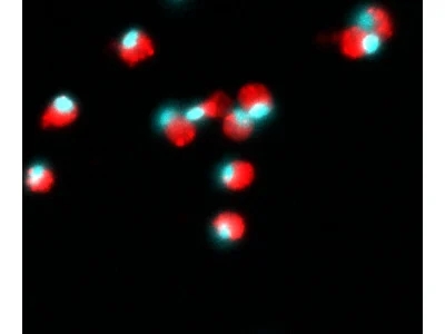
Anti-phospho Lyn (Y396) Rabbit Monoclonal Antibody

Excellent, submitted by on

Source: Biocompare.com

SKU P01424-1
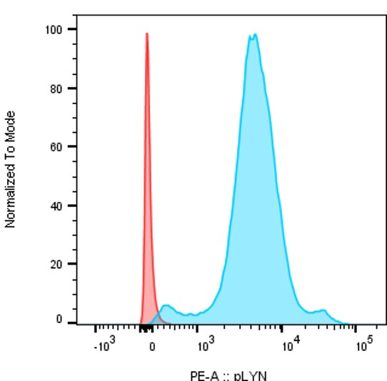
Application Flow Cytometry
Sample B cells
Primary Incubation 0.25 µl/10^6 cells 50min RT
Blocking Agent FBS 5%
Secondary Incubation F(ab')2-Donkey anti-Rabbit IgG (H+L), PE 0.1 µl/10^6
Detection Flow Cytometry
Results Summary Staining of B cells from mice bone marrow after cytoplasmic permezbiliation. The cells were first fixed and permeabilized with intracellular fix and perm set from ebioscience and then stained with 0,25 µl/ million cells with pLYN antibody (P01424-1) during 50min After, a seconde staining was performed with 0,1µl/million cells of F(ab')2-Donkey anti-Rabbit IgG (H+L), PE, Secondary Antibody from invitrogen. In red secondary antibody only and in blue primary anti-phospho lyn + secondary antibody.

Mettl23 Detection in Cell Lines

Excellent, submitted by on

Source: Biocompare.com

SKU DZ41142
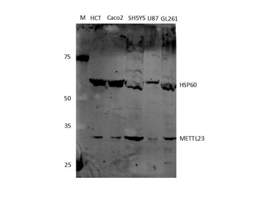
Application Western Blot
Sample Human cell lines
Primary Incubation 1:500 with 1% BSA in TBS, incubated overnight, at 4oC, with shaking
Blocking Agent 5% BSA in TBS
Secondary Incubation Licor IRDye 800 Goat anti Rabbit diluted 1:10000 with 1% BSA in TBS, incubated for 1 hour at RT , with shaking
Detection Licor Odyssey IR Scanner
Additional Notes The antibody has very good stability at room temperature (transit took over 2 weeks and the ice packs had long melted before delivery).
Results Summary The antibody gave a detectable band at the expected size for all cell lines tested with no non-specific bands.

"This antibody was used to determine the level of protein expression in various cell lines in order to set up two groups (high vs low expression). This antibody was custom made because numerous suppliers have either stopped their production for this antibody or quality/specificity was low."

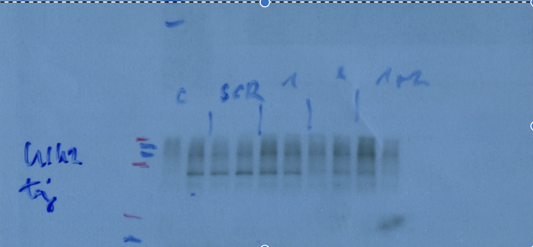
Anti-ULK2 Antibody Works Great for Western Blotting

Excellent, submitted by on

Source: Biocompare.com

SKU A05219
Application Western Blot
Sample HEK293T
Primary Incubation 1:5000 for 24hours at 4degrees celsius.
Blocking Agent Skim milk
Secondary Incubation 1 hour room temperature 1:3000
Detection ECL
Results Summary We silenced ULK1 and ULK2 genes in HEK293T cells, the antibody could differentiate the two proteins.

"Used for western blotting. The antibody worked good, used skim milk for blocking. It was used for investigating silenced HEK293T cells."

Beautiful Imaging of Zebrafish Eosinophils

Excellent, submitted by on

Source: Biocompare.com

SKU DZ41383
Application Immunofluorescence
Sample Zebrafish WKM, IPEX and FFPE tissue sections
Primary Incubation Overnight
Blocking Agent 3% goat serum in PBS
Secondary Incubation 1 hour
Tertiary Incubation None
Detection Fluorescence microscopy
DOI or PMID # https://doi.org/10.1101/2024.04.29.591640
Results Summary Antibody detects eosinophils and stains eosinophil granula. It is highly specific and can be used in dilutions such as 1:1000 for IF.

"We ordered this antibody as a custom antibody from Boster Bio to stain for zebrafish eosinophils in FFPE tissue sections and fixed cells. It works wonderfully: very bright, with no relevant background or off-target staining, and highly reproducible."

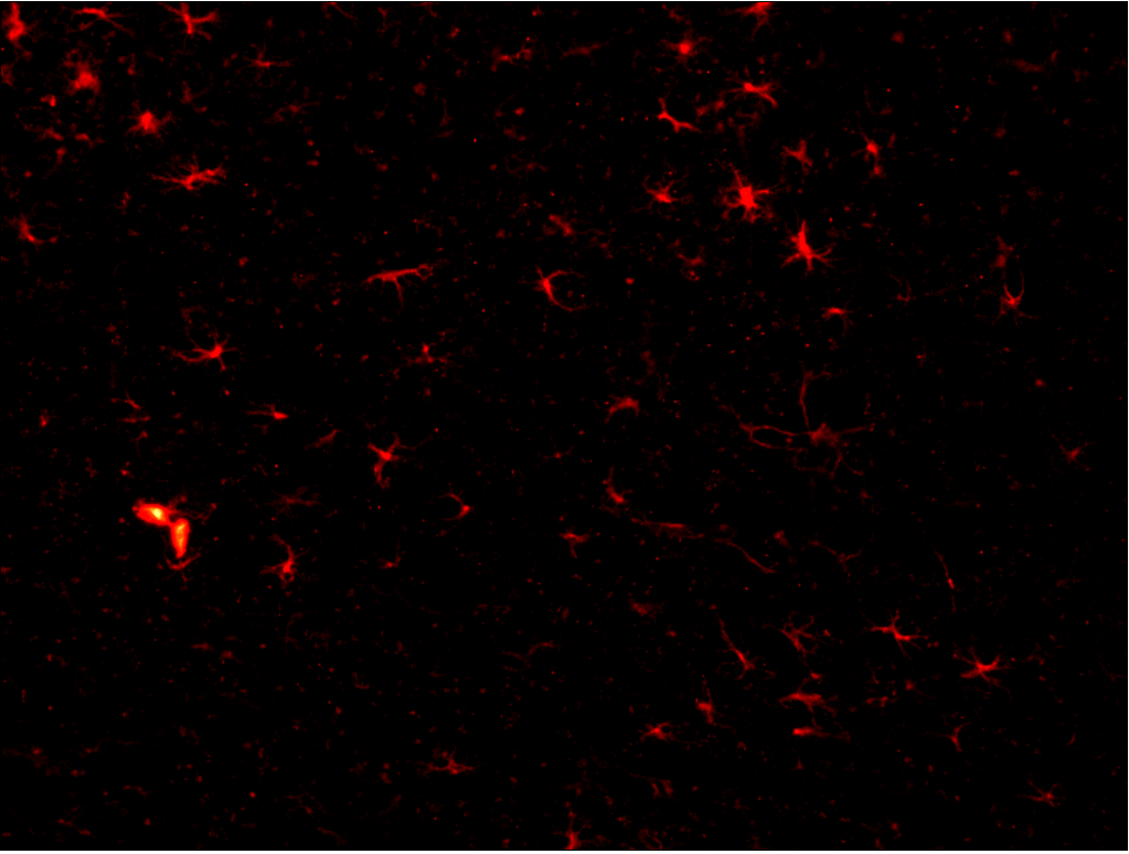
The antibody worked well even though we used fresh frozen brain sections cut at 20 micron thickness thaw-mounted onto microscope slide that were then post-fixed with 4% paraformaldehyde. The antibody worked equally well when used on fixed brains sectioned

Excellent, submitted by on

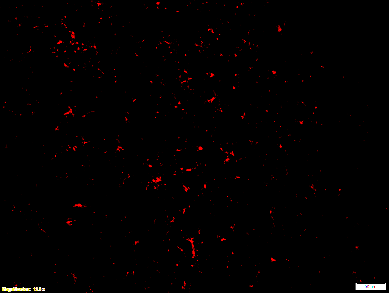
Boster bio GFAP 1:100 PFA (7-2) - cortex 12x - with scale bar


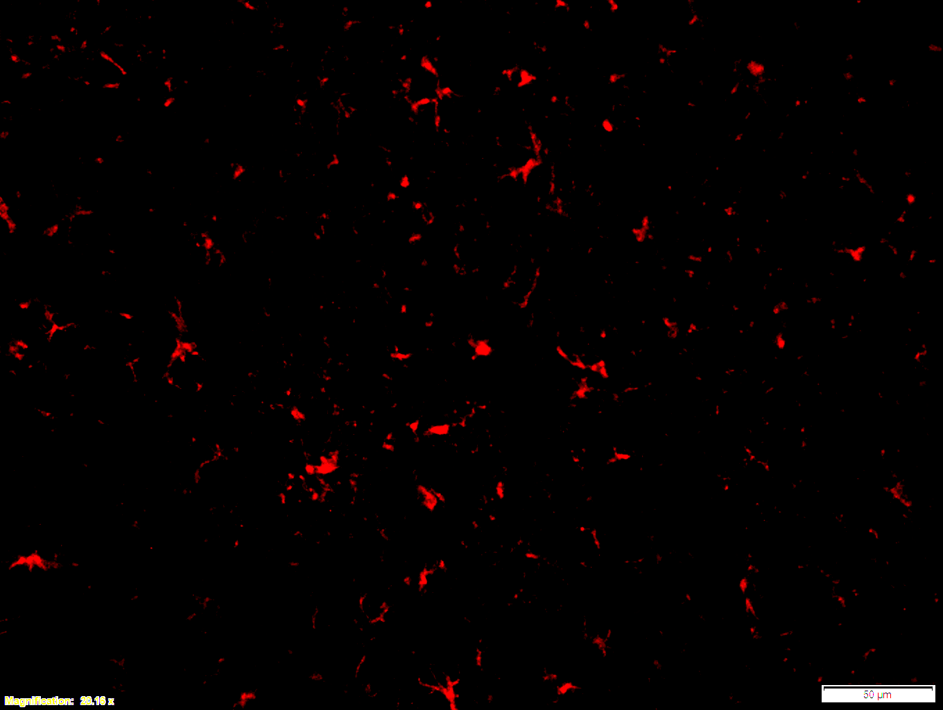
Boster bio GFAP 1:100 PFA (7-2) - cortex 20x - with scale bar


Boster bio GFAP 1:250 12x (free-floating)

SKU PB9082
Application Immunofluorescence
Sample Mouse brain
Primary Antibody Dilution 1:100

Images that were made from fresh frozen cryostat sections, were thaw-mounted onto microscope slides. Mounted sections were later post-fixed with 300 µL of 4% paraformaldehyde (PFA) in PBS at room temperature for 10 minutes. After fixation and prior to incubation with antibodies, slides were washed 3 times with 300 µL of 1X PBS with 0.01% sodium azide for 3 minutes per rinse, followed by permeabilization with 300 µL of 0.3% Triton X-100 in 1X PBS with 0.01% sodium azide for 30 minutes at room temperature, then blocking with 700 µL of 4% donkey serum diluted in 1X PBS with 0.01% sodium azide + Triton X-100 (blocking buffer) for 30 minutes at room temperature. Slides were incubated with 325 µL of GFAP (1:100). Concentrations of 1:100 were achieved by diluting 10 µL of antibody in 1,000 µL of blocking buffer; concentrations of 1:250 were achieved by diluting 4 µL of antibody in 1,000 µL of blocking buffer. Sections were incubated overnight at 4 ˚C. On the next day, slides were washed 3 times with 300 µL of 1X PBS with 0.01% sodium azide for 3 minutes per rinse, then incubated with the secondary antibodies at room temperature, in the dark, for 1 hour. Washing step was repeated then slides were left in the dark to dry. Mounting media was added to cover slip the sections. Slides were kept in the dark at 4 ˚C prior to imaging.

Immunohistochemistry was also performed with free-floating sections, which were exposed to the same primary antibodies diluted (1:250) in blocking buffer overnight and, subsequently, exposed to secondary antibodies diluted in blocking buffer for 1 hour, following the same procedure as the mounted sections. After incubation, sections were washed 3 times with 300 µL of 1X PBS with 0.01% sodium azide for 3 minutes per rinse and transferred with a painting brush to a container filled with 1X PBS with 0.01% sodium azide, from which they were mounted onto slides. Slides were left to dry in the dark, after which mounting media was added to cover slip the sections. As with slide-mounted sections, these slides were kept in the dark at 4 ˚C prior to imaging.


"Works well for immunofluorescence"

Excellent, submitted by on

Source: Customer Feedback Submission

"Works well for immunofluorescence"

--Jason Tennessen

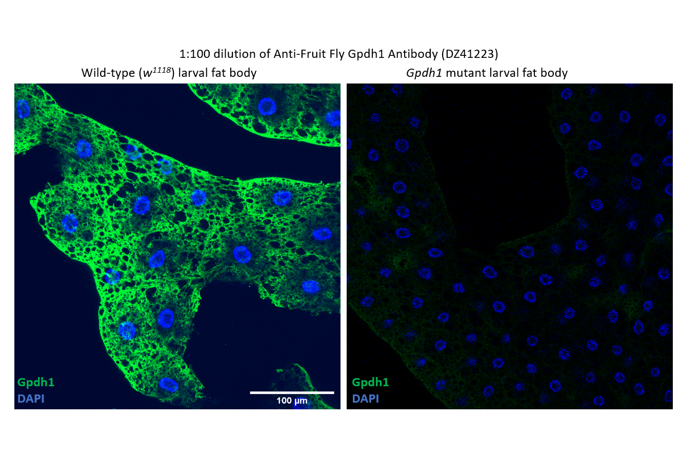
SKU DZ41223
Application IF
Sample Drosophila larval fat body

"We’ve verified the specificity of this custom antibody that Boster generated for us. The antibody "Anti-Fruit Fly Gpdh1 Antibody (DZ41223)” works well for immunofluorescence. A 1:100 dilution of this antibody stains fat body tissues of wild-type larvae but not Gpdh1 mutant. I’m happy to advertise this antibody to the fly community."

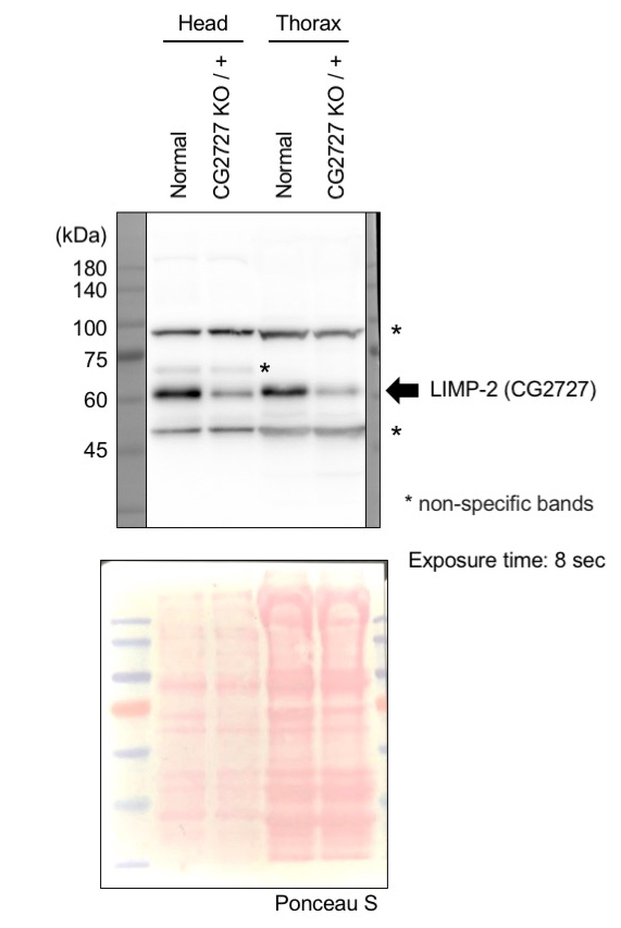
Western blot for Polyclonal Anti-Drosophila melanogaster (Fruit fly) emp Antibody

--Yuzuru Imai, Department of Research for Parkinson's Disease Juntendo University Graduate School of Medicine

Excellent, submitted by on
SKU DZ41152
Application Western blot
Sample Drosophila melanogaster (head and thorax)
Detection Immobilon Forte Western HRP substrate

"There are non-specific bands and immunostaining may not be possible, but Western blotting is sufficient with this antibody."