Customer Testimonials

Peer Reviews from Real Labs

Browse peer-submitted results linked to specific products—ratings, images, and practical notes included. Contribute your own result to strengthen the evidence base and claim rewards after verification.

Video Interviews

See real workflows and decision points—what researchers tried, what worked, and why. Share your story in a short interview and claim rewards!

Get Interview Rewards

Filter by category, reactivity, and host to find the most relevant peer results.

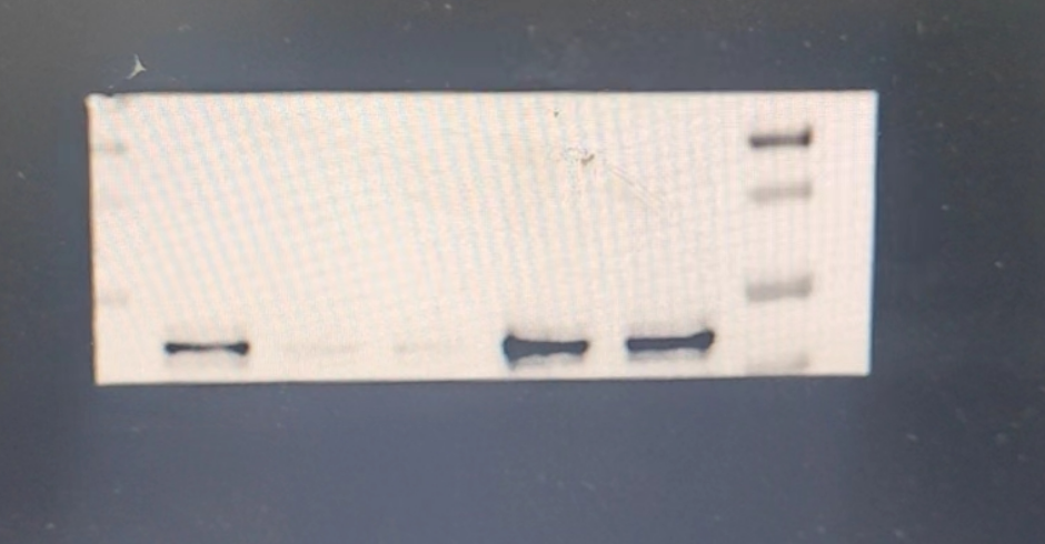
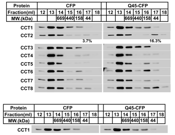
In this study, these two antibodies were primarily used for Western blot experiments. In the Western blot results, the CCT1 antibody showed a strong specific band and maintained a robust signal even after multiple reuses.

Excellent, submitted by on
SKU M02389
Application Western Blot
Sample U2OS cell
Sample Processing Description Cells were lysed with NP-40 lysis buffer to extract proteins. After quantification using the BCA assay, 5× loading buffer was added, and the samples were boiled for denaturation. Then, 20 µg of protein was loaded into each lane.
Primary Antibody Anti-TCP1 alpha Antibody Picoband® (monoclonal, 2E7)
Primary Incubation 1:1000, overnight at 4 ℃
Blocking Agent BSA
Secondary Antibody Goat Anti-Rabbit IgG (H+L) Secondary Antibody, Unconjugated (BA1039)
Secondary Incubation Incubate at room temperature for 1 hour
Detection Substrate: ECL substrate, Imaging system: ChemiDoc MP (Bio-Rad)
Results Summary I will purchase Boster products again and recommend them to my classmates and colleagues.

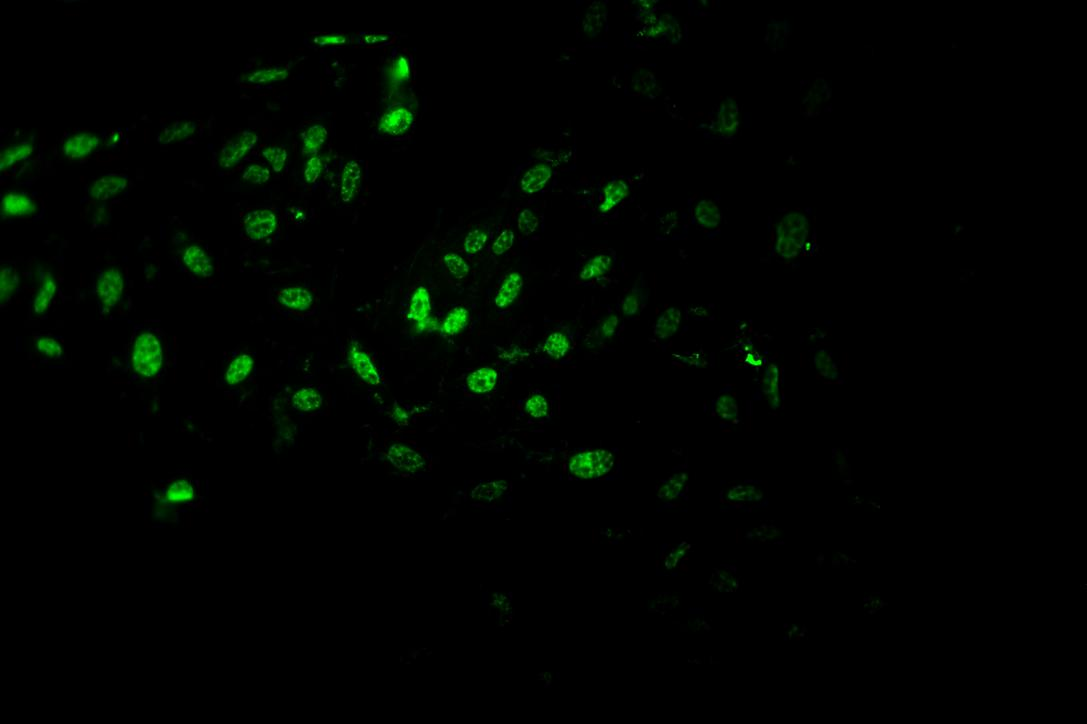
Highly prone to positive signals, with precise localization of expression

Excellent, submitted by on
MA1083 Immunofluorenscence
SKU MA1083
Application Immunofluorenscence
Sample Mouse MC-8 cells
Sample Processing Description 4% paraformaldehyde for 15 minutes
Primary Antibody Anti-PCNA Antibody (Monoclonal, PC 10)
Primary Incubation 1:1000, overnight at 4 ℃
Blocking Agent Goat serum
Secondary Antibody DyLight 550-conjugated goat anti-rabbit antibody.
Secondary Incubation Incubate at room temperature for 1 hour
Detection Laser confocal microscopy
Results Summary I will purchase Boster products again and recommend them to my classmates and colleagues.

This antibody is highly efficient and specific, with virtually no non-specific bands.

Excellent, submitted by on
A00284-1 Immunofluorenscence
SKU A00284-1
Application Immunofluorenscence
Sample Mouse cell climbing slides
Sample Processing Description Cells were directly lysed in NP40 lysis buffer, mixed with loading buffer at the appropriate ratio, and denatured by heating at 98°C. The samples were then loaded onto SDS-PAGE, with 20 μl of cell protein sample per lane.
Primary Antibody Anti-NF-kB p65/RELA Antibody Picoband®
Primary Incubation 1:2000, overnight at 4 ℃
Blocking Agent 5% Non-fat milk
Secondary Antibody HRP-conjugated Goat Anti-Rabbit IgG
Secondary Incubation Incubate at room temperature for 1 hour
Detection Imaging system: Tanon, ECL substrate
Results Summary I will purchase Boster products again and recommend them to my classmates and colleagues.

Highly prone to positive signals, with precise expression localization

Excellent, submitted by on
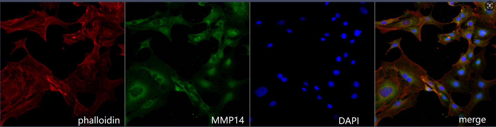
SKU M00656
Application Immunofluorenscence
Sample Mouse MC-8 cells
Sample Processing Description 4% paraformaldehyde for 15 minutes
Primary Antibody Anti-MMP14/Mt1 Mmp Rabbit Monoclonal Antibody
Primary Incubation 1:200, overnight at 4 ℃
Blocking Agent Goat serum
Secondary Antibody DyLight 488-conjugated Goat Anti-Rabbit IgG.
Secondary Incubation Incubate at room temperature for 1 hour
Detection Laser confocal microscopy
Results Summary I will purchase Boster products again and recommend them to my classmates and colleagues.

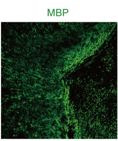
BOSTER’s rabbit anti-MBP antibody (catalog BA0094) exhibits high specificity and low background, enabling sensitive detection of demyelination and myelin regeneration processes, greatly facilitating this study.

Excellent, submitted by on
SKU PA1050
Application Immunofluorenscence
Sample Mouse spinal cord
Sample Processing Description Mouse spinal cord fixed with 2% paraformaldehyde for 6–8 hours, dehydrated in 30% sucrose, embedded in OCT, and sectioned using a cryostat.
Primary Antibody Anti-MBP9 antibody
Primary Incubation 1:100, overnight at 4 ℃
Blocking Agent 3% BSA
Secondary Antibody Anti-rabbit IgG-CY3 conjugated antibody.
Secondary Incubation Incubate at room temperature for 1 hour
Detection Immunofluorescence images were acquired using a confocal laser microscope (Leica SP8, Zeiss LSM 880 Airyscan).
Results Summary BOSTER’s rabbit anti-MBP antibody (catalog BA0094) has high specificity and low background, enabling sensitive detection of demyelination and myelin regeneration processes, greatly facilitating this study.

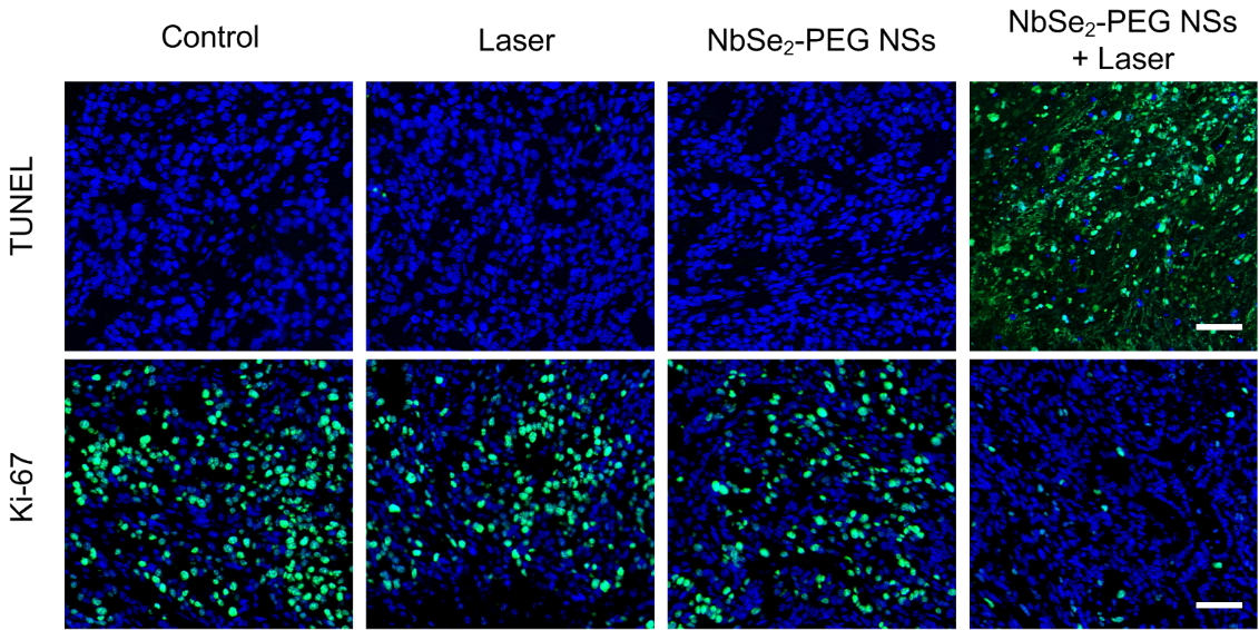
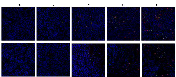
BOSTER’s Ki-67 antibody can effectively mark the proliferative activity of tumor cells. After treatment, a significant decrease in Ki-67 expression in tumor tissues can be clearly observed, indicating that the proliferative activity of tumor cells is mark

Excellent, submitted by on
SKU PB9026
Application Immunofluorenscence
Sample Cells in nude mouse tumor tissue
Sample Processing Description Sections of nude mouse tumor tissues fixed and embedded under different treatment conditions (Scale Bar = 100 μm)
Primary Antibody Anti-Ki67/MKI67 Antibody Picoband®
Primary Incubation 1:200, overnight at 4 ℃
Blocking Agent Goat serum
Secondary Antibody DyLight 488-conjugated goat anti-rabbit antibody.
Secondary Incubation Incubate at room temperature for 1 hour
Detection Fluorescence microscope
Results Summary The antibodies used in the experiment demonstrated good sensitivity and high cost-effectiveness, providing strong support for the smooth progress of the study.

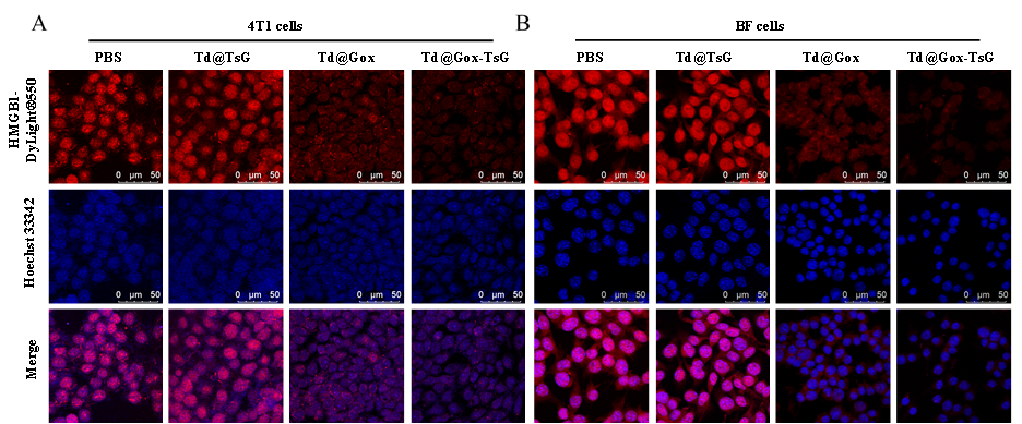
Accurate localization and good imaging quality

Excellent, submitted by on
SKU A00066-1
Application Immunofluorenscence
Sample Mouse 4T1 cell climbing slides
Sample Processing Description Mouse 4T1 cells fixed with 4% paraformaldehyde for 15 minutes
Primary Antibody Anti-HMGB1 Antibody Picoband®
Primary Incubation 1:500, overnight at 4 ℃
Blocking Agent Goat serum
Secondary Antibody DyLight 550-conjugated goat anti-rabbit antibody.
Secondary Incubation Incubate at room temperature for 1 hour
Detection Laser confocal microscopy
Results Summary The ordering and delivery cycle of the antibodies is very short — I was able to receive the products within a week, which saved a lot of time. The pre-sales and after-sales services are convenient and efficient. Since my background is in chemistry, I consulted Boster’s technical team about many details of biological experiments. The technical specialists were patient and thorough in their analysis and explanations, which helped me avoid many detours during the actual experimental procedures. Most importantly, these antibodies offer a high cost-performance ratio, with good binding performance, excellent imaging quality, and a high experimental success rate, providing a solid foundation for conducting related experiments.

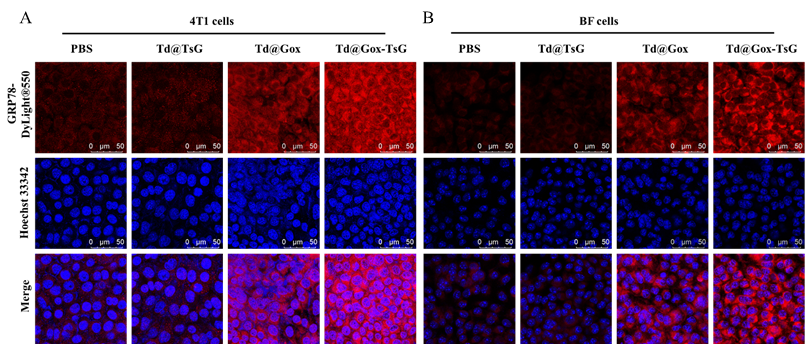
Accurate localization and good imaging quality

Excellent, submitted by on
SKU PB9640
Application Immunofluorenscence
Sample Mouse 4T1 cell climbing slides
Sample Processing Description Mouse 4T1 cells fixed with 4% paraformaldehyde for 15 minutes
Primary Antibody Anti-GRP78 antibody
Primary Incubation 1:400, overnight at 4 ℃
Blocking Agent Goat serum
Secondary Antibody DyLight 550-conjugated goat anti-rabbit antibody.
Secondary Incubation Incubate at room temperature for 1 hour
Detection Laser confocal microscopy
Results Summary The ordering and delivery cycle of the antibodies is short — I was able to receive the products within a week, which saved a lot of time. The pre-sales and after-sales services are convenient and efficient. Since my background is in chemistry, I consulted Boster’s technical staff about many details of biological experiments. The technical specialists were patient and helpful in their analysis and explanations, which allowed me to avoid many detours during the actual experimental procedures. Most importantly, these antibodies offer a high cost-performance ratio, with good binding performance, excellent imaging results, and a high experimental success rate, providing a solid foundation for conducting related experiments.

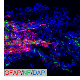
Accurate localization and good imaging quality

Excellent, submitted by on
SKU PA1239
Application Immunofluorenscence
Sample Rat Brain
Sample Processing Description Fixed in 4% paraformaldehyde for 48 hours, followed by paraffin embedding and sectioning.
Primary Antibody Anti-GFAP antibody
Primary Incubation 1:500, overnight at 4 ℃
Blocking Agent Goat serum
Secondary Antibody DyLight 550-conjugated goat anti-rabbit antibody.
Secondary Incubation Incubate at room temperature for 1 hour
Detection Laser confocal microscopy
Results Summary During the experiment, this primary antibody can be conveniently used with the complimentary antibody diluent provided. The results obtained showed good quality and reproducibility, with no false-positive signals. Overall, this primary antibody offers excellent cost performance and provides key data and strong support for publication.

Accurate localization and good imaging performance.

Excellent, submitted by on
SKU PB9093
Application Immunofluorenscence
Sample Mouse 4T1 cell xenograft tumor
Sample Processing Description Fixed in 4% paraformaldehyde for 48 hours, followed by paraffin embedding and sectioning.
Primary Incubation 1:200, overnight at 4 ℃
Blocking Agent Goat serum
Secondary Antibody DyLight 550-conjugated goat anti-rabbit antibody.
Secondary Incubation Incubate at room temperature for 1 hour
Detection Laser confocal microscopy
Results Summary The delivery time for antibodies is impressively fast — I usually receive the products within a week, which saves me a lot of valuable time. Both the pre-sales and after-sales support are efficient and reliable. Since my background is in chemistry, I often consulted Boster’s technical specialists about various details of biological experiments. They were always patient and thorough in their explanations, helping me avoid many detours during my experiments. Most importantly, these antibodies offer excellent value for money — they have strong binding performance, produce clear imaging results, and ensure a high experiment success rate, which has provided a solid foundation for my research.