Customer Testimonials

Peer Reviews from Real Labs

Browse peer-submitted results linked to specific products—ratings, images, and practical notes included. Contribute your own result to strengthen the evidence base and claim rewards after verification.

Video Interviews

See real workflows and decision points—what researchers tried, what worked, and why. Share your story in a short interview and claim rewards!

Get Interview Rewards

Filter by category, reactivity, and host to find the most relevant peer results.

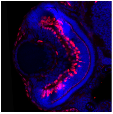
Was able to detect tfap2a expression in the retinal ganglion cells and anterior segment at 3dpf.

Excellent, submitted by on
SKU DZ41119
Application Immunofluorescence
Sample Zebrafish retinal cryo-section
Sample Processing Description Embryos fixed in 4% PFA for 4h. Embryos washed in PBST and 30% then 50% sucrose. Embedded in OCT and cryo-sectioned at 20nm
Primary Antibody Zebrafish Tfap2a Antibody
Primary Incubation 1:100, overnight at 4 ℃
Detection Used a Nikon C2+ confocal microscope
Results Summary Was able to detect tfap2a expression in the retinal ganglion cells and anterior segment at 3dpf.

This antibody exhibits high specificity and clear bands, and the resulting experimental data are reliable, providing important support for elucidating the molecular mechanisms underlying the role of PTN in this study.

Excellent, submitted by on
SKU P30433
Application Western Blot
Sample Mouse Spinal
Sample Processing Description Tissue samples were directly lysed in RIPA lysis buffer, mixed with loading buffer at the appropriate ratio, and denatured by heating at 98°C. Then, 20 μl of each cell protein sample was loaded into each lane of the SDS-PAGE gel.
Primary Antibody Phospho GRF-1 (Tyr1105) Antibody
Primary Incubation 1:1000, overnight at 4 ℃
Blocking Agent 5% non-fat milk
Secondary Antibody HRP-conjugated anti-rabbit IgG
Secondary Incubation Incubate at room temperature for 1 hour
Detection Substrate: ECL substrate, Imaging system: Sangon Biotech
Results Summary I will purchase Boster products again and recommend them to my classmates and colleagues.

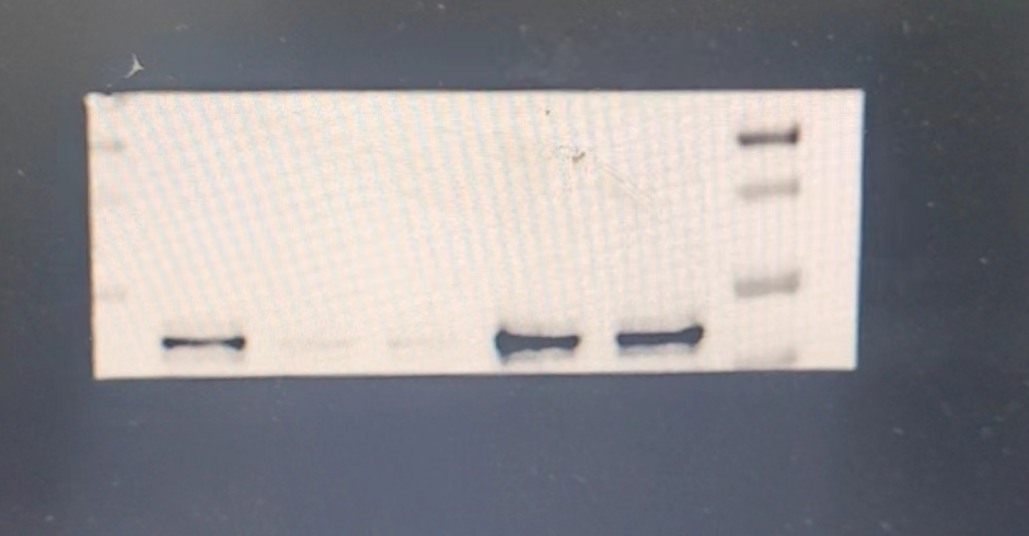
The staining is clear, highly specific, with almost no non-specific bands.

Excellent, submitted by on
SKU PA2290
Application Western Blot
Sample Protein extracts from LA795 and 16HBE cells.
Sample Processing Description After collecting the cells, lyse them with RIPA buffer containing protease inhibitors, quantify the protein using the BCA assay, and add loading buffer proportionally. Denature by heating at 98 °C. Load the samples onto SDS-PAGE, with 30 μg of total protein per lane.
Primary Antibody Anti-TMEM16A/ANO1 Antibody Picoband®
Primary Incubation 1:1000, overnight at 4 ℃
Blocking Agent 5% non-fat milk
Secondary Antibody HRP-conjugated goat anti-rabbit IgG
Secondary Incubation Incubate at room temperature for 1 hour
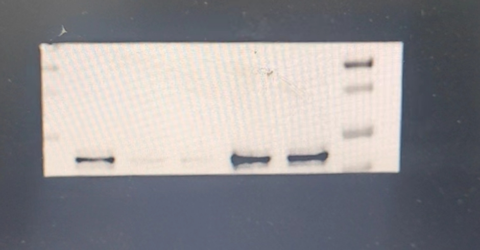
Detection Substrate: ECL substrate, Imaging system: Tanon
Results Summary The TMEM16A antibody we previously used was never ideal; despite long-term optimization, it was difficult to obtain clear bands. This time, by using BOSTER’s polyclonal antibody, we achieved much better experimental results. The bands were successfully exposed in essentially one attempt, with clear signals and no non-specific bands. We are very satisfied with Boster’s antibody.

I was so thrilled that I took the photo below — it felt like a true treasure.

Excellent, submitted by on
SKU PB9234
Application ICC/IF
Sample Endothelial cells
Sample Processing Description Endothelial cells were seeded in collagen gel-treated chips for 12 hours.
Primary Antibody Anti-ZO1 tight junction protein/TJP1 Antibody Picoband®
Primary Incubation 1:200, overnight at 4 ℃
Blocking Agent Ready-to-use Goat Serum
Secondary Antibody Goat Anti-Rabbit IgG (H+L) Secondary Antibody, DyLight®488 Conjugated(BA1127)
Secondary Incubation Incubate at room temperature for 1 hour
Other reagents DAPI (AR1176), Anti-fade mounting medium
Detection Fluorescence microscope
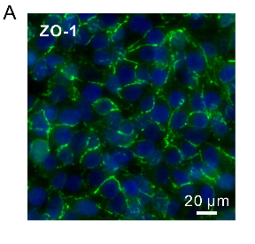
Results Summary At a critical stage of our manuscript preparation, we needed to stain tight junction proteins in endothelial cells. After trying primary antibodies from many companies without achieving satisfactory results, it was ultimately Boster’s product that solved the problem (Figure 5A). I was so excited that I took the photo below — treating it like a true treasure.

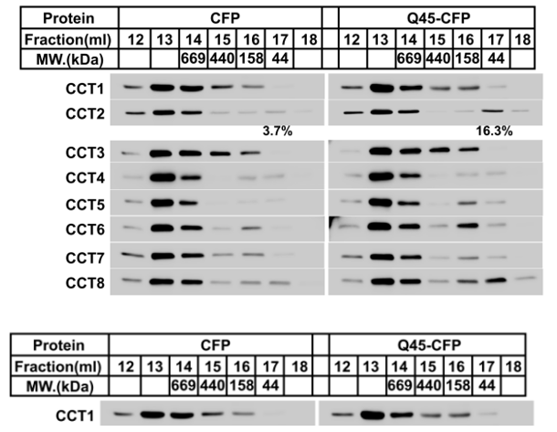
In this study, these two antibodies were primarily used for Western blot experiments. In the Western blot results, the CCT1 antibody showed a strong specific band and maintained a robust signal even after multiple reuses.

Excellent, submitted by on
SKU M02389
Application Western Blot
Sample U2OS cell
Sample Processing Description Cells were lysed with NP-40 lysis buffer to extract proteins. After quantification using the BCA assay, 5× loading buffer was added, and the samples were boiled for denaturation. Then, 20 µg of protein was loaded into each lane.
Primary Antibody Anti-TCP1 alpha Antibody Picoband® (monoclonal, 2E7)
Primary Incubation 1:1000, overnight at 4 ℃
Blocking Agent BSA
Secondary Antibody Goat Anti-Rabbit IgG (H+L) Secondary Antibody, Unconjugated (BA1039)
Secondary Incubation Incubate at room temperature for 1 hour
Detection Substrate: ECL substrate, Imaging system: ChemiDoc MP (Bio-Rad)
Results Summary I will purchase Boster products again and recommend them to my classmates and colleagues.

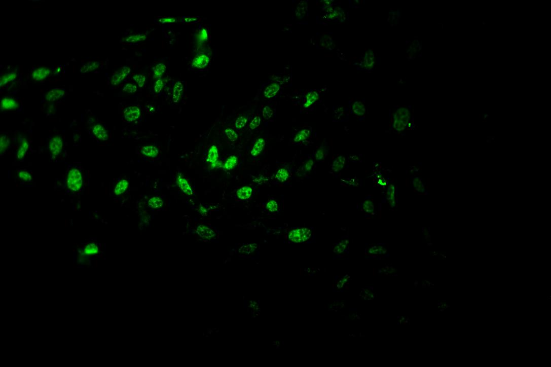
Highly prone to positive signals, with precise localization of expression

Excellent, submitted by on
MA1083 Immunofluorenscence
SKU MA1083
Application Immunofluorenscence
Sample Mouse MC-8 cells
Sample Processing Description 4% paraformaldehyde for 15 minutes
Primary Antibody Anti-PCNA Antibody (Monoclonal, PC 10)
Primary Incubation 1:1000, overnight at 4 ℃
Blocking Agent Goat serum
Secondary Antibody DyLight 550-conjugated goat anti-rabbit antibody.
Secondary Incubation Incubate at room temperature for 1 hour
Detection Laser confocal microscopy
Results Summary I will purchase Boster products again and recommend them to my classmates and colleagues.

This antibody is highly efficient and specific, with virtually no non-specific bands.

Excellent, submitted by on
A00284-1 Immunofluorenscence
SKU A00284-1
Application Immunofluorenscence
Sample Mouse cell climbing slides
Sample Processing Description Cells were directly lysed in NP40 lysis buffer, mixed with loading buffer at the appropriate ratio, and denatured by heating at 98°C. The samples were then loaded onto SDS-PAGE, with 20 μl of cell protein sample per lane.
Primary Antibody Anti-NF-kB p65/RELA Antibody Picoband®
Primary Incubation 1:2000, overnight at 4 ℃
Blocking Agent 5% Non-fat milk
Secondary Antibody HRP-conjugated Goat Anti-Rabbit IgG
Secondary Incubation Incubate at room temperature for 1 hour
Detection Imaging system: Tanon, ECL substrate
Results Summary I will purchase Boster products again and recommend them to my classmates and colleagues.

Highly prone to positive signals, with precise expression localization

Excellent, submitted by on
SKU M00656
Application Immunofluorenscence
Sample Mouse MC-8 cells
Sample Processing Description 4% paraformaldehyde for 15 minutes
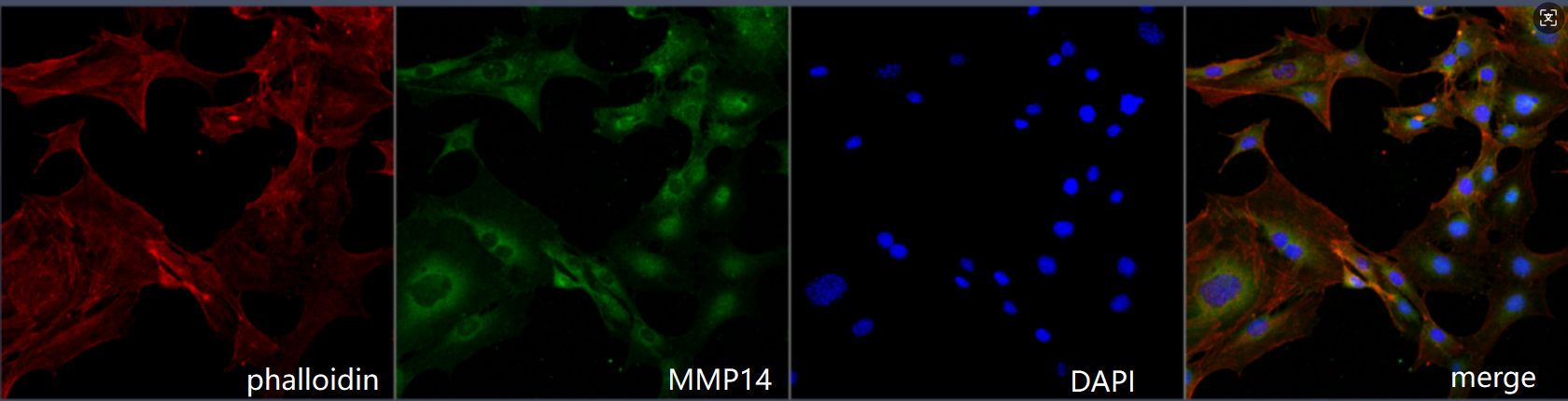
Primary Antibody Anti-MMP14/Mt1 Mmp Rabbit Monoclonal Antibody
Primary Incubation 1:200, overnight at 4 ℃
Blocking Agent Goat serum
Secondary Antibody DyLight 488-conjugated Goat Anti-Rabbit IgG.
Secondary Incubation Incubate at room temperature for 1 hour
Detection Laser confocal microscopy
Results Summary I will purchase Boster products again and recommend them to my classmates and colleagues.

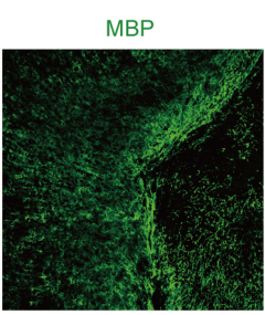
BOSTER’s rabbit anti-MBP antibody (catalog BA0094) exhibits high specificity and low background, enabling sensitive detection of demyelination and myelin regeneration processes, greatly facilitating this study.

Excellent, submitted by on
SKU PA1050
Application Immunofluorenscence
Sample Mouse spinal cord
Sample Processing Description Mouse spinal cord fixed with 2% paraformaldehyde for 6–8 hours, dehydrated in 30% sucrose, embedded in OCT, and sectioned using a cryostat.
Primary Antibody Anti-MBP9 antibody
Primary Incubation 1:100, overnight at 4 ℃
Blocking Agent 3% BSA
Secondary Antibody Anti-rabbit IgG-CY3 conjugated antibody.
Secondary Incubation Incubate at room temperature for 1 hour
Detection Immunofluorescence images were acquired using a confocal laser microscope (Leica SP8, Zeiss LSM 880 Airyscan).
Results Summary BOSTER’s rabbit anti-MBP antibody (catalog BA0094) has high specificity and low background, enabling sensitive detection of demyelination and myelin regeneration processes, greatly facilitating this study.

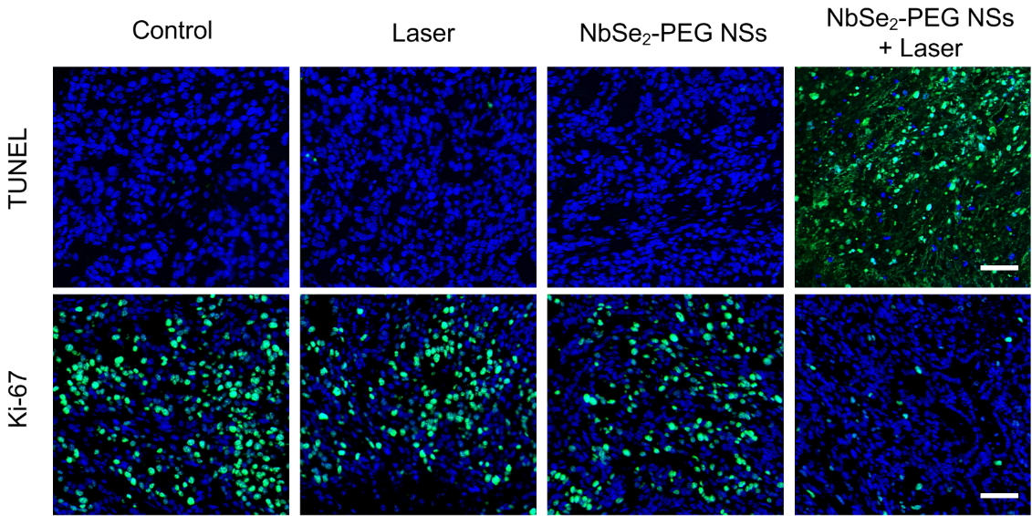
BOSTER’s Ki-67 antibody can effectively mark the proliferative activity of tumor cells. After treatment, a significant decrease in Ki-67 expression in tumor tissues can be clearly observed, indicating that the proliferative activity of tumor cells is mark

Excellent, submitted by on
SKU PB9026
Application Immunofluorenscence
Sample Cells in nude mouse tumor tissue
Sample Processing Description Sections of nude mouse tumor tissues fixed and embedded under different treatment conditions (Scale Bar = 100 μm)
Primary Antibody Anti-Ki67/MKI67 Antibody Picoband®
Primary Incubation 1:200, overnight at 4 ℃
Blocking Agent Goat serum
Secondary Antibody DyLight 488-conjugated goat anti-rabbit antibody.
Secondary Incubation Incubate at room temperature for 1 hour
Detection Fluorescence microscope
Results Summary The antibodies used in the experiment demonstrated good sensitivity and high cost-effectiveness, providing strong support for the smooth progress of the study.