| SKU | PB9093 |
|---|---|
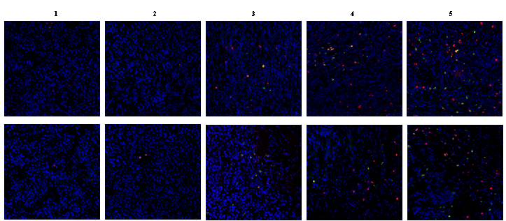
| Application | Immunofluorenscence |
| Sample | Mouse 4T1 cell xenograft tumor |
| Sample Processing Description | Fixed in 4% paraformaldehyde for 48 hours, followed by paraffin embedding and sectioning. |
| Primary Incubation | 1:200, overnight at 4 ℃ |
| Blocking Agent | Goat serum |
| Secondary Antibody | DyLight 550-conjugated goat anti-rabbit antibody. |
| Secondary Incubation | Incubate at room temperature for 1 hour |
| Detection | Laser confocal microscopy |
| Results Summary | The delivery time for antibodies is impressively fast — I usually receive the products within a week, which saves me a lot of valuable time. Both the pre-sales and after-sales support are efficient and reliable. Since my background is in chemistry, I often consulted Boster’s technical specialists about various details of biological experiments. They were always patient and thorough in their explanations, helping me avoid many detours during my experiments. Most importantly, these antibodies offer excellent value for money — they have strong binding performance, produce clear imaging results, and ensure a high experiment success rate, which has provided a solid foundation for my research. |