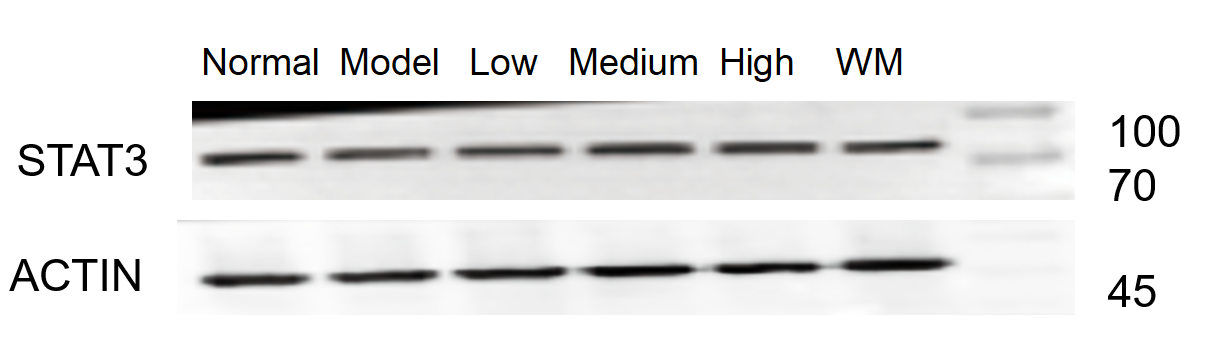
| SKU | M01394-4 |
|---|---|
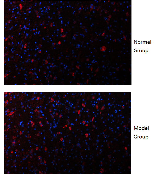
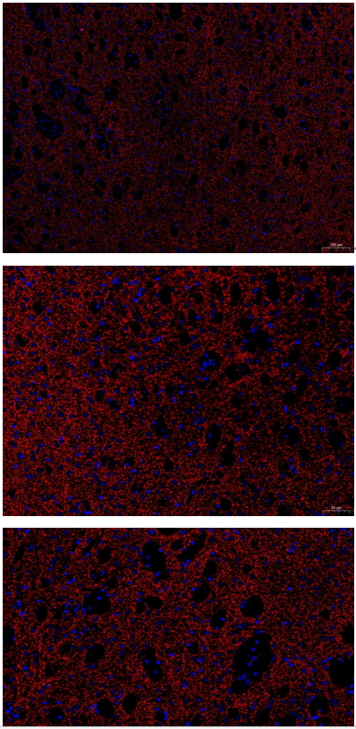
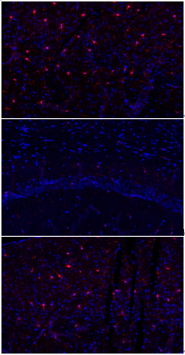
| Application | Immunofluorescence |
| Sample | mouse brain tissue |
| Sample Processing Description | Mouse cerebral infarction model; brain tissues were collected, fixed in formaldehyde for 48 hours, and then sagittally paraffin-embedded. |
| Other Reagents | Goat serum, DAPI Staining Solution, Antifade fluorescence mounting medium. |
| Primary Antibody | Iba1 Rabbit Monoclonal Antibody |
| Primary Incubation | 1:100, overnight at 4 ℃ |
| Secondary Antibody | Goat Anti-Rabbit IgG (H+L) Secondary Antibody, Fluoro594 Conjugated (BA1142, Boster) |
| Secondary Incubation | 45 min at 37℃ |
| Detection | Imaging system:Leica DMi3000 |
| Results Summary | IBA-1 is a well-established marker of microglia in the nervous system. Microglia are the primary immune effector cells in the central nervous system and respond rapidly to CNS injury by proliferating, upregulating or re-expressing MHC antigens, migrating, and adopting a phagocyte-like morphology. In this study, IBA-1 was used to label microglia in cerebral infarction samples, showing clear staining and accurate cellular morphology. |