| SKU | M01917 |
|---|---|
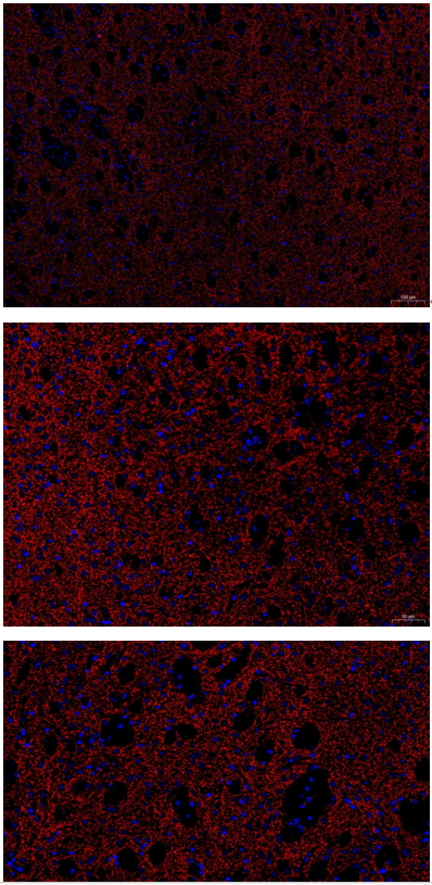
| Application | Immunofluorescence |
| Sample | mouse brain |
| Sample Processing Description | Sagittal sections of mouse striatum were fixed in formaldehyde for 48 hours and paraffin-embedded. |
| Other Reagents | Goat serum,DAPI,Anti-fade mounting medium |
| Primary Antibody | Iba1 Rabbit Monoclonal Antibody |
| Primary Incubation | 1:200, overnight at 4 ℃ |
| Secondary Antibody | 1:500, Goat Anti-Rabbit IgG (H+L) Secondary Antibody, Fluoro594 Conjugated |
| Secondary Incubation | 45 min at 37℃ |
| Detection | Imaging system:Leica DM2500 |
| Results Summary | TH is a marker of catecholaminergic neurons and can specifically label and identify dopaminergic, noradrenergic, and adrenergic neurons. In this experiment, TH immunofluorescence was performed to observe the distribution of dopaminergic neurons in the striatum. The results show clear staining and precise localization. |